Introduction to Body Cavities Flashcards
what plane is this?
sagittal
what plane is this?

transverse
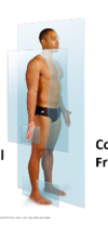
what is the name of this plane?

coronal plane
how do u look at an axial CT or MRI?
is if standing by bedside at feet

SO: Left hand site of CT = right hand side of patient
what is the cranial cavity continious with?
cranial cavity continues into vertebral canal

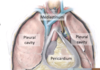
which subcavities are found in the thoracic region?
thoracic cavity:
- pleural cavity (lungs)
- mediastinum (heart, esophagus, trachea, thoracic nerves and systemic blood vessels)
- pericardial cavity (inside the mediastinum) (heart)

what surrounds and protects the brain (inside the skull) - where do they go?
3 membranous layers: meninges - continue down vertebral column:
- dura mater (outer layer)
- arachnoid mater (middle layer - spider leg layer)
- pia meter (closest to brain - follows the contours of the brain and spinal cord

what is found betwen the arachnoid and pia meter layers
role of ^?
sub arachnoid space: filled with cerbrospinal fluid
- role: buffers and protects the brain and spinal scord
label this correctly


what is meningitis?
how test?
inflammation of the meninges
test: test CSF via lumbar puncture
what are serous membranes?
serous membranes: sealed, two layered internal cavities of the body. filled with serous fluid:
2 layers are continous:
a) partietal: line body cavity and share nerve supply to body wall: somatic
b) visceral: cover the organ and share same NS to organ: autonomic
which are the main three serous membranes?
- pleural cavity: serous membranes of lung
- pericardium: serous membrane o fheart
- peritoneum: serous membrane of (continuous membrane which lines the abdominal cavity and covers the abdominal organs)
how many pleural cavities are there?
whats in the mediastinum?
whats the pericardium
- 2 - 2 lungs lol
- mediastinum: where following are collected: heart, great vessels, trachea, oesophagus etc
- peridcardium: a thin sac that surrounds your heart.
what is inbetween the viesceral and parietal pleura?
function of ^?
- pleural fluid
-
function:
- reduces friction of expansion / depression
- help stick viseral pleura to parietal pleura

describe the structure of the pericardium
- viseral pericardium (surface of heart)
- parietal pericardium (ourtside of visc)
- parietal cavity: space between ^. filled with fluid
- fibrous pericardium: fibrous sack

why is it bad if fibrous pericardium gets fluid in it?
bc cannot expand - so fluid drains into sac and builds up and compress the heart = cardiac tampondade
what are these?

A - left lung
B - pleural cavity
C - oesophagus
D - thoracic aorta
E - peridcardium

how do you split up the regions of the abdomen?

what is the serous membrane that lines the abdominal cavity?
peritoneum: has a visceral and parietal layer. BUT bc visercal layer folds to contain lots of different organs - get lots of different folds in peritoneum
label these

phyloric sphincter controls movement of food into duodenum

which organ does duodenum curl around?
head of the pancreas - pancreatic and bile ducts drain into duodenum

which ducts drain into the duodenum?
- pancreatic duct
- bile duct
where is pancreas found?
is the pancreas an exocrine or endocrine gland?
location: posterior abdomen wall
both !


