Muscle Histology Flashcards
Name the three muscle types.
skeletal muscle, cardiac muscle, smooth muscle
skeletal muscle
attaches to bone, ligaments, the dermis of facial, skin, and the eyeballs
also called striated muscle because of actin and myosin filaments
muscle cells are a very long syncytium with peripheral nuclei
10-100 microns in diameter with
motor unit
number of muscle cells innervated by one neuron
cardiac muslce
striated and restricted to the heart
differs from skeletal muscle in that cells branch, have central nuclei, and are attached to each other with intercalated discs
contraction is initiated within the conduction system but regulated by the autonomic nervous system
smooth muscle
lines the walls of blood vessels and hollow organs
controls blood pressure, contractions of the stomach, peristalsis in the GI tract, and childbirth
smaller than skeletal and cardiac muscle
spindle shaped, central nuclei
contraction is slower, requires less energy, and can result form nerve impulses, hormones, and response to stretch

molecular mechanism of muscle contraction
the rachetlike sliding of actin and myosin myofilaments alongside each other
role of calcium in muscle contraction
required for binding of actin and myosin filaments in all three muscle types
mechanism is different for smooth muscle
fascicles
groups of muscle fibers, which are the individual cells
endomysium
thin connective tissue layer surrounding the fibers
perimysium
surrounds fascicles and forms larger septa within muscles
epimysium
connective tissue surrounding the whole muscle
myofibrils
filamnets of thick and thin myofilaments that pack muscle fibers
three components of thin filaments
actin, tropomyosin, and a troponin complex
f-actin
a long double-stranded helix formed from the polymerization of g-actin
tropomyosin
a double helix that runs in the groove between F-actin molecules
troponin complex and its three components
plays an important role in the attachment of the myosin thick filaments
made up of troponin-C, tropnon-T, and troponin-I
Troponin-C
binds calcium and allows myosin to bind with actin
troponin-T
anchors the complex to tropomyosin
troponin-I
binds actin to inhibit myosin-actin interactions
Describe the structure of the sarcomere.
I band = actin myofilaments
H band = isolated myosin in A band
M line = myosin cross-linked
A band = kength of myosin molecules, here overlapping with actin
Z line = boundaries of a sarcomere

I band
actin filaments by itself
Z line bisects the I band
H band
isolated myosin in the A band
M line
myosin-cross linked with each other by myomesin and C protein
stabilizes the myosin and keeps them in register
A band
length of myosin molecules, overlapping with actin
length remains constant during contraction of the sarcomere
Z line
boundaries of sarcomere
composed of alpha-actinin
thin filaments are anchored to the Z line and thick filaments are attached indirectly by titin
titin
a molecules that connects myosin to the Z-line
coiled appearance, has elastic properties
excitation-contraction coupling
the linking of the external neural signal to the internal mobilization of calcium to initiate a contraction cycle
dystrophin protein/glycoprotein complex
links f-actin/cell interior with extracellular proteins laminin and agrin
just under the cell membrane, transmembrane glycoproteins connect it with the extracellular proteins
sarcoplasm
cytoplasm that surrounds myofibrils
contains mitochondria, glycogen, myoglobin, and and extensive network of ER
transverse T tubules
invaginations of muscle cell membranes that are flanked two cysterns of ER
brings the depolarization into the interior of the cell at the level of the A-I junction
triad
the combination of a T-tubule and two adjacent terminal cisternae of sarcoplasmic reticulum that responds to depolarization of the membrane and releases calcium into the sarcoplasm
How is calcium released in response to membrane depolarization?
L-type Ca2+ channels open in response to membrane depolarization
done through mechanical coupling

major steps of the contraction cycle
depolarization of the sarcolemma, mobilization of intracellular calcium, and the hydrolysis of ATP
Describe the contraction cycle.
two important effects of P released form myosin head:
head binds more tightly to actin
head unbends to its original conformation, which is the power stroke

rigor configuration
the orientation of the myosin heads without the presence of ATP or ADP
bound tightly to actin
power stroke
the process of ADP and phosphate detaching from the myosin head, which moves the head to its original conformation
motor unit
the number of muscle fibers supplied by one neuron
force of contraction is determined in part by how many motor units are recruited by the central nervous system
neuromuscular junction
characterized by a motor end plate, the terminal portion of an axon branch that sits in a recess of the sarcolemma called the synaptic cleft
myelin sheath ends, and only a thin layer of Schwann cell cytoplasm overlies the end plate
What is the neurotransmitter used in motor innervation?
acetylcholine
Describe the process of depolarization of the muscle cell.
acetylcholine is released from the neurons
ACh-gated Na+ channels open
Volatage-gated Na+ channels open
Na+ enters the cell and depolarization spreads across the plasma membrane
myasthenia gravis
an autoimmune disease where antibodies block the acetylcholine receptor sites on the sarcolemma
sensory innervation in skeletal muscle
consists of general sensation including pain, but also encapsulated sensory receptors that sense stretch and tension in muscle
proprioception
sensory input that helps the brain determine movement and positions int he body
components of the muscle spinder
nuclear bag fiber
nuclear chain fiber
afferent nerve endings
internal capsule
nuclear bag fiber
specialized muscle cell with an expanded section containing many nuclei
nuclear chain fiber
specialized muscle cell with a section containing a series of aligned nuclei
golgi tendon organs
encapsulated receptors similar in function that are found in tendons
organization of heart muscle
similar to skeletal muscle but the nucleus is central, myofibrils are not discrete, mitochondria are large, T-tubules are lare at the Z line, and there are diads instead of triads
interalatred disks
dense-staining junctions that have step-like interdigitations with fascia adherens junctions, macula adherens junctions, and gap junjunctions
also serve as anchors for the thin actin filaments
How does the depolarization of the heart differ from that of skeletal muscle?
T-tubules penetrate group sof myofibrils at the level of the Z line
myofibrils and sarcoplasmic reticulum are less organized
calcium-triggered calcium release
calcium-triggered calcium release mechanism
calcium passing from the lumen of the T-tubule into sarcoplasm is an absolute requirement for contraction
depolarization of the cell and T-tubule membranes open voltage-sensitive calcium channels that open the SR calcium channels nearby
the amount of calcium released in cardiac muscle is dependent on the amount of calcium that enters the cell, whereas in skeletal muscle, it depends on the frequency of membrane polarizations
purkinje fibers
specialized cells in the heart that can carry an impulse faster than heart muscle cells
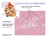
Where does a heartbeat start?
in pacemaker cells in the sinuatrial node in the right atrium
two hormones secreted by heart muscle cells and their functions
atrial natriuretic factor (ANF) and brain natriuretic factor (BNF) - both are diuretics that affect sodium excretion, act on kidney, adrenal gland, and vascular smooth muscle to lower blood pressure
dense bodies
the equivalent of Z-lines in smooth muscle cells
they are the attachment sites for thin filaments and contain alpha-actinin within a reticulum of intermediate filaments (desmin and viementin)
caveolae
numerous invaginationso f the cell membranes that replace T-tubules in smooth muscle cells along with vesicles that provide a route of entry of calcium into the cell
initiation of actin-myosin interactions in smooth muscle
result from the phosphorylation of myosin heads rather than calcium acting on the troponin complex, which is absent in smooth muscle
three mechanisms that initiate contraction in smooth muscle cells
voltage-sensitive calcium channels in the cell membrane that open from autonomic impulses, no neuromuscular junctions so neurotransmitters diffuse to initiate depolarization
stretching of smooth muscle opens mechanosensitive calcium channels
hormone receptors and other signals trigger the formation of second messengers that act on gated channels in the ER to release calcium

Ca++-calmodulin-myosin light chain kinase
in the presence of calcium, regulates contraction by phosphorylating regulatory light chains on the myosin heads to expose actin-binding sites
latch state
where smooth muscles remains contracted for long periods of time with minimal ATP expenditure
after myosin attaches to actin, the heads are dephosphorylated, which results in a decrease in aATPase activity, and the myosin head is unable to detach from actin
smooth muscle relaxation
results from the synergistic action of calcium efflux from the cell, membrane hyperpolarization, and reduced sensitivity to calcium
can be triggered through specific receptors with their own G protein and second messenger pathways that results in the phosphorylation of many proteins
epinephrine
activates a beta2-receptor cAMP pathway in skeletal muscle arterioles
relaxes smooth muscles in blodo vessels of the GI tract to contract and skeletal muscle vasculature to relax
nitric oxide
an important vasodilator
diffuses into vascular smooth muscle cells to act via a cGMP pathway
