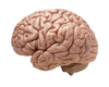
Introduction and basic topography Flashcards
What are the basic components of the central nervous system?
- Cerebral hemispheres
- Brainstem and cerebellum
- Spinal cord
What are the basic components of the peripheral nervous system?
- Dorsal and venral roots
- Spinal nerves
- Peripheral nerves
- Cauda Equina
How does the cell type that myelinates neurones differ in the CNS vs PNS?
CNS: oligodendrocites myelinate - cannot regenerate - multiple neurones myelinated by one cell
PNS: Schwaan cell - have some capacity to regenerate

What makes up grey matter?
Cell bodies and dendrites
Highly vascular
What makes up white matter?
White matter is composed of axons and their supporting cells
How does grey matter communicate with white matter?
Grey matter contains axons allowing it to communicate with white matter
Why does white matter appear white?
Due to presence of fatty myelin
What is the PNS equivalent of grey matter and white matter?
Grey matter = a Ganglion
White matter = peripheral nerve
How is white and grey matter arranged in the spinal cord?
Grey matter is central with an outer layer of white matter
How many segments of the spinal cord are there?
31
What is a funiculus?
What are the 3 divisions
A segment of white matter containing multiple distinct tracts
Impulses travel in multiple directions
3 divisions: Dorsal, Lateral and Ventral

What is a tract?
An anatomically and functionally defined white matter pathway that connects two distinct regions of grey matter
Impulses travel in one direction
What is a fasciculus?
A subdivision of a tract supplying a distinct region of the body
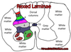
How is grey matter in the spinal cord organised?
Organised in cell columns (Rexed’s Laminae)
What is a nucleus (grey matter)?
A collection of functionally related cell bodies

What is the cortex (grey matter)?
A folded sheet of cell bodies found on the surface of a brain structure
Typically 1-5 mm thick

What is a fibre (white matter)?
A term relating to an axon in association with is supporting cells
What are the 3 types of fibre (white matter)?
- Association - connect cortical regions within the same hemisphere
- Commissural- connect left and right hemispheres or cord halves
- Projection - connect cerebral hemispheres with the cord/ brain stem and vice versa

What are the 3 components of the brainstem and the basic functions of each?
-
Midbrain (misencephalon)
- Eye movement and reflex responses to sound and vision
-
Pons
- Feeding and sleep (particularly REM)
-
Medulla
- Cardiovascular and respiratory centres
- Contrains major motor pathways (medullary pyramids)
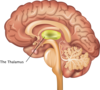
What components make up the Diencephalon?
Thalmus and Hypothalmus

What are colliculi?
Reflex centres giving rapid response at the back of the midbrain
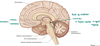
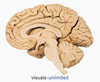
Identify the central sulcus


Locate the pre central gyrus and post central gyrus and give the function of each

Precentral- primary motor cortex
Postcentral- primary sensory cortex

Identify the lateral/ sylvian fissure