Week 8: Nerves of Thigh and Leg Flashcards

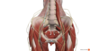
Structure: Iliohypogastric n.
Lateral cut. branches.
Anterior cut. branches.
Innervates the:
- Transversus abdominis.
- Internal oblique.
- Posterolateral gluteal and skin above pubis

Structure: Ilioinguinal N.
Origin: L1, ventral division
Innervates:
- Interal Oblique
- skin of superomedial thigh and genital area
Structure: Genitofemoral N.
Origin: L1, L2 ventral divisions
Innervates: cutaneous skin over femoral triangle and genital area

Structure: Lateral cutaneous nerve of the thigh
aka lateral femoral cutaneous n.
Origin: L2, L3 dorsal divisions
Innervates: Cutaneous skin over the lateral aspect of the thigh down to the knee.

Structure: Femoral Nerve
Origin: L2, L3, L4
Supplies:
- Iliacus, psoas major, sartorius, pectineus, rectus femoris, vastus lateralis, medialisand intermedialis. It also gives articular branches to the hip and knee joints.
- skin over the anterior and lateral thigh and the medial leg and foot.
Structure: Obturator N.
Origin: L2, L3, L4
Supplies:
Motor: adductors of the thigh: adductor magnus, adductor longus, adductor brevis, obturator externus, and gracilis.
Sensory: hip and knee joints and cruciate ligaments.
Cutaneous: skin of the medial side of the thigh.

Structure: Pudendal N.
Origin: S2, S3, S4: ventral divisions.
Innervates:
Motor: perineal muscles.
Cutaneous: skin of the perineum.

Structure: Saphenous N.
Origin: Femoral N.
Supplies: skin of anteromedial aspect of leg and knee
Structure: Superior gluteal nerve
Origin: Sacral plexus (post. divisions of ant. rami of L4-S1 spinal nerves)
Supplies:
- gluteus medius
- gluteus minimus
- tensor fasciae latae
Structure: Inferior gluteal nerve
Origin: Sacral plexus L5-S2
Supplies: gluteus maximus

Structure: Sciatic N.
Origin: sacral plexus L4-S3
Supplies:
semitendinous
semimembranosus
biceps femoris
adductor magnus
skin of leg and foot
all muscles of leg and foot

Structure: Posterior femoral cutaneous n.
Origin: Ventral spinal rami S1-S3
Supplies:
- skin of posterior thigh and knee
- scrotum
- labia majora

Structure: Tibial nerve
Origin: sciatic nerve
Supplies:
- posterior muscles of leg and knee joint
- skin of plantar foot and toes

Structure: common fibular nerve
Origin: sciatic nerve
Supplies:
- knee joint via its articular branch
- skin on lateral part of posterior aspect of leg via lateral sural cutaneous nerve

Structure: Deep Fibular N.
Origin: common fibular nerve
Supplies:
- tibialis anterior
- fibularis tertius
- extensor digitorum longus
- extsor hallucis longus

Structure: Superficial fibular nerve
Origin: common fibular nerve
Supplies:
- fibularis longus and brevis
- skin on distal 1/3 of anterior surface of leg and dorsum of foot
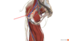
Identify the nerve highlighted in orange and indicated by the red arrow

Structure: Sural Nerve
Origin: branches of both tibial and common fibular nerves
Supplies:
- skin on posterior and lateral aspects of leg
- lateral side of foot
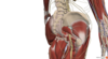
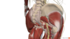
Identify the nerve highlighted in orange and indicated by the red arrow

Structure: Lateral Plantar nerve
Origin: smaller terminal branch of tibial nerve
Supplies:
- quad. plantae
- abductor digiti minimi
- digiti minimi brevis
- deep branch supplies dorsal interossei, lateral three lumbricals and adductor hallucis,
- skin on sole lateral to a line splitting 4th digit

Structure: Medial plantar nerve
Origin: Larger terminal branch of tibial nerve
Supplies:
- abd. hallucis
- flex. digitorum brevis
- flex. hallucis brevis
- first lumbrical
- skin of medial side of sole of foot and sides of first three digits
