W15 - Lower GI pathology Flashcards
What does congenital atresia mean?
atresia = parts are not fully formed. for example duodenal atresia!
Congenital hirschsprung’s disease
Explain the pathophysiology
Absence of ganglion cells in myenteric plexus = distal colon fails to dilate!
Congenital hirschsprung’s disease
Explain the symptoms (4)
- Constipation
- Abdominal distension
- Vomiting
- overflow diarrhoea
5.
Congenital hirschsprung’s disease
- Male:Female ratio?
- Associated diseases?
- 80:20 M:F
- Associated with Down’s syndrome (2%)
What is the treatment of Hischsprung’s disease? How to know if fully treated?
Treatment = resection of affected (constricted) segment until we reach segment that have ganglion
affected region is = hypertrophied nerve fibers but no ganglia
Define a volvulus
–Complete twisting of a loop of bowel at mesenteric base, around vascular pedicle, causing intestinal obstruction +/- infarction
________ volvulus is more common in infants, and _________ volvulus is more common in elderly
small bowel volvulus is more common in infants, and sigmoid volvulus is more common in elderly
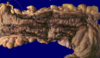
What do you see?

This is an example of cecal volvulus.
Volvulus is a twisting of the bowel. Volvulus is most common in adults, where it occurs with equal frequency in small intestine (around a twisted mesentery) and colon (in either sigmoid or cecum which are more mobile). In very young children, volvulus almost always happens in the small intestine.
What is the pathogenesis of diverticular disease?
Low fibre diet + other factors lead to high intraluminal pressure, and lead to weak points in wall of bowel
What does this barium enema show?

Outpouchings from side of the colon = diverticular disease
What does this endoscopy show?

All of these smaller holes are the divertulae and some can get filled with food/debris/pus
What do the gross specimen and the histo slide show?

gross specimen = outpouchings
histo: outpouching circled
(diverticular disease)
In which part of the GI tract does diverticular disease usually occur?
90% occurs in left colon
What does this endoscopy show?

inflamed diverticulum = diverticulitis
What does this gross pathology of the colon show?

large bowel and mucosa:
- very oedematous and red
- wet corn flakes = pseudomembranes
pseudomembranous colitis
What does this show?

colonic mucosa shows volvanic eruption + puss moving to surface of mucosa = pseudomembranous colitis
What are 5 complications of diverticular disease?
- Pain
- Diverticulitis
- Gross perforation
- Fistula (bowel, bladder, vagina)
- Obstruction
Inflammatory disorders of large bowel can be divided into acute and chronic colitis. Name causes under each category
•Acute colitis
–Infection (bacterial, viral, protozoal etc.)
–Drug/toxin (esp.antibiotic)
–Chemotherapy
–Radiation
•Chronic colitis
–Crohn’s
–Ulcerative colitis
–TB
What does this histo slide of the colon show?

inflammation + haemorrhage = ISCHAEMIC colitis!
What is pseudomembranous colitis? What causes it?
antibiotic associated colitis with acute onset, cused by protein exotoxins of C diff
What 2 investigations can be used to confirm pseudomembranous colitis?
- Histology - characteristic microscopic features (volcanic eruption)
- Lab - C diff toxin stool assay
What do you see?

2 skip lesions = Crohn’s Disease!
What do you see?

some parts of the colon are inflamed and others are healthy = CD
This is a histo slide of the colon - what do you see?

non-caseating granuloma = CD!
Ischaemic colitis usually happens in segments in ___________
watershed zones = splenic flexure (1) and rectosigmois region (2)
Name the arteries associated with each:
- Splenic flexure
- Rectosigmoid region
- Splenic flexure = SMA, IMA
- Rectosigmoid region = IMA, internal iliac artery
Name 5 aetiology for ichaemic colitis
–Arterial Occlusion: atheroma, thrombosis, embolism
–Venous Occlusion: thrombus, hypercoagulable states
–Small Vessel Disease: DM, cholesterol emboli, vasculitis
–Low Flow States: CCF, haemorrhage, shock
–Obstruction: hernia, intussusception, volvulus, adhesions
What do you see in this gross specimen?

Right bottom is anus
all inflamed and haemorrhaged
no skip lesion
= UC!
What do you see in this gross specimen?

All inflamed, no skip lesions = UC!
Crohn’s disease - Risk factors (5)
- Western populations
- White 2-5x > non-white
- teens-20s
- Jewish population
- Smoking
In terms of the inflammation in CD:
- area of GI tract affected?
- thickness of inflammation?
- Whole of GI tract can be affected (mouth to anus)
- Transmural inflammation
What does this endoscopy and histo slide show?

hyperplastic polyps = a non-neoplastic polyp of the colon
Name 8 charactersitic features of CD
- Skip lesions
- Non-caseating granulomas
- sinus/fistula formation
- fat wrapping
- Thick rubber-hose like wall
- Narrow lumen
- Cobblestone mucosa
- Fissures/abscesses
Does CD have extra-intestinal symptoms? If yes, name them
Yes
- Arthritis
- Uveitis
- Stomatitis/cheilitis
- Skin lesions
–Pyoderma gangrenosum
–Erythema multiforme
–Erythema nodosum
What do these endoscopy images show?

adenoma polyps of colon
What does this gross pathology show?

Adenoma polyp of the colon
What do these gross specimens and histo slide show?

Normal mucosa, a stalk, and a polyp = TUBULAR ADENOMA
What does this histo slide show?

stalk, polyp = tubular adenoma!
What does this histo slide show?

villious adenoma (sea cloral looking)
What do these show?

Villious adenoma = looks like a carpet
Left (low magnification), right (high magnification)
What does this gross specimen show?

This adenocarcinoma is arising in a villous adenoma. The surface of the neoplasm is polypoid and reddish pink. Hemorrhage from the surface of the tumor creates a guaiac positive stool. This neoplasm was located in the sigmoid colon, just out of reach of digital examination, but easily visualized with sigmoidoscopy
UC - in what ethnic group and age group does it present?
Whites > non-whites
peak 20-25 years but can affect any age
In terms of the inflammation in UC:
- area of GI tract affected?
- thickness of inflammation?
- involves rectum and colon in contiguous fashion
- inflammation confined to mucosa = shallow ulcers
Name 3 complications of UC
- Severe haemorrhage
- Toxic megacolon
- Adenocarcinoma (20-30x risk)
What does this gross specimen show?

1000s of polyps = Familial adenomatous polyposis (FAP)
Does UC have extra-intestinal symptoms? If yes, name them
Yes
- Arthritis
- Myositis
- Uveitis/iritis
- Skin changes:
- Erythema nodosum
- pyoderma gangrenosum
5. Primary sclerosing cholangitis (PSC)
What does this endoscopy show?

colon growth, red/angry looking, bleeding, irregular = colorectal carcinoma
What do these histo slides of the colorectal region show?

Lots of purple, lots of activity and cellular infiltration = colorectal carcinoma
remember: pink is good, purple is bad!
tumours of the colon and rectum - polyps => can be either ______________ or _____________
tumours of the colon and rectum - polyps => can be either non-neoplastic or neoplastic
Name 3 types of non-neoplastic polyps of colon/rectum
- Hyperplastic
- Inflammatory (pseudo-polyps)
- Hamartamatous (juvenile, Peutz Jeghers)
hyperplastic polyps - can they develop into cancer?
no!
Name the 3 types of neoplastic polyps of the colon/rectum
- Tubular adenoma
- Tubulovillious adenoma
- Villious adenoma
What sort of colon polyps most commonly predispace to adenocarcinoma of the colon?
Adenomas
(doesn’t matter if tubular or villious)
What are the risk factors (3) for a colonic polyp to develop to cancer?
- Size of polyp (> 4 cm approx 45% have invasive malignancy)
- Proportion of villous component
- Degree of dysplastic change within polyp
Can colon adenomas develop into carcinomas?
yes!
What are the symptoms of colon adenomas?
- None (hence why there’s screening for over 60s)
- Bleeding/anaemia
What 3 familial syndromes predispose to colon polyps?
- Peutz Jeghers
- Familial adenomatous polyposis (FAP)
- Hereditary non-polyposis colon cancer (HNPCC)
FAP:
- inheritance mode?
- age of onset?
- nature of polyps + # polyps + distrubution of polyps?
FAP:
- inheritance mode = autosomal dominant
- age of onset = average 25 years
- nature of polyps + # polyps + distrubution of polyps = adenomatous polyps + 1000s on average (min 100) + mostly colorectal
Hereditary non-polyposis colorectal cancer ( HNPCC):
- inheritance mode?
- age of onset?
- nature of polyps + # polyps + distrubution of polyps?
- extracolonic cancers?
Hereditary non-polyposis colorectal cancer ( HNPCC):
- inheritance mode = autosomal dominant (uncommon disease)
- age of onset = early age (40s?) colorectal cancer
- nature of polyps + distrubution of polyps = poorly differentiated and mucinous carcinoma + high frequency proximal to splenic flexure
- extracolonic cancers = endomtrium, prostate, breast, stomach
Colorectal carcinomas:
- type of cancer?
- Age of onset?
- 98% are adenocarcinomas
- age: 60-79 years
Risk factors (5) for colorectal carcicnoma?
- Diet (low fibre, high fat etc)
- Lack of exercise
- Obesity
- Familial
- Chronic IBD
Symptoms (6) of colorectal carcinoma
- Change of bowel habit
- Bleeding
- Anaemia
- Weight loss
- Pain
- Fistula
What is grading and staging in terms of colorectal carcinoma?
What staging system is used?
Grade = level of differentiation
Stage = how fat has it spread?
Duke’s staging
–A = confined to wall of bowel
–B = through wall of bowel
–C = lymph node metastases
–D = distant metastases
*NB: TNM also used.
Impaired blood supply to the colon/bowel most commonly causes what?
Ischaemic colitis
A patchy inflammatory bowel condition affecting the stomach, small bowel and colon is most likely to be what?
Crohn’s Disease
A 76 year old man presents with rectal bleeding. The diagnosis that must be exluded first is what?
Colorectal carcinoma