Ventricular System Flashcards
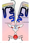
What is tela choroidae?
Penetrating choroidal artery with invaginating pia mater, vascular bundle and efferent choroidal vein
From what is the choroid plexus formed?
Tela choroidae covered by ependymal cells
How does choroid plexus functionally produce CSF?
Ependymal cells lining tela choroidea have active secretory Na pumps
Cl follows the Na passively to maintain electroneutrality.
H2O is pulled with it.
Ependymal cells also possess glucose transporters and can transport glucose from blood but these are less effective, therefore CSF [glucose] < [serum glucose]
Rough [csf glucose] : [serum glucose]
0.66 : 1
Which substances are transported from csf to the vascular tuft of the tela choroidae?
K
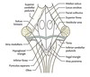
What is the name for the distal dilatation of the spinal canal?
Terminal ventricle
What is the name of the dilatated subarachnoid space into which foramen luschke open?
(Cerebello)Pontine cistern
Into which subarachnoid swelling does the foramen Magendie open?
Cerebellomedullary cistern (cisterna magna)
In which portion of the lateral ventricle is the foramen of Monro found?
Body of lateral ventricle
How low does the subarachnoid space extend?
S2
What are the names of the two layers of dura mater?
Periosteal layer
Meningeal layer
What cell type lines dural venous sinsus?
Endothelium
What is an arachnoid granulation
Macroscopic view of arachnoid mater projecting into dural venous sinuses as arachnoid villi.
How does CSF move through arachnoid villi to venous drainage?
Via vesicular channels between endothelial cells
What are the key functions of CSF?
Cushion of protection
Buoyancy
Reservoir regulating intracranial contents/pressure
Nourishment
Metabolic waste removal
Hormone transport
What is the weight of brain when suspended in CSF?
50g
What is the mechanism through which raised ICP causes projectile vomiting?
Traction on vagus nerves
Leptomeninges=
Pia and arachnoid mater
Colour of normal CSF
Clear
Cellular composition of normal CSF
Lymphocytes- <5/mm^3
No RBC
No neutrophils
Glucose in normal CSF
[2/3 serum]
As less efficiently transported
Normal level of protein in CSF
<0.4g/L
Colour of CSF
in pyogenic bacterial meningitis
Yellow/turbid
Colour of CSF
in TB meningitis
Turbid +/- fibrin web
Colour of CSF
in viral meningitis
Clear
Cell count in
Pyogenic bacterial meningitis
High levels of neutrophils
Low lymphocytes (acute rather than chronic inflammation)
Cell profile in TB meningitis
Low neutrophils (chronic rather than acute inflammation)
High lymphocytes
Cell profile in viral meningitis
Low neutrophils (chronic rather than acute inflammation)
High lymphocytes
Glucose level in pyogenic meningitis
[<50% serum]
Bacteria use glucose
Glucose level in TB meningitis
[<50% serum]
Bacteria use glucose
Glucose level in viral meningitis
[>50% serum glucose]
Protein in pyogenic bacterial meningitis
Increased significantly
Protein in TB meningitis
Significantly increased
Protein in viral meningitis
<=1g/L
Def: Hydrocephalus
Excessive CSF within cranial cavity
Causes of hydrocephalus in simple terms
Excess production
Abnormal circulation
Impaired drainage
Causes of excessive CSF production?
Choroid plexus papilloma
Non-communicating hydrocephalus
(Obstructive)
CSF from within the ventricular system is not able to communicate with subarachnoid space
Communicating hydrocephalus
CSF able to flow from ventricular system to subarachnoid space
Queckenstedt’s Test
Test for spinal blockage of CSF using lumbar manometer
With occlusion of IJV manually, there should be increase in CSF pressure.
If there is no increase this indicates blockage of the spinal subarachnoid space
Positive if abnormal- i.e. no increase in CSF pressure with IJV occlusion
What constitutes blood-CSF barrier?
Tight junctions between choroidal epithelial cells (in contrast to BBB which is due to tight junctions between endothelial cells)
Macroscopic appearance of ependyma
The distinctive, shiny white appearance
Embryological associations of the CSF ventricles
Lateral-> telencephalon
Third-> diencephalon
Fourth- rhombencephalon
What is the embryological precursor to the aqueduct of Sylvius?
The lumen of the mesencephalon
Def: Frontal horn
The portion of the lateral ventricle anterior to the foramen of Monro continuous posteriorly with the body of the lateral ventricle.
If no choroid plexus is seen on entering the lateral ventricle, what is the likely location of the endoscope?
Anterior horn as there is no choroid plexus
Location of the body of the lateral ventricle
Predominantly in the parietal lobe
Continuous anteriorly with the frontal horn and posteriorly with the atrium
Extends from the foramen of Monro anteriorly to the limit of the septum pellucidum posteriorly
What is the limit of the septum pellucidum posteriorly
Where the fornix fuses with the corpus callosum
What divides the caudate from the thalamus in the lateral ventricle?
The caudothalamic grooove (striothalamic sulcus)
Contains the stria terminalis and thalamostriate vein
What is the benefit of identifying the choroid plexus when in the lateral ventricle?
Following it anteriorly will reliably lead to the foramen of Monro
Comminucations of the atrium (trigone)
Anterosuperiorly-> body
Anteroinferiorly-> temporal horn
Posteriorly-> occipital horn
Medial wall of the atrium
Superiorly the bulb of the corpus callosum
inferiorly the calcar avis (which overlies the calcarine sulcus)
Lateral wall of the atrium
Caudate nucleus and the tapetum of the corpus callosum
Floor of the atrium
Collateral trigone, which overlies the collateral sulcus
Roof of the atrium
Corpus callosum
Where is the most voluminous colloid plexus in the brain found?
In the atrium and is referred to as the choroid glomus which may contain benign, incidental choroid cysts and often becomes calcified with age
Boundaries of the temporal horn
Floor
Made up of the hippocampus medially and the collateral eminence laterally
Floor of the third ventricle
Bounder anteriorly by the optic chiasm and posteriorly by the opening of the cerebral aqueduct.
From A->P:
Optic chiasma, infundibulum, tuber cinereum, mamillary bodies, posterior perforated substance and mesencephalon.
Recesses of the third ventricle
Chiasmatlc recess into the angle formed by the attachment of the chiasm to the anterior wall
Infundibular recess
Bulging or rounding out of these recesses is an early indicator of HCP
What is the surgically important area of the floor of the third
Between the hypothalamus and the tuber cinereum
Roof of the third
Curves from the foramen of Monro to the suprapineal recess
Most superior layer of the roof is formed by the fornix,
tela choroidea and blood vessels form the inferior layers
Tela choroidea
Two thin layers of pia mater sandwiching the medial posterior choroidal arteries and the internal cerebral veins in a space called the velum interpositum
Anterior wall of the third
Foramen of Monro superiorly
Anterior commissure
Lamina terminalis
Optic recess
Optic chiasm
Posterior wall of the third
Suprapineal recess
The commissure of the habenula
Body of the pineal and its associated recess
Posterior commissure and the mesencephalic opening of the aqueduct.
Lateral wall of the third
Made up of the thalamus and hypothalamus which are demarcated by the hypothalamic sulcus
What is the prevalence of the massa intermedia?
Absent in up to 25%
Vertical divisions of the floor of the fourth
Divided into two lateral halves by the median sulcus
Sulcus limitans is lateral to median sulcus and the medial eminences run between these
Three triangles of the lower part of the fourth
Hypoglossal triangle
Vagal triangle
Area postrema
Location of the vestibular area in the floor of the fourth?
Lateral to medial eminence and thus sulcus limitans
What demarcates the superior and inferior triangles in the floor of the fourth?
Striae medullares which contains cochlear fibres
What makes up the cranial portion of the roof of the fourth?
Superior cerebellar peduncles and their interconnecting superior medullary velum
What makes up the caudal roof of the fourth ventricle?
Consists of ependymal and non-neural inferior medullary velum with a layer of tela choroidea
Use of anterior transcallosal approach
The anterior portion of the lateral ventricle
Access to third ventricle
Advantages of the anterior transcallosal approach
Avoids cortical incision
Reduces risk of seizures and cortical deficit
Feasible even with small or normal ventricles
Allows access to both ventricles
Disadvantages of anterior transcallosal approach
Potential injury to falcine bridging veins
Relatively narrow working corridor
Disconnection-type syndromes are extremely rare
Beta-transferrin
Transferrin isomer found in CSF and inner ear perilymph
Positive in CSF leak (though may also suggest perilymph leak if otorrhoea)
Ions in CSF compared to plasma
Same Na, osmolarity
Increased Cl
Reduced K, Ca, uric acid and glucose
Froin’s Syndrome
CSF xanthochromia
High protein
Coagulation (due to presence of fibrinogen)
Occurs when CSF loculated, usually in the lumbar thecal sac
Can be caused by dural irritation, blockage of CSF flow by tumour or abscess
Normal CSF glucose
2/3rds of serum
normal CSF glucose levels lie between 2.5 and 4.4 mmol/L
CSF specific gravity
1.007
CSF pH
7.33-7.35
Traumatic tap cell count
700 RBCs per 1 WBC
Sites of CSF production
70% choroid plexus
18% ultrafiltrate
12% H2O production from metabolism
Control of CSF production
Raphe nuclei send axons (5HT) to the periependymal vessels
Factors causing decreased CSF production
Carbonic anhydrase inhibitors
Noradrenaline
Factors causing increased CSF production
Volatile anaesthetics
Increased CO2
CSF volume within ventricles
25mL
Location of choroid plexus
Roof of fourth
Inferior medullary velum and lateral recess to the foramen of Luschka
Posterior roof of third
Floors of the bodies and roofs of the temporal horns of the lateral ventricles
CSF production ml/min
0.3-0.37
Normal ICP in adults
5-15mmHg
Normal ICP in younger children
3-8mmHg
Normal ICP in infants
1.5-6mmHg
Constituents of BBB
Capillary endothelial tight junctions
Pinocytic activity in endothelial cells
Astrocytic foot processes
Mechanisms of molecular movement across the BBB
Diffusion
Carrier-mediated transport
Active transport
BBB highly permeable to which substances?
H2O
CO2
O2
Lipid-soluble drugs e.g. ETOH, barbiturates, heroin, anaesthetics
BBB slightly permeable to which substances
Ions
Na, Cl, K
BBB Impermeable to which substances?
Plasma proteins
Protein bound molecules
Large organic molecules
L-glucose
Def: Circumventricular organ
Midline ventricular system structures of specialised tissues with absent BBB due to fenestrated capillaries
Circumventricular organs from rostral->caudal
Organum vasculosum (lamina terminalis)
Neurohypophysis
Median eminence of hypothalamus
Subfornical organ
Subcommisural organ
Pineal gland
Area postrema
Function:
Organum vasculosum
Outlet for hypothalamic peptides
Detects peptides, amino acids and proteins in blood
Function:
Neurohypophysis
Outlet for hypothalamic hormones (vasopressin and oxytoxin)
Function:
Median eminence of hypothalamus
Release hypothalamic releasing factors
Function:
Subfornical organ
May be involved in body fluid regulation
Located between the foramina of Monro
Connected to choroid plexus
Function;
Pineal gland
Melatonin production
Role in circadian rhythm
Function:
Subcommissural organ
Unknown
Only circumventricular organ with intact BBB
Function:
Area postrema
Chemoreceptor that induces emesis when stimulated by digitalis or apomorphine
Floor of fourth ventricle
Only paired circumventricular organ
Taenia choroidea
2 narrow bands of WM one on either side, which complete the lower part of the roof of the fourth ventricle.
Each consists of a vertical and a horizontal part.
The vertical part is continuous below the obex with the gracile nucleus, to which it is adherent by its lateral border.
The horizontal portion extends transversely across the inferior peduncle, below the striæ medullares, and roofs in the lower and posterior part of the lateral recess
Attached by its lower margin to the inferior peduncle, and partly encloses the choroid plexus, which, however, projects beyond it like a cluster of grapes; and hence this part of the tænia has been termed the cornucopia.
CSF xanthochromia
High protein
Coagulation (due to presence of fibrinogen)
Occurs when CSF loculated, usually in the lumbar thecal sac
Can be caused by dural irritation, blockage of CSF flow by tumour or abscess
Froin’s Syndrome
Boundaries of the frontal horn of the lateral ventricle
Anterior wall
Anterior wall: genu of the corpus callosum
Boundaries of the frontal horn of the lateral ventricle
Floor
Rostrum of corpus callosum
Boundaries of the frontal horn of the lateral ventricle
Roof
Genu of corpus callosum
Boundaries of the frontal horn of the lateral ventricle
Lateral wall
Head of caudate nucleus
Boundaries of the frontal horn of the lateral ventricle
Medial wall
Septum pellucidum
Border of body of lateral ventricle
Roof
Body of the corpus callosum
Borders of the body of lateral ventricle
Medial wall
Continuation of the septum pellucidum in upper part and fornix in the lower
Borders of the body of lateral ventricle
Floor
Sloping from lateral to medial:
Body of caudate
Thalamus
Choroid plexus
Body of fornix
Borders of the body of lateral ventricle
Lateral wall
Caudate nucleus
Boundaries of the temporal horn
Roof
Lateral roof formed by tapetum
Medial roof is the inferior thalamus, caudate tail and intervening caudothalamic groove
Boundaries of the temporal horn
Lateral wall
Tapetum
Boundaries of the temporal horn
Medial
Choroidal fissure
Boundaries of the temporal horn
Anterior
Amygdaloid nucleus
Location of crural cistern
Between uncus and crus cerebri at ventrolateral aspect of midbrain
Contents of crural cistern
Anterior choroidal artery
Medial posterior choroidal artery
Basal vein of Rosenthal
Contents of cisterna magna
VA and PICA origin
9/10/11/12
Choroid plexus
Prepontine cistern contents
BA
AICA origin
SCA origin
CN6
CP cistern contents
5
7/8
AICA
Petrosal vein
Communication of interpeduncular cistern
With Ambient
With PComm cistern
Prepontine and cerebellopontine cisterns
Division of the interpeduncular cistern
BA bifurcation divides cistern into deep and superficial portions
Superficial portion adjoins CN3 and deep communicates with ambient cistersn
Deep subdivided by posterior perforated membrane- anterior and posterior areas
Contents of interpeduncular cistern
Arteries:
BA bifurcation
PCA
SCA
PCA perforators
PComm
Veins:
Basal vein of Rosenthal
Nerves:
3
Contents of chiasmatic cistern
Anterior aspect of chiasm and optic nerves
Hypophyseal stalk
ACA origin
Contents of carotid cistern
ICA
Anterior choroidal
PComm
Sylvian cistern contents
MCA
Middle cerebral veins
Fronto-orbital veins
Collaterals to vein of Rosenthal
Contents of lamina terminalis cistern
ACA (A1 and A2)
AComm
Heubner’s
Hypothalamic
Fronto-orbital arteries
Quadrigeminal cistern contents
Arteries:
The third portion of SCA
Posterior pericallosal
PCA perforators
P3
Medial posterior choroidal
Veins:
VoG
Contents of the ambient cistern
Supratentorial portion:
Basal vein of Rosenthal
PCA
Infratentorial portion:
SCA
4
Anterior border of atrium + occipital horn of lateral ventricle
Crus of fornix medially
Pulvinar of thalamus laterally
Medial border of atrium + occipital horn of lateral ventricle
Forceps major of corpus callosum superiorly
Calcar avis inferiorly
Lateral border of atrium + occipital horn of lateral ventricle
Caudate nucleus
Fibres of tapetum
Inferior border of atrium + occipital horn of lateral ventricle
Collateral trigone
Blood supply of choroid plexus in the body of lateral ventricle
From medial posterior choroidal arteries passing forward through velum interpositum
4 layers of tissue between body of lateral ventricle and third
fornix, upper layer of tela, velum interpositum (containing medial posterior choroidal arteries, internalcerbral veins), lower layer of tela
Which side does Rhoton suggest opening when opening choroidal fissure
Forniceal side- allow thalamus and choroid to on other side to reduce traction on thalamus and risk of thalamic venous infarction
What are the 5 layers of the roof of the third
Neural layer- fornices
2nd: Sup layer of tela choroidea
Velum interpositum
3rd: Vascular ayera: ICV and Post choroidal
4th: inf layer of tela choroidea
5th: choroid plexus of lateral ventricle which extends posterosuperiorly from foramen of monro
Attachments of the chorodi plexus of the lateral ventricle
Ependymal attachment to the fornix (taenia fornicis) and the thalamus (taenia thalmi)
What is the operative significance of the choroid plexus attachments
The taenia fornicis is avascular whereas the taenia thalami contains tributaries of the ICV and the medial PChA
Opening the taenia fornicis thus avoids these structures
Floor of third from A->P
Anterior half: diencephalic
Optic chiasm, infundibular recess, median eminence, tuber cinereum, mamillary podies and posterior perforated substance
Posterior:
Mesencephalic
Tegmentum of the midbrain, aqueduct of sylvius
Anterior wall of the third ventricle
Columns of fornix, foramina of Monro, Anterior commissure, lamina terminalis, optioc recess and chiasm
Posterior wall of. thethird ventricle
Suprapineal recess
Habenular commisure
Sylvian aqeuduct
What delineates the walls of the thid
Hypothalamic sucus- thalamus superiorly and hypothalamus inferiorly
CP: chorodi plexus
MI: massa intermedia
VI: velum interpositum
HC: habenular commisure
PC: posterior commisure
SC: superior colliculus
MB: mamillary body
OC: chiasm