Peripheral Nerves COPY Flashcards

1
Q

Radial nerve origins

A

C5-8

Pass through upper, middle and lower trunks then posterior cord of brachial plexus

2
Q

Radial nerve anatomy in upper arm

A

As it winds around the humerus or proximal to this it innervates the tricpes.

After course in spiral groove it supplies brachioradialis and ECRL and brevis.

Bifurcates into a superifical (sensory) and deep (motor) branches

3
Q

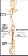
Muscles supplied by proximal branches of radial nerve

A

Triceps

Brachioradiailis

ECRL

ECRB

4
Q

Superficial branch of radial nerve

A

In forearm, passes distally into the hand where it supplies skin of the radial aspect of the dorsum of the hand and dorsum of first four fingers.

5
Q

Deep branch of radial nerve

A

Passes deep through the fibrous arch of supinator (arch of Froshe) to enter the posterior compartment of forearm

Continuous in this compartment as the purely motor posterior interosseous branch

Innervates

Supinator

Extensor digitorum

Extensor digit mini

ECU

Abductor pollicis longus

Extensor pollicis longus

Extensor pollicis brevis

Extensor indicies

6
Q

Forearm muscles dupplied by posterior interosseous nerve

A

Supinator

Extensor digitorum

Extensor digiti minimi

ECU

Abductor pollicis longus

Extensor pollicis longus

Extensor pollicis brevis

Extensor indicis

7
Q

Supinator function

A

Forearm supinator

Test with resisted supination

8
Q

Extensor digitorum

A

Extensor of 2nd to 5th metacraophalangeal joints

9
Q

Extensor digiti minimi function

A

Extensor if fifth MCP

10
Q

ECU function

A

Ulnar extenor of the wrist

11
Q

APL function

A

Abductor of carpometacarpal joint of thumb

12
Q

EPL function

A

Extension of thumb interphalangeal joint

13
Q

EPB function

A

Extensor of the MCPJ of thumb

14
Q

Extensor indicies function

A

Extensor of index finger

15
Q

Saturday night palsy

A

AKA radial nerve palsy

Classically associated with a drunkard who falls asleep with arm hyper abducted across a park bench

Site of compression is in the region of the spiral groove

16
Q

Why is the triceps preserved in radial nerve palsy (Saturday night palsy)

A

Because branches of the tricpes originate proximal to spiral groove

17
Q

Wrist drop

Inability to extend fingers at MCPJ

Supinator weakness

Triceps spared

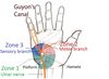
Absent sensation first dorsal interosseuous

A

Radial nerve palsy

Compression in the spira groove of humerus/humeral fracture

18
Q

Why is weakness of supination only partial in Radial nerve palsy

A

Because it may be accomplished with either biceps or supinator

19
Q

Posterior interosseous nerve syndrome

A

Most common syndrome caused by compression at the arcade of Frohse (fibrous arch at the origin of supinator) which may pathologically constrict nerve

Inability to extend fingers at MCPJ, absence of wrist drop and normal sensation

Supintaor spared as branches are given off proximal to PIN entering the arcade of Frohse

20
Q

Why is wrist drop absent in PIN palsy

A

The ECR is presreved

The ECU is innervated by PIN so there may be radial deviation of the hand on extension

21
Q

Why is there no senosry deficit in PIN syndrome

A

It is purely motor

22
Q

Inability to extend fingers at MCPJ

No wrist drop but radial deviation on extension of wrist

Presrved sensation

Preserved supinator and triceps

A

Posterior interosseus nerve syndrome

23
Q
A

Arcade of Frohse associated with PIN syndrome/supinator syndrome

24
Q

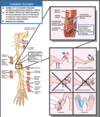
Roots of median nerve

A

C6 to T1

Pasing through upper middle and lower trunks and the lateral and medial cords of the brachial plexus

25
Median nerve branches proximal to the elbow
None
26
Median nerve at elbow Muscles supplied
Pronator teres Flexor carpi radialis Palmaris longue FDS
27
Prontaor teres function
Forearm pronator
28
FCR function
Radial wrist flexor
29
PL
Wrist flexor
30
FDS
Flexor at the IPJ for 2nd-5th fingers
31
Contents of the cubital fossa Really Need Beer To Be At My Nicest
L-\>M Radial nerve Biceps tendon Brachial artery Median nerve
32
Under what is the median nerve found at the elbow?
Behind the biccipital aponeurous (lacertus fibrosus)
33
How does the median nerve enter the forearm
Between the two heads of pronator teres
34
Where is the AIN given off?
As it the median nerve passes deep to pronator teres
35
Anterior interosseous nerve function
Purely motor Supplies FPL, Pronator quadratus, FDP 1 and 2
36
FPL function
Flexion of thumb at interphalangeal joint, key function for grip
37
Pronator quadratus
Pronation of forearm
38
Function of FDP
Flexion of DIP in 2-4th fingers
39
Median nerve in the hand
Passes deep to the flexor retinaculum to innervate the LOAF muscle Lumbricals Opponens pollicis Abductor pollicis brevis Palmar digital nerves-\> palmar aspect of thumb, second, third and half of fourth fingers. Raedial aspect of the palm and dorsl aspect of the distal middle phalanges of the sconed, third and half of fourth fingers Palmar cutaneous bracnh given off proximal to the carpal tunnel and supplies the median eminence and proximal palm on radial aspect of hand
40
Three major median nerve entrapment syndromes
Pronator teres syndrome AIN syndrome CTS
41
Pronator teres syndrome
Results from entrapment of the median nerve as it passes between the two heads of the pronator teres and under the fibrous arch of the FDS Compression may be caused by thickened lacertus fibrosus, hypertrophied pronator teres, tight fibrous band of the FDS Pain in forearm with weakness in hand grip and numbness and tingling in the index finger and thumb. Similar symptoms to CTS with hand assuming a "benedicton" attitude in severe cases. Nocturnal exacerbation is absent normally Weakness is variable but often there is no measurable weakness in median nerve innervated muscles
42
How to differentiate between Hand of Benediction and Ulnar Claw
Ulnar claw usually seen at rest whereas in Hand of Benediction there is weakness on active flexion in the radial three digirs
43
Pain in forearm Weakness in hand attitdue Numbness in median nerve distribution without nocturnal exacerbation
?Pronator teres syndrome
44
DDx
Ulnar claw or Hand of Benediction
45
AIN snyfrome
Most commonly due to a constricting band causing entrapment neuropathy near the origin of the nerve FDP and FPL weakness Abnormal pinch attitude of the hand Weakness in pronator quadratus is usually clinically insignificant due to more powerful pronator teres. No sensory deficit
46
Abnormal pinch attitude
Extension of hyperextension of the terminal phalanges of the thumb and index finger when the thumb and index finger are opposed
47
Abnormal pinch attitude of the hand due to weakness in FDP and FPL Normal sensation
AIN syndrome
48
CTS
Caused by compression of the median nerve as it passes through the carpal tunnel Pain anad paraesthesia in wrist and hand awakening patient from sleep Sensory loss in te ditsirubiton of the palmar digital branches Isensory loss easiest to discern along vola tips of index and middle fingers) Pain may also involve forearm, elbow or shoulder- classically this pain may radiate from distal to proximal and may be associated with shaking the hand to alleviate pain in contrast to cervical radiculopathy in which movement exacerbates pain. Motor symptoms are usually late- LOAF muscles affected- weakness in abduciton, opposition and flexion of thumb. Phalen's positive
49
Function of lumbricals
Flex fingers at the MCPJ
50
Function of opponens pollicis
Opposition refers to the rather complex movement of the thumb which is a combination of flexion, adduction and medial rotation at the first carpometacarpal joint
51
Function of Abductor pollicis brevis
Abduction of thumb at MCPJ
52
Phalen's test
Forcibly dosriflexing affected hand for 60 seconds Positive test will reproduce symptoms
53
What is the most reliable assessment of thenar muscle function?
Abductor pollicis brevis
54
Nocturnal pain and paraesthesia in median distirubtion Weakness and atrophy in LOAF muscless Sensory loss
Carpal tunnel syndrome
55
Contents of carpal tunnel 9 tendons, 1 nerve
4 tendons of the FDS superifically Median nerve 1 tendon of FPL 4 tendons of FDP
56
Roots of the ulnar nerve
C7/8/T1 Medial cord of the brachial plexus
57
Branches of ulnar nerve in arm
None
58
Ulnar nerve branches at the elbow
Ulnar nerve enters a groove between the medial humeral epicondyle and olecranon process Groove covered by an aponeurosis formng an osseofibrous canal (cubital tunnel) Two motor branches- FCU and FDP to fourth and fifth fingers
59
Boundaries of the cubital tunnel
Roof: Cubital tunnel retinaculum (band of Osbourne) Laterally: Olecranon Medially: Medial epicondyle Floor: elbow joint capsule, posterior band of medial collateral ligament of elbow joint
60
Ulnar nerve in forearm
Passes between two heads of FCU to take place superifical to FDP Two sensory branches- palmar cutaneous branch-\> skin over hypothenar eminence Dorsal cutaneous bramnch-\> dorsal ulnar aspect of the hand and dorsal aspect of the fifth finger and half of fourth finger
61
Sensory branches of ulnar nereve in forearm
Palmar cutaneous branch Dorsal cutaneous branch Superficial sensory branch is given off as the ulnar nerve enters Guyon's canal at the wrist
62
63
How does the ulnar nerve enter the hand?
Through Guyon's canal
64
Borders of Guyon's canal
Floor- transverse carpal ligament, hypothenar msucles Roof- Volar carpal ligament Ulnar border- Pisiform and pisohamate ligament, abductor minimimuscle belly Radial border- Hook of hamate
65
Branches of the ulnar nerve in hand
Superficial sensory branch given off proximally in the canal- supplies the distal part of theulnar aspect of th eplam and the palmar aspect of the fifth and half of the fourth finger Contineus as deep motor branch supplying: Abductor digiti minimi Opponens digiti minimi Flexor digiti minimi Lumbricals 3 and 4 Interosseous muscles Addcutor pollicis Flexor pollicic brevis
66
Action of abductor digiti minimi
Abducts and flexes 5th finger
67
Action of opponens digiti minimi
Opposition of the little finger with the tip of the thumb Deepends hollow of the palm
68
Action of the flexor digiti minimi
Flexes little finger
69
Lumbricals action
Flex MCPJ and extend interphalangeals
70
Interosseous muscle actions function
PAD- palmar adduct DAB- dorsal abduct
71
Action of adductor pollicis
Adducts CMCJ of the thumb
72
Flexor pollicis brevis function
Flexes thumb at MCPJ
73
Two major syndromes assocaited with ulnar nerve
Cubital tunnel syndrome Guyon's canal syndrome
74
What is a key differentiator between Cubital tunnel and Guyon's canal syndrome
Dorsal cutaneous nerve which is spared in Guyon's canal but affected in cubital tunnel
75
Cardinal features of cubital tunnel syndrome
Numbness and tingling of the ulnar aspect of hand Weakness variously described as imapirment of hand grip, clumsiness or difficulty buttoning shirt Atrophy in hypothenar eminence and first interosseous space. Sensory loss most easily observed in the distal two phalnges of the little finger Claw hand deformity Froment's sign
76
Claw hand
Results from simultaenous hypextension at the MCPJ and flexion at IPJ Hyperextension at the MCPJ is due to wekness of rthe lumbricals with unopposed action of the extensor digotorum Flexion at the IPJ is due to passive tethering pull of the flexor digitorum which occurs when the MCPJ are in extension
77
Froment's sign
Due to weakness in adductor pollicis On grasping a piece of paper between thumb and index finger and pulling Positive when the patient attempts to compensate for lack of thumb adduction with extension of the PIPJ and felxion of the distal phalangex
78
Why are the FCU and FDP often spared in cubital tunnel syndrome?
Because they are situated deeply within the nerve and may be spared the more suprifically localted fibres which are compressed first
79
Differneces between cubital tunnel and CTS
Cubital tunnel less frequently associated with pain with weakness and atrophy early rather than late
80
Hypothenar wasting
81
Paraesthesia on medial border of hand Weakness and clumsiness in hand Atrophy of the dorsal interossei and hypothenar eminence Claw hand Froment's sign
?Cubital tunnel syndrome
82
Classification of Guyon's canal syndrome
Three types based on the site of the compression Zone 1 to 3 based on site of compression
83
Guyon's Canal Syndrome Zone 1
Mixed motor and sensory symptoms Proximal to bifurcation of the nerve Common causes of compression: Ganglia and hook of hamate fractures
84
Guyon's Canal Syndrome Zone 2
Surrounds deep motor branch Predominantly motor symptoms Common causes include ganglia and hok of hamate fractures
85
Guyons Canal Syndrome Zone 3
Surrounds superifical sensory branch Senosry symptoms only Common caauses include ulnar artery thrombosis or aneursym
86
Loss of abductir digiti minimi, abductor pollicis, FDM, dorsal. interossei Froment's sign and claw hand Loss of palmar sensation but not dorsal
Zone 1 Guyon's canal syndrome Sensory pattern is due to superifical sensory branch being affected but sparing of the dorsal cutaneous branch
87
Hypothenar muscles spared (abductor digiti minimi and flexor digiti minimi) Loss of adductor pollicis and dorsal interossei Froment's sign and claw hand Preserved sensation
Zone 2 Guyon's canal syndrome Hypothenar muscle sparing due to early origin Sensation spared
88
All hand muscles spared Loss of palmar but not dorsal sensation over ulnar border Froment's and Claw hand absent
Zone 3 Guyon's canal syndrome
89
LFC
Direct branch of the lumbar plexus Provides sesnosry innervation to the skin of the ventrolateral aspect of the thigh
90
Anatomy of the LFC
Purely sensory nerve dervied from the second and third lumbar nerves of the lumbar plexus Emerges from lateral aspect of psoas to run obliquely and forward across iliacus Passes across the iliac fossa medial to ASIS, enters the thigh beneath the inguinal ligament piercing the fascia lata of te lateral thigh. Supplis the skin of the ventrolateral aspect of the thigh
91
Meralgia paraesthetica
Syndrome of the LFC Usually caused by compression of the nerve in the inguinal region. Characterised by paraesrthesia involving the ventrolateral of th e thigh. Common in obese individuals whose abdominal girth causes excessive strain on the inguinal ligament
92
Fat No motor signs Paraesthesia in ventrolateral thigh
Meralgia paraesthetica
93
Saphenous nerve
Terminal branch of femoral nerve in lower limb, provides sensory innervation to the ventromedial aspect of the knee leg and foot
94
Anatomy of the saphenous nerve
Purely sensory branch of femoral nerve Originatse below the inguinal ligament and enters Hunter's canal, crossing the femoral artery from medial to laterally and exits by piercing its root Divides into two terminal branches- infrapatellar branch supplying ventromedial aspect of the knee and a descneding branch which supplies the ventromedial aspect of the leg and ankle
95
Saphenous nerve compression syndrome
Occurs at exit point from adductor canal Characterised by intense pain along medial aspect of the knee with associated numbness in this area and down medial leg. No motor signs or symptoms
96
Intense pain along medial aspect of knee and leg Numbness along medial aspect of knee and leg Intact motor function
Saphenous nerve entrapment
97
What is the largest peripheral nerve in the body?
Saphenous
98
Anatomy of the sciatic nerve
Deirves from L4-S3 Leaves pelvis through the GSF behind the tendinous origin of piriformis Courses laterally and downward to innervate semitendinous, semimembranosus, biceps femoris and adductor magnus. Proceeds to popliteal fossa where it divides into terminal branches- tibial nerve and common peroneal nerve
99
Piriformis syndrome
Compression of the proximal segment of the sciatic nerve as it passes underneath the piriformis muscle Cardinal features are; Weakness in any or all of the knee flexors, ankle flexors, foot intrinsices. Sensory loss that may involve all of the foot Can be confiused with lumbosacral radiculopathy
100
Weakness in any or all of the knee felxors, ankles flexors or extensors and foot intrinsics Sensory loss involving all or part of the feet
?Piriformis syndrome
101
Peroneal nerve roots
L4 - S2
102
Anatomy of the common peroneal nerve
One of two terminal branches of the sciatic nerve taking origin in popliteal fossa. Courses laterally winding round the neck of the fibula to divide into superficial and deep peroneal nerves
103
Superificial peroneal nerve
Branch of common peroneal nerve Motor innervation to peroneus longus and brevis (foot everters) Sensory to lower lateral leg and dorsum of the foot excluding the first dorsal webspace
104
Sensory supply of superficial peroneal
Lower lateral leg and dorsum of foot excluding first dorsal webspace
105
Deep peroneal nerve
Descends deep in anterior compartment of leg Motor to: Tibialis anterior, EHL, EDL, EDB Sensory to: First dorsal webspace
106
Tibialis anterior function
Deep peroneal nerve Foot dorsiflexion and eversion
107
EHL function
Great toe extension Foot dorsiflexion Deep peroneal nerve
108
EDL foot function
Extensor of four lateral toes Foot dorsiflexion
109
Function EDB in foot
Extenros of
110
Division of deep peroneal nerve
Proximal to ankle the nerve becomes superificial and divides into medial and lateral branches Lateral (motor branch)-\> EDB Medial (sensory)-\> first dorsal webspace
111
Most common site of peroneal nerve compression
Fibular head Most superificial location and thus most susceptible to injury Can be due to extrinsic compression Deep peroneal nerve most commonly affected but common and superficial may also be
112
Peroneal nerve syndrome
Depends on affected branch Ranges from common peroneal, to ddep or superficai palsy
113
Common peroneal palsy
Weakness of foot eversion and toe and foot dorsiflexion Loss of sensation over dorsum of foot, toes and lateral aspect of lower leg
114
Deep peroneal nerve palsy
Weakness of foot and toe dorsiflexion as well as sensory loss involving the first dorsal webspace of foot
115
Superficial peroneal nerve palsy
Weakness of foot eversion Loss of sensation in lateral aspect of lower leg and dorsum of foot and toes excluding first webspace
116
Weakness of foot eversion and toe and foot dorsiflexion Loss of sensation over dorsum of foot, toes and lateral aspect of lower leg
Common peroneal palsy
117
Weakness of foot and toe dorsiflexion as well as sensory loss involving the first dorsal webspace of foot
Deep peroneal nerve palsy
118
Cardinal features of peroneal nerve syndrome
Weakness in foot eversion and dorsfilexion of foot and toes Senosry loss involving lateral lower leg and dorsum of foot an toes
119
Weakness of foot eversion Loss of sensation in lateral aspect of lower leg and dorsum of foot and toes excluding first webspace
Superficial peroneal nerve palsy
120
Tibial nerve roots
L4-S3 Terminal branch of sciatic nerve
121
Tibial anatomy at popliteal fossa
Distal popliteal fossa-\> medial sural cutaneous nerve Branches to gastrocnemius and then soleus passing distally in the plane between these muscles
122
Formation of sural nerve
Medial sural cutaneous nerve (branch of tibial at popliteal fossa) joins the lateral sural cutaneous nerve (branch of the common peroneal nerve) at the level of the Achilles tendon Sensory to skin on the lateral heel and lateral aspect of the foot and the small toe
123
Tibial nerve anatomy in upper thid of leg
Motor branches to tibialis posterior, FDL, FHL
124
TP function
Foot invresion
125
FDL function
Plantar flexion
126
FHL
Plantar flexion of great toe
127
Tibial nerve anatomy at ankle
Passes caudal and dorsal to the medial malleolus and under the flexor retinaculum (tarsal tunnel) Gives off medial calacaneal branch variably, proximal or distal to the tarsal tunnel, pure sesnory branch supplying skin of medial heel Two terminal branches distally in tarsal tunnel: Medial plantar nerve- motor to medial intricis, sensory to medial three and a half toes Lateral plantar nerve- sensory to lateral one and a half toes, and lateral intrinsccs
128
Tarsal tunnel syndrome
Burning pain and paraesthesia in any combination of heel (calcaneal), medial sole (medial plantar), lateral sole (lateral plantar) Weakness of foot intrinsics
129
Draw the cervical plexus
https://www.youtube.com/watch?v=xdrzbM8ESNA
130
Suprahyoid muscles My Gravy Spoon, Darling
Mylohyoid Geniohyoid Stylohyoid Digastric
131
Infrahyoid muscles TOSS
Thyrohyoid Omohyoid Sternothyroid Sternohyoid
132
Mylohyoid innervation
Supplied by the nerve to mylohyoid, a branch of the inferior alveolar nerve From V3
133
Geniohyoid innervation
Innervated by a branch of ventral ramus of C1 from cervical plexus, which courses with the hypoglossal nerve into the floor of the mouth
134
Stylohyoid innervation
CNVII
135
Innervation of diagstric
anterior belly- Trigeminal Posterior velly- CN VII
136
Which is the only suprahyoid muscle not supplied by a cranial nerve?
Geniohyoid, supplied by ventral rami of C1 running with hypoglossal
137
Location of ansa cervicalis
Behind SCM, closesly related to spinal accessory and hypoglossal nerves
138
Division of cervical plexus
Superificial sensory branches Deep motor branches
139
Superficial sensory branches of cervial plexus
Greater occipital nerve Lesser occipital nerve Greater auricular nerve Transverse cervical nerve Supraclavicular nerve
140
Greater occipital nerve
C2 Supplies skin of the posterior scalp
141
Which segment of ansa cervicalis provides no sensory branches
C1 as it has no dorsal root
142
Lesser occipital nerve
C2 spinal root Supplies skin overlying the mastoid process extending just above and below the mastoid process to include part of the lateral head and lateral neck
143
Greater auricular nerve
C2-3 Supplies skin overlying external ear, parotid and angle of mandible
144
Transvere cervical nerve
C2-3 Supplies skin overlying anterior and lateral aspects of the neck from the body of the mandible to the sternum
145
Supraclavicular nerve
C3-4 Supply skin just above the clavicle
146
Deep motor branches
Branches to the accessory nerve Ansa cervicalis Branches to adjacent neck muscles Phrenic nerve
147
Cervical plexus Branches to accessory nerve
Travel with CN XI to supply SCM (C2-3) and trapezius (C3-4) muscles
148
Ansa cervicalis
Loop formed by a superior (C1/2) and inferior root (C2/3) Superior root fibres run with the hypoglossal enrve for a short distance Supplies the infrahyoid muscles- head flexors including thyrohyoid, omohyoid, sternohyoid, sternothyroid and geniohyoid (suprahyoid muscle)
149
Cervical plexus- branches to adjacent neck mucles
Small muscular branches innervate adjacent muscles of the neck which are flexors and rotators Include the longus anteriorly, middle scalene laterally and levator scapulae posteriorly
150
Phrenic nerve
C3-5 Diahphragm
151
Injuires to cervical plexus
Involvement of superficial senosry roots-partial numbess of head or neck Deep motor- weakness of forward or lateral neck flexion (infrahyoid/scalenes), rotation of head (SCM), rotation of scapula (levator scapulae) Include penetrating trauma or iatrogenic lesions or mass lesions
152
Unilateral phrenic nerve injury
Tolerated whilst patient at rest but may result in SOBOE
153
Bilateral phrenic nerve injury
Usually associated with severe ventilatory compromise at rest unless it receives an anastomotic branch from subclavian nerve Penetrating injury, surgical injury, intrathoracic mass
154
Weakness in: Lateral neck flexion Head rotation Head flexion Shoulder shurg Respiration
Deep motor branches of ansa cervicalis Scalene (deep motor) SCM (spinal accesory) Infrahyoid (ansa cervicalis) Diagphragm (Phrenic) Trapezius (spinal accessory)
155
Etymology phrenic
Phren- diaphragm
156
Draw the brachial plexus
https://www.youtube.com/watch?v=Z\_Y\_kVdH9zE
157
Where are the roots of the brachial plexus found?
In the posterior traingle of the neck
158
Bordres of posterior triangle
SCM anterioly Trapezius posteriorly Inferiorly middle third of clavicle
159
Where do the branchial trunks become their divisions
In the supraclavicular fossa the three drinks give rise to anterior and posterior divisions The posterior divisions unite to form the posterior cord
160
Which trunks of the brachial plexus form the lateral cord?
anterior divsions of the upper and middle trunk
161
Which trunks form the medial cord
Anterior division of the lower trunk
162
How do the brachial plexus cords leave the posterior traingle of the neck
Through te otulet between the first rib and clavicle to enter the axilla
163
What are the branches from the roots of the brachial plexusD
Dorsal scapular LTN
164
Function of LTN
Serratus anterior Pulls scapula forward around thorax. Palsy results in winging of scapula
165
Function of dorsal scapular nerve
Supplies rhomboids Scapular retraction around the scapularthoracic joint Levator scapulae: The levator scapulae functions to elevate the scapula and tilt the glenoid cavity inferiorly by rotating the scapula downward. If the scapula is fixed, a contraction of the levator scapulae leads to the lateral flexion of the cervical vertebral column to the side and stabilizes the vertebral column during rotation.
166
Nerve roots dorsal scapular
C5
167
Nerve roots LTN
C5, 6, 7
168
Branches from the trunk of the brachial plexus
Nerve to subclavius Suprascapular nerve Both from upper trunk
169
Nerve to subclavius
From upper trunk of brachial plexus Supplies subclavius muscle but may also contain accessory nerve fibres that join the phrenic nerve in the mediastinum
170
Suprascapular nerve
Infraspinatus- external rotation Supraspinatus- abduction
171
Branches from divisions of brachial plexus
None
172
Branches of lateral cord of brachial plexus
Lateral pectoral Muscultocutaneous Lateral root of median nerve
173
Lateral pectroal nerve
Supplies pec major
174
Musculocutaneous nerve
BBC and Lateral cutaneous nerve of forearm
175
Branches of medial cord of brachial plexus
Medial pectroal Medial brachial cutaneous Medial antebrachial cutnaeous Ulnar Medial root of median nerve
176
Medial pectroal nerve
Pectoralis major and minor
177
Medial brachial cutaneous nerve
Sensory to skin on medial aspect of forearm
178
Medial antebrachial cutaneous nerve
Skin on medial aspect of the forearm
179
Branches of posterior cord of brachial plexus
Upper subscapular Thoracodorsal Lower subscapular Termiantes as axillary and raidal nerves
180
Upper subscapular
Supplies upper part of subscapularis
181
Lower subscapular nerve
Lower part of subscapularis
182
Thoracodorsal nerve
Supplies lat dorsi
183
Axillary nerve
Supplies delotid and skin overlying muscle (regimental patch)
184
Common brachial plexus klesions
Traumatic: Erb-Duchenne Dejerine Klumpke Entire plexus Isolated lateral, medial, posterior cords Non-traumatic: Throacic outlet syndrome Radiation brachial plexoparhy Neuralgic amyotrophy
185
Describe Erb-Ducheenne Brachial plexus injury
Upper brachial plexus, traction on the C5/6 roots, excessive lateral displacement of the head to contralateral side or downward displacement of ipsilateral shoulder e.g. fall on shoulder Arm internally rotated and adducted, forearm extend and pronated. Palm faces out and backwards-\> waiter's tip Impairment of shoulder abduction (deltoid and supraspinatius) Impariment of elbow flexion (Biceps, BR and brachialis involvement) Impairment of external rotation of arm (infraspinatus involvement) Impairment of supination (biceps) Sensory loss limited to sin over deltoid Depressed or absent biceps and BR reflexes
186
Arm internally rotated and adducted, elbow extended and pronated, palm faces out and back. Numbness over regimental patch
Upper brachial plexus injury Erb-Duchenne Impairment of shoulder abduction (deltoid and supraspinatus) Impariment of elbow flexion (Biceps, BR and brachialis involvement) Impairment of external rotation of arm (infraspinatus involvement) Impairment of supination (biceps) Sensory loss limited to sin over deltoid Depressed or absent biceps and BR reflexes
187
Describe Dejerine-Klumpke brachial plexus injury
Lower brachial plexus injury involving traction on C8 and T1 roots, associated with hyperabduction of the arm. Claw hand deformity with sesnosry loss in the ulnar distribution of hand and forearm +/- ipsilateral Horners Deep tendon reflexes intact
188
Claw hand deformity with sesnosry loss in the ulnar distribution of hand and forearm +/- ipsilateral Horners Deep tendon reflexes intact
?Klumpke's
189
Explain the ulnar claw
Fingers extended at MCPJ due to unopposed action of the extensor digitorum (radial), flexed and IPJ due to unopposed action of the FD muscles (median) Lumbricals and interossei normally provide flexion atthe MCPJ and extension at teh IPJ
190
Describe complete brachial plexus injury
Rare High speed vehicular accident Completely paralysed, asensate, areflexic limb
191
Describe lateral cord palsy
Loss of function of the median and musculocutaneous nerves Impairment in elbow flexion and supination and impairment of forearm pronation, radial wrist flexion, wrist flexion, PIP flexion, a distal phalanx of thumb flexion, 2nd and 3rd DIP flexion impairment, impaired forearm supination. Numbness in the lateral forearm Biceps hyporeflexia
192
Impairment in elbow flexion and supination and impairment of forearm pronation, radial wrist flexion, wrist flexion, PIP flexion, a distal phalanx of thumb flexion, 2nd and 3rd DIP flexion impairment, impaired forearm supination. Numbness in the lateral forearm Biceps hyporeflexia
Lateral cord palsy
193
Describe medial cord palsy
Primarily affect the ulnar and median nerve Impairment of ulnar wrist flexion, impairment of DIPJ flexion 3 and 4, finger abduction Impairment of thumb abduction, opposition, proximal phalanx flexion Sensory loss in the medial aspect of arm and forearm. Deep tendon reflexes intact
194
Impairment of ulnar wrist flexion, impairment of DIPJ flexion 3 and 4, finger abduction Impairment of thumb abduction, opposition, proximal phalanx flexion Sensory loss in the medial aspect of arm and forearm. Deep tendon reflexes intact
Medial cord palsy
195
Describe posterior cord palsy
Subscapular, thoracodorsal, axillary and radial nerves Impairment of internal rotation of humerus (subscapular) Impaired adduction of elevated arm (TD) Impaired abduction (supraspinatus) Impairment of elbow extension, wrist extension, forearm supination, finger extension Sensory loss over entire extensor surface of the arm and forearm, dorsum of hand and first four finger Absent or depressed triceps reflex
196
mpairment of internal rotation of humerus (subscapular) Impaired adduction of elevated arm (TD) Impaired abduction (supraspinatus) Impairment of elbow extension, wrist extension, forearm supination, finger extension Sensory loss over entire extensor surface of the arm and forearm, dorsum of hand and first four finger Absent or depressed triceps reflex
Posterior cord lesion
197
Vascular thoracic outlet syndrome
Compression of subclavian artery or vein
198
Neurogenic thoracic outlet syndrome
Medial or lateral tunrk of brahcial plexus compression
199
Causes of thoracic outlet syndrome
Usually due to several anatomic anomalies in the region, most common is an incomplete cervical rib with a fascial band extending from the tip to the first rib. Elongated C7 transverse process Complete cervical rib Anomalous insertion of anterior and medial scalene muscles
200
Shoulder and arm pain Slight wasting and weakness of the hypothenar, interosseous, adductor pollicis and deep flexor muscles of fourth and fifth fingers Numbness and tingling along the medial aspect of the forearm and hand
Thoracic outlet syndrome
201
Describe thoracic outlet syndrom
Vascular or neurogenic compression of the subclavian artery/vein or medial cord or lateral trunk of brachial plexus Shoulder and arm pain Slight wasting and weakness of the hypothenar, interosseous, adductor pollicis and deep flexor muscles of the fourth and fifth fingers Reflexes usually preserved May complain of numbness and tingling along medial aspect of forearm and hand
202
Apical lung tumour syndrome
Pancoast's tumour, usually SCC in the superior sulcus of lung May compress or envelope lower brachial plexus May have symptoms similar to thoracic outlet syndrome with posterior cord involvement including weakness of triceps May precede diagnosis of tumour
203
Pevious Breast Ca, RTx Delayed onset intrinsic hand weakness associated with distal paraesthesias and sensory loss. Atrophy
Radiation plexopathy Severe pain as a presenting symptom may point to malignant infiltration of the plexus Tends to be \>1y post-irradiation.
204
Parsonage-Turner Syndrome
Abrupt onset of shoulder girdle or scapular pain Followed by prominent weakness and atrophy of the upper arm muscle. Movement or activity of the shoulder muscles tends to aggravate the pain, which is usually quite severe May be preceded by infectious events Pain persists for up to 2/52 5then abates, weakness and atrophy usually appear when pain disppaers.
205
Abrupt onset of shoulder girdle or scapular pain Followed by prominent weakness and atrophy of the upper arm muscle. Movement or activity of the shoulder muscles tends to aggravate the pain, which is usually quite severe May be preceded by infectious events
Parsonage-Turner Syndrome
206
Draw the lumbosacral plexus
https://www.youtube.com/watch?v=T\_GlJu0dxkA
207
Anatomy of lumbar plexus
Union of the ventral rami of T12 to L4 Upper part- T12-L2, 3 nerves with sensory branches Lower part L2-4, 2 mixed nerves femoral and obturator, 1 sensory nerve LFC) After leaving the psoas muscle, the upper nerves of the lumbar plexus each run parallel to the lower intercostal nerves where they help supply the transverse and olbique abdominal muscles
208
Iliohypogastric
T12-L2 Two cutaneous branches, anterior and lateral Anterior- skin over anterior wall about pubis Lateral- skin over outer buttock and hip
209
Ilioinguinal nerve
L1 Skin of medial thigh below the IL and skin of symphysis pubis and external genitalia
210
Genitofemoral nerve
L1-2 Two branches Genital- Skin over scrotum Femoral- skin over femoral triangle Motor to cremaster
211
Nerve roots of femoral nerve
L2-L4
212
Anatomical course of femoral nerve
Mixed motor/sensory nerve that arises within psoas Runs in the groove between the psoas and iliacus (hip flexors), which it supplies Descends behind the inguinal ligament to enter the femoral triangle and divide into anterior and posterior divisions
213
Innervation of psoas major
Direct branches of ventral rami from lumbar plexus L1-3
214
Innervation of iliacus
Femoral nerve L2-4
215
Anterior division of the femoral nerve
Supplies a muscular branch to sartorius (external rotator of thigh) Sensory to skin of the anterior and medial aspects of thigh- the anterior cutaneous nerve of thigh
216
Supplies a muscular branch to sartorius (external rotator of thigh) Sensory to skin of the anterior and medial aspects of thigh- the anterior cutaneous nerve of thigh
Anterior division of femoral nerve
217
Posterior division of femoral nerve
Supplies a muscular branch to quadriceps (extensor) Sensory to skin over medial aspect of leg and foot (saphenous nerve)
218
Supplies a muscular branch to quadriceps (extensor) Sensory to skin over medial aspect of leg and foot (saphenous nerve)
Posterior division of femoral nerve
219
Function of femoral nerve
Hip extension External rotation Leg extension Sensation of anteromedial thigh and medial leg and foot
220
Obutrator nerve roots
L2-4
221
Anatomical course of obturator
Mixed motor/sensory nerve that arises within psoas and passes through the obturator canal, descending into the medial thigh. Motor to adductor muscles of the thigh Sensory to the skin over the medial aspect of the thigh
222
Mixed motor/sensory nerve that arises within psoas and passes through the obturator canal, descending into the medial thigh. Motor to adductor muscles of the thigh Sensory to the skin over the medial aspect of the thigh
Obturator nerve
223
What is the only muscle of the medial compartment of thigh not supplied by obturator
Hamstring portion of adductor magnus (tibial portion of sacral)
224
What are the four groups of nerves formed by the sacral plexus
Motor gluteal nerves Posterior femoral cutaneous nerve (sensory) Pudendal (mixed) Sciatic (mixed)
225
Superior gluteal nerve roots
L4-S1
226
Anatomical course of superior gluteal nerve
Passes above piriformis to supply glkuteus medius, gluteus minimus and TFL which are abductors and internal rotators of the thigh
227
Inferior gluteal nerve roots
L5-S2
228
Anatomical course of inferior gluteal nerve
Passes below piriformis to supply gluteus maximus, major hip extensor
229
Posterior femoral cutaneous nerve roots
S1-S3
230
Anatomical course of PFC
Sensory nerve leaves the pelvis to enter buttock via the greater sciatic notch Supplies skin of posterior thigh and popliteal fossa
231
Pudendal nerve roots
S2-4
232
Anatomical course of pudendla nerve
mixed leaves pelvis to enter perineum via the greater sciatic notch Motor to perineal muscles and external anal sphincter Sensory to skin of perineum, penis, scrotum and anus
233
Features of lesions of the lumbar plexus
Usually incomplete, abdominal pathology Motor: weakness in hip flexion, leg extension, thigh external rotation and thigh adduction Sensory loss to the inguinal and genital region, lateral thigh, anterior and medial thigh, medial leg and foot Absent femoral or genitofemoral reflexes
234
Usually incomplete, abdominal pathology Motor: weakness in hip flexion, leg extension, thigh external rotation and thigh adduction Sensory loss to the inguinal and genital region, lateral thigh, anterior and medial thigh, medial leg and foot Absent femoral or genitofemoral reflexes
Lumbar plexus lesion
235
Sacral plexus lesions
Usually incomplete Pelvic Weakness in the abduction and internal rotation of the thigh, hip extension, knee flexion and all muscles of leg and foot Sensory loss in posterior thigh and most of leg and foot except medial aspect Absent achilles Bowel and bladder control compromised
236
Neurotransmitter of postganglionic SNS
NAdr except for adrenal medulla (Adr) and sweat glands (NACh)
237
What causes the functional divergence of the symapthetic nervous system
Preganglionic SNS fibres characteristically synapse on several postganglionic neurones In contrast to PNS in which the ganglionic innervation is relatively discrete
238
Afferent sympathetic fibres
Pass through sympathetic ganglia without synapsing Enter spinal nerves via white communicating rami and synapse in thoracolumbar DRG
239
Afferent fibres carrying visercal pain related impulse run in
Sympathetic fibfes In contrast to other visceral afferent stimuli which travel with parasympathetics
240
Hering-Bruer reflex
Ventilatory reflex protecting lung from hyperinflatoin As lung inflates afferent impulses are sent to expiratory centre through relay in nucleus of the solitary tract expiratory centre in turn inhibits the inspiratory centre and promotes passive expirration.
241
Termination of autonomic afferent fibres from baroreceptors
In the nucleus solitarius
242