Neurosurgical Anatomy Flashcards
Surface anatomy of the pterion?
2.5cm above the zygomatic arch and 1.5cm behind the frontal process of the zygomatic bone.

Which bones contribute to the pterion?
Frontal
Parietal
Temporal
Greater wing of the sphenoid
Def: Asterion
Junction of the lamboid, occipitomastoid and parietomastoid sutures

What does the asterion overlie?
The junction of the transverse and sigmoid sinuses
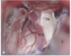
Surface anatomy of the Sylvian fissure?
Marked by a line drawn from the lateral canthus to a point 75% of the distance from the nasion to the external occipital protuberance

Surface anatomy of the central sulcus
4-5cm posterior to the coronal suture
This is also at a point approximately 2cm posterior to the mid-position of the arc joining the nasion and the external occipital protuberance

Surface anatomy of the SSS?
Runs posteriorly from the nasion to the external occipital protuberance in the midline
Surface markings of the transverse sinus?
From the level of the occipital protuberance towards the mastoid at the same level as a line projected posteriorly from the zygomatic arch.

Subarachnoid cisterns:
1

Olfactory cistern
Subarachnoid cisterns:
2a
2b

Callosal cistern
Lamina terminalis cistern
Subarachnoid cisterns:
3
4

Chiasmatic cistern
Carotid cistern
Subarachnoid cisterns:
5
6
7
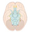
Sylvian cistern
Crural cistern
Interpeduncular cistern
Subarachnoid cisterns:
8
9
10

Ambient cistern
Superior CP cistern
Pre-pontine cistern
Subarachnoid cisterns:
11
12
13

Inferior CP cistern
Anterior spinal
Posterior spinal
Cistern contents:
Lamina terminalis
AComm and branches
Cistern contents:
Chiasmatic
Precommunicating ACA
Optic nerves
Cistern contents:
Carotid
ICA
Pcomm origin
Anterior choroidal
Cistern contents:
Sylvian
MCA
Cistern contents:
Crural
Anterior choroidal
Medial posterior choridal
Cistern contents:
Interpeduncular
Basilar bifurcation
PCAs
III
Cistern contents:
Ambient
PCA
SCA
Basal veins
IV
Cistern contents:
Quadrigeminal
Vein of Galen
Distal pericallosal arteries
Distal PCA and SCA
IV
Cistern contents:
Prepontine
Basilar
AICA
SCA
VI
Cistern contents:
Premedullary
Vertebral
PICA
XII