Overview and Embryology COPY Flashcards
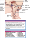
Efferent neurone columns
3
Somatic
Branchial
Visceral
Arrangement of efferent cell columns from M->L
SBV
Somatic
Branchial
Visceral
Afferent neurone columns
Special visceral
General visceral
Special somatic
General somatic
Arrangement of afferent cell columns from M-> L
VG VS, SG, SS
Visceral General
Visceral special (taste)
Somatic general
Somatic special (hearing, vestibular)
To what do the colours correspond
Blue- sensory
Orange- intermediant visceral (i.e. autonomic)
Red motor
Pure corticospinal tracts in humans
Thought to be exceedingly rare due to close proximity to other corticofugal tracts (corticoreticular, corticopontine, reticulospinal, vestibulospinal).
Thought to result in deficits in delicate fractionate movement and Babinski
Fucntion of ventral rami of spinal nerves
Innervates limbs and anterior skin and muscles of trunk
Function of dorsal rami of spinal nerves
Postvertebral muscles and skin of back
nAChR
Inotropic receptor
mAChR
Metabotropic receptor (GPCR)
Formation of the cerebelleum
Forms from dorsolateral thickenings of metencephalon which overgrow the root of the fourth ventricle (rhombic lips)
These lips fuse in the midline to form the cerebellar vermis.
Peripheral neuroblasts contribute to cerebellar cortex, central-> deep nuclei
Origin of tectal nuclei
Neuroblasts from the alar plates migrate into the tectum to form the superior and inferior colliculi
Origin of central gray matter around aqueduct
Neurobalsts of the alar plates
What are the three major commissures beign to develop in the lamina terminalis
Anterior commissure
Hippocampal commissure
Corpus callosum
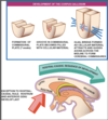
Describe the development of the corpus callosum
7th week
The dorsal aspect of the lamina terminalis thickens into the commissural plate which becomes thickened with cellular material that forms a glial bridge across the groove
Develops in rostral to caudal fashion.
The exception is the rostral most portion of the corpus callosum- rostrum and anterior genu which develop last
What might cause violation of the corpus callosal front to back development
Secondary destructive processes might damage the corpus callosum after it is already formed
How does the presentation of corpus callosum agenesis occur?
Due to front to back development, then developmental arrest will normally result in an intact genu with a partially or completely formed body and small or absent splenium or rostrum.
Small or absent genu but intact splenium and rostrum in corpus callosal agenesis suggests?
Secondary destructive process
What is the corpus callosum abnormality which is an exception to the front to back and secondary destructive lesion
The callosal abnormality associated with holoprosencephaly in which the corpus callosum demonstrates and intact splenium in the absence of a genu or body.
Agenesis of the corpus callosum
Cardinal features of corpus callosum agensis
Hypogensis of the corpus typically produces an intact genu and body with absent splenium and rostrum.
Other patterns suggest secondary destructive process
Associated anomalies, corpus callosum agenesis?
Dandy-Walker
Disorders of neuronal migration, organisation
Encephaloceles
Symptoms of callosal agenesis
Seizures, mental retardation.
Commonly also related to associated brain abnormalities
Aicardi’s syndrome
X-linked disorder
Infantile spasms, callosal agenesis or hypogensis, chorioretinopathy
Abnormal EEG
Cranium bifidum
Defect of neural tube closure
Cranial defect with herniation of meniniges (meningoceole) or meninges and brain (meningoencephalocoele) or meninges, brain and ventricles (meningohydroencephalocoele)
Varies from no functional impairment to severe motor and mental impairment with seizures
Lissencephaly
Absence of cortical gyri
Mental retardation and hypotonia or spasticity
Lissencephaly
Polymicrogyria
Overabundant, undersized, cortical gyri
Mental retradation and hypotonia
Pathophysiology of intracranial lipomas
Mesenchyme gives rise to the leptomeninges
Abnormal differentiation of this mesenchyme may lead to the formation and deposition of fat in subarachnoid space
Location of intracranial lipomas from most to least common
Deep interhemispheric fissure
Quadrigeminal plate cistern
Interpeduncular cistern
CPA
Sylvian cistern
Interhemispheric lipomas
Also known as lipomas of the corpus callosum, associated with hypogensis or agenesis of the corpus callosum.
May also be evidence of punctate or curvilinear midline calcifications or the presence of other anomalies such as encephaloceles and cutaneous lipomas
Interhemispheric lipoma
Quadrigeminal plate lipoma
Interpeduncular lipoma
Def: Cephalocoele
Skull base or calvarial defect assocaited with herniaton of intracranial defects
Aetiology of cephalocoeles
Skull base cephalocoeles are defects of enchondral bone- defects in induction of bone or disunion of basilar ossification
Calvarial- defects of membraneous bone either caused by defect of bone induction or mass effect and pressure erosion by an expanding intracranial lesion or failure of neural tube closure.
Most common locations of cepalocoeles
Occipital
Frontoemthmoidal
Parietal
Nasopharyngeal
Associations with occipital cephalocoele
Callosal anomalies
Anomalies of neuronal migration
Chiari malformation
Dandy-Walker
Poor Px include HCP, microcephaly, meningoencephalocoele
Which type of cephaloceole has least favourable prognosis
Occiptal
Frontoethmoidal cephalocole
Results from failure in normal regression of dura that extends from cranial cavity to the skin through persistent foramen caecum or fonticulus frontalis
Persistence may give a dermal sinus tract which can give rise to dermoid or epidermoid tumour
Examination reveals superficial skin-covered mass or nasal dimple with hypertelorism
Frontoethmoidal cephalocoele
Subtypes of frontoethmoidal cephalocoele
Frontnasal ephalocle
Frontoethmoidal cephalocoele
naso-orbital cephalocele
Parietal cephalocel
Uncommoon
Poor prognosis as commonly associated with major anomalies including Dandy-Walker, callosal agensis, Chiari II and holoprosencephaly
SSS involvement is common
Atretic cephaloclee
Small, hairless, midline masses associated with sharply marginated calvarial defect and high incidence of midline anomalies e.g. porencephalies, interhemispheric cysts and callosal agenesis
Nasopharyngeal cephalocele
Very uncommon
Occult, often not presenting until 10y/o where patient presents with nasal sutffiness of excessive mouth breathing
O/E: Nasopharyngeal mass that increases with Valsalva.
Associated with callosal agensis and may include tethering of hypothalmaus and optic chiasm resulting in endocrine and visual dysfunction
Dermal sinus
3-5th week
Defect in separation of neuroectodem from surface ectoderm
Sinus commonly contains elements of both dermal and epidermal tissue
Midline found anyway between nasion and coccyx but commonly between the glabella andanion
May have associated cysts along tract
Dermal sinus
Clinical presentation of dermal sinus
Benign cutaneous cosmetic blemish to serious intracranial infection or tumourlike process due to mass effect from dermoid or epidermoid cyst
May have associated angiomata, abnormalities of pigmentation, hypertrichosis, abnormal hair pattern, subcutaneous lipomata, skin tags
Def: Arachnoid cysts
CSF containing lesions covered by membranes that consist of arachnoid cells and collagen fibres that are continuous with the surrounding arachnoid.
Pathophysiology of arachnoid cysts
Anomalous splitting and duplication of endomeninx which normally forms a loose extracellular substance in the future subarachnoid space.
Common locations of arachnoid cysts
2/3rds supratentorial most commonly Sylvian cistern, others include suprasellar, interhemispheric, intraventricular
1/3rds infraentroial- divdied between CPA, posterior to vermis and superior to quadirgeminal plate
Presentaiton of arachnoid cysts
Grading of middle cranial fossa arachnoid cysts
Galassi classification
Galassi 1
Small,spindle shaped
Limited to the anterior portion of the middle cranial fossa below sphenoid ridge
Free communication with subarachnoid space
Galassi 2
Superior extent along sylvian fissure
Displacement of temporal lobe
Slow communication with subarachnoid space
Galassi 3
Large
Fills whole middle cranial fossa
Dispalcement of temporal, frontal and parietal lobes
Often results in MLS
Little communication with subarachnoid space
What is the exception to the inside out pattern of neuronal migration
The neurones that form the most superficial layer- the molecular layer which seem to migrate first
Anomalies as a result of defect in neuronal migration
Tend to cause malformations in which cortical neurones are residing in abnormal locations or patterns.
Lissencephaly
Heterotopia
Polymicrogyria
Schizencephaly
Lissencephaly
Associated with severe mental retardation
Defective migration of cerebral neurones results in failure of cortical gyri to develop.
Cerebral hemispheres are smooth with absent cortical sulci and cerebral fissures are shallow.
Microscopically aberrant cortical cell layers
Lissencephaly etymology
Derives from greek lissos- “smooth”
Heterotopia
Collections of normal cortical neurones that fail to reach the cortex as a defect in radial neuronal migration.
May occur in isolcation or in association with other brain anoamlies.
Subtypes based on location and pattern of organisation
Heterotopia
Heter- other
Topia- place
Subtypes of heterotopia
Subependymal heterotopia
Focal subcortical heterotopia
Band heterotopia
Normal development, normal motor function.
Onset of seizures in second decade of life
Subependymal heterotopia
Dependant on size can present with normal to severely abnormal developmental delay
Motor disturbances in association with seizure disorders
Focal subcortical heterotopia
Moderate to severe developmental delay
Medically intractable seizures
Band heterotopia (AKA diffuse gray matter heterotopia)