The Posterior Abdominal Wall Flashcards
Some specific bones create the framework for the posterior abdominal wall (PAW). What are 4 bones (groups) labelled 1-4 in the figure below that create the framework for the PAW?

1 - vertebral column
2 - ilia (pleural of iliac)
3 - sacrum
4 - ribs 11-12
Where does the posterior abdominal wall (PAW) begin in relation to the ribs?
- ribs 11 and 12
Which vertebrae contribute the majority of the posterior abdominal wall?
- lumbar vertebrae
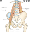
There are 4 main muscles that contribute towards the posterior abdominal wall, labelled 1-4 in the image below. What are there names?
- diaphragm is also included
1 - quadratus Lumborum
2 - iliacus
3 - psoas minor
4 - psoas major

The diaphragm is one of the 4 major muscles that contributes towards the the posterior abdominal wall. Where does it originate from?
- cura (left and right tendons) from lumbar vertebrae & discs
- costal cartilages of ribs 7-10
- xiphoid process

What are the 2 major componenets of the diaphragm?
- muscle and tendon
- muscle become aponeurotic and forms central tendon

What are the 3 passageways of the diaphragm? This mnemonic may help: I Eight 10 Eggs At 12 oclock.
- I Eight = Inferior Vena Cava at T8
- 10 Eggs = Oesophagus at T10
- At 12pm = Aorta at T12

The posterior aspect of the diaphragm contributes the majority of the of the diaphragm towards the posterior abdomainl wall. The are 2 parts of the diaphragm that anchor it to the vertebrae, what are these called?
- crus (means anchor or root)
- thick attachment
- right is thicker and more extensive

The posterior aspect of the diaphragm contributes the majority of the of the diaphragm towards the posterior abdomainl wall through the left and right crus (means anchor or root). The right is thicker and more extensive, why is this important?
- forms a loop around the oesophagus
- forms a physiological sphinter for the oesophagus
- reduces reflux on stomach into oesophagus

The quadratus lumborum is one of the 4 major muscles that contributes towards the the posterior abdominal wall. Where does it originate and insert?
- origin = iliac crest
- insertion = rib 12 and T12 down to L5 vertebrae
- merges laterally to transversus abdominis muscle

The quadratus lumborum is one of the 4 major muscles that contributes towards the the posterior abdominal wall. What is the function of the quadratus lumborum?
- abdominal stability/lateral flexion
- ensures rib cage doesnt shift too much during respiration
The psoas major is one of the 4 major muscles that contributes towards the the posterior abdominal wall. Where does the psoas major originate and insert?
- origin = T12 down to L5 on transverse processes & vertebral bodies
- insertion = lesser trochanter of femur

The iliacus is one of the 4 major muscles that contributes towards the the posterior abdominal wall. Where does the iliacus originate and insert?
- insertion = trochanter minor femoris
- origin = iliac surface/fossa of hip (pelvic) bone on each side

The iliacus and the psoas major are 2 of the major muscles that contributes towards the the posterior abdominal wall. These muscles merge in the pelvis and connect to which bone?
- femeur
In addition to the 4 main muscles that make up the posterior abdominal wall PAW), what other large muscle group that allows flexion and extension of the spine completes the PAW?
- erector spinae

The erector spinae, quadratus Lumborum and psoas major all form part of the posterior abdominal wall (PAW). But are they in direct contact or are they seperated by something?
- seperated by a thick deep fascia
- fascia is called the thoracolumbar fascia

The erector spinae, quadratus lumborum and psoas major all form part of the posterior abdominal wall (PAW). The thoracolumbar fascia is a thick deep fascia that seperates the erector spinae and the quadratus lumborum. How many layers of the thoracolumbar fascia are there?
- 3
- anterior
- middle
- posterior

The erector spinae, quadratus lumborum and psoas major all form part of the posterior abdominal wall (PAW). The thoracolumbar fascia is a thick deep fascia that seperates the erector spinae and the quadratus lumborum. Which layers of the thoracolumbar fascia surround the quadratus lumborum?
- anterior and medial

Ther fasica, such as the thoracolumbar fascia that contribute to the posterior abdominal wall have 2 main functions, what are they?
1 - maintain muscle shape
2 - protect muscles from infection
Is the psoas major surrounded by the thoracolumbar fascia?
- no
- has its own fascia
In patients who have tuberculosis (TB), there is a risk that the infection can spread to the vertebral bones. This can infect the muscles attaching to the vertebrae. How does the fascia of the psoas major protect other muscles and tissues within the abdomen from the TB infection?
- fascia ensure within the fascia only affecting the psoas major
- eventially collects in groin as an abscess

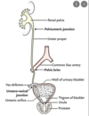
There are 3 sites of constrcition in the urinary system. The first site is located where the renal pelvis meets the uereter. Why is there a constriction at this site?
- the renal pelvis narrows to facilitate flow into the ureter

There are 3 sites of constrcition in the urinary system. The second site is located where the ureter crosses over the iliac, called the iliac vessels/pelvic brim. Why is there a constriction at this site?
- ureter is crossing a ridge in the bone which compresses the ureter

There are 3 sites of constrcition in the urinary system. The third site is located where the ureter meets the bladder/ischial spine. Why is there a constriction at this site?
- the ureter narrows to enter the bladder

Why are the 3 contraction areas of importance in the urinary tract?
- these sits are where renal stones are most likley to occur

What are uteric calculi more commonly known as?
- kidney stones

On radiology where would the 3 site on constriction of the urinary system Pelvi-ureteric junction/L2-L3, Crossing iliac vessels/pelvic brim and Entering bladder/ischial spine. Where would these come up on radiography?
- 1st site = L2-L3 (transverse process)
- 2nd site = sacroiliac joint
- 3rd site = ischial spine
The abdominal aorta has unpaired, paired, paired somatic and terminal blood vessels. What are the unpaired visceral blood vessels?
- visceral arteries supply the gut (ventral)
- coeliac, superior mesenteric artery, inferior mesenteric artery
The abdominal aorta has unpaired, paired, paired somatic and terminal blood vessels. Lebel the unpaired visceral blood vessels in the image?

1 - superior mesenteric artery
2 - inferior mesenteric artery
3 - coeliac artery
The abdominal aorta has unpaired, paired, paired somatic and terminal blood vessels. What are the paired visceral that originate from the abdominal aorta?
- supply the gut in pairs
- suprarenal (adrenal) aerteries
- renal arteries
- gonadal arteries

The abdominal aorta has unpaired, paired, paired somatic and terminal blood vessels. Label the paired visceral arteries that originate from the abdominal aorta in the image?

1 - right renal artery
2 - right gonadal artery
3 - left renal artery
4 - left gonadal artery
The abdominal aorta has unpaired, paired, paired somatic and terminal blood vessels. What are the paired somatic arteries that originate in the abdominal aorta?
- somatic means not supplying organs but tissue of abdomen
- inferior phrenic arteries
- lumbar arteries
The abdominal aorta has unpaired, paired, paired somatic and terminal blood vessels. What are the terminal arteries that originate in the abdominal aorta, and label them in the image below?

1 = right common iliac artery
2 = left common iliac artery
At what verterbrae does the abdomainal aorta bifurcate into the right and left iliac arteries?
- L4-5

At what verterbrae do the renal and gonadal arteries come away from the abdomainal aorta?
- L2

At what verterbrae does the superior and inferior mesenteric arteries come away from the abdomainal aorta?
- superior = L1
- inferior = L3

At what verterbrae does the inferior vena cava form from the right and left iliac veins
- L5

At what verterbrae does the coeliac artery originate from the abdomainal aorta?
- T12

Do the veins of the abdominal wall have different names, or are they mirrors of the arteries?
- same as arteries

What are pre-aortic lymph nodes?
- means infront of the aorta
- examples include coeliac lymph nodes
What are para-aortic lymph nodes?
- located on either side of the aorta in pairs generally
- example include renal lymph nodes
What do the lymph nodes follow in the abdominal wall and get their names from?
- follow blood vessels
- names are generated from blood vessels

What is an aortic aneurysm and is it likely to occur above or below the kidneys?
- weakening of blood vessesl that causes them to expand in increase risk of rupture
- below renal arteries

Anywhere in the systematic blood vessels can become occluded. If occlusion occurs at the aortic bifurcation, what can patients experience?
- claudication (pain) is felt generally on the lower limbs
- can lead to impotence in men as well if gonads are affected

What is somatic innervation?
- innervation responsible for voluntary control
- includes sensory receptors
Innervation of somatic structures of the posterior abdominal wall comes under the control of what?
- thoracic and lumbar spinal nerves
- provided by the lumbar and sacral plexus

What are dermatones?
- area of skin supplied by a single spinal nerve
- clinicians can determine whether there is damage to the spinal cord
- importantly where the damage in the spine is

What does the autonomic control of the posterior abdominal wall innervate?
- involuntary control
- visceral/ splanchnic / organs (controls peristalsis of stomach, gall bladder emptying etc)
What is the chain controlling the sympatheticv activity of abdomen?
- sympathetic trunk
- lots of ganglia lining the trunk

Pre-vertebral refers to infront of the vertebrae, muscles of the posterior abdominal wall and aorta. What are the 3 ganglia of the prevertebral ganglia that we need to know that are linked to blood supply for the fore, mid and hindgut?
-supplies the 3 main ventral arteries of the abdominal aorta
1 - celiac ganglia
2 - superior mesenteric ganglia
3 - inferior mesenteric ganglia

Where does parasympathetic innervation of the abdomen and its contents come from?
- vagus nerves
- pelvic splanchnic nerves originating in S2-S4

The vagus nerve provides a large proportion of the parasympathetic innervation of the abdomen and its contents. Where does the vagus nerve innervate from and to?
- stomach down to splanchnic flexure (transverse colon)
The vagus nerve provides a large proportion of the parasympathetic innervation from the stomach down to splanchnic flexure (transverse colon). What nerve supplies the rest of the hind gut?
- pelvic splanchnic nerves
- originate from S2-4 supplying the pelvic organs

There are 3 paired sympathetic splanchnic nerves that feed into the celiac, superior and inferior mesenteric ganglion, what are they?
- greater splanchnic nerves
- lesser splanchnic nerves
- least splanchnic nerves

There are 3 paired sympathetic splanchnic nerves, the greater, lesser and least splanchnic nerves. Which vertebrae does the greater splanchnic nerve originate from and innervate?
- T5-9
- foregut

There are 3 paired sympathetic splanchnic nerves, the greater, lesser and least splanchnic nerves. Which vertebrae does the lesser splanchnic nerve originate from and innervate?
- T9-10/11
- Midgut

There are 3 paired sympathetic splanchnic nerves, the greater, lesser and least splanchnic nerves. Which vertebrae does the least splanchnic nerve originate from and innervate?
- T11-12/L1
- Hindgut

There are 3 paired sympathetic splanchnic nerves, the greater, lesser and least splanchnic nerves. Do all of these enter the sympathetic chain?
- no
- great and lesser bypass
- least comes from sympathetic trunk

The greater and lesser splanchnic nerves bypass the sympathetic trunk, but synapse at what instead?
- celiac ganglia

Which nerves supply the kidneys out of the greater, lesser and least splanchnic nerves?
- lesser and least splanchnic nerves
- MOSTLY least though
Which nerves supply the suprarenal (adrenal) glands out of the greater, lesser and least splanchnic nerves?
- coeliac plexus and greater and lesser splanchnic nerves

What is somatic pain?
- pain felt from the somatic structures
- skin, fascia, muscle and/or parietal peritoneum
Somatic pain is pain felt from the somatic structures, such as the skin, fascia, muscle and/or parietal peritoneum. What characteristic is the pain described as?
- very precisely localised and sharp
Somatic pain is pain felt from the somatic structures, such as the skin, fascia, muscle and/or parietal peritoneum. This type of pain is very precisely localised and sharp. If this pain is felt on only one side, what is this called?
- lateralised
What is visceral pain and where does it come from in the abdomen?
- pain from the organs
- abdominal organs, mesenteries and visceral peritoneum
Visceral pain in the abdomen comes from organs and could be from the abdominal organs, mesenteries and visceral peritoneum. What causes visceral pain?
- stretching viscus or mesentery (e.g. stones) (swelling of an organ)
- impaired blood supply to viscus (ischaemia)
- chemical damage to viscus (e.g. ulcers)
Visceral pain in the abdomen comes from organs and could be from the abdominal organs, mesenteries and visceral peritoneum. How is visceral pain generally characterised?
- dull and poorly localised
- pain is referred to midline (due to the embryological origin)
- colic pain is a form of visceral pain
What is another name for the arcuate line?
- linea semicularis
- douglas line


