Imaging of the Abdominal Viscera Flashcards
Is radiography or CT more accurate in abdominal imaging?
- CT
What are the 4 key benefits of using X-ray imaging?
- cheap and quick
- easy for patient
- high spatial resolution (good for bone)
- low radiation dose
What are the 2 key negatives of using X-ray imaging?
- poor contrast resolution (poor for soft tissue)
- 2D imaging (superimposes structures)
What are the 3 key benefits of using CT scanners?
- quick and widely available
- cross sectional images instead of 2D
- high contrast resolution (good for soft tissue)
What are the 2 key benefits of using CT scanners?
- radiation dose
- IV Contrast die increases risks
What are the 3 key benefits of ultrasound in imaging?
- cheap, quick, no radiation
- cross sectional images
- offers US guided interventions
What are the 2 key negatives of ultrasound in imaging?
- operator dependent
- saved images are only a snapshot of examination
What are the 3 key benefits of MRI in imaging?
- contrast resolution
- specific applications (e.g. small bowel)
- no radiation
What are the 4 key negatives of MRI in imaging?
- limited availability
- patient experience (duration, claustrophobia)
- expense
- magnet / contrast die risks

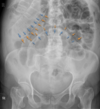
Where should the X-ray cover in an abdominal X-ray?
- pubis bone up to diaphragm
- ilias of hips

In an abdominal X-ray should patients inhale or exhale on an X-ray?
- image is captured during exhalation
If a patient presents with abdominal pain only, is an X-ray, CT or ultrasound more appropriate?
- erect ultrasound or CT
- not an X-ray
If a patient has a clinical obstruction, would an abdominal X-ray be suitable?
- yes
If a patient has an acute exacerbation of inflammatory bowel disease, would an abdominal X-ray be suitable?
- yes
If a patient has a -alpable mass in the abdomen, would an abdominal X-ray be suitable?
- yes, but only specific cases
Constipation is generally not an indication for an abdominal X-ray. What patient group with constipation would an abdominal X-ray be suitable for?
- elderly patients
In acute and chronic pancreatitis is an abdominal X-ray always suitable?
- not always
- suitable in specific circumstances
If a patients has a sharp or potentially poisonous foreign body inside the abdomen, is an abdominal X-ray always suitable?
- yes
If a patients has a suspected smooth and small foreign body (e.g. battery) trapped in the abdomen, is an abdominal X-ray always suitable?
- yes
If a patients has a suspected blunt or stab injury in the abdomen, is an abdominal X-ray always suitable?
- yes
Solid organs and tissue can be detected on an abdominal X-ray. What are the key solid organs we should be able to identify?
- liver
- kidneys
- ilios muscles

When using abdomainl X-rays, what makes it difficult to identify all the organs of the abdomen?
- trapped gas in organs
Some organs in an abdominal X-ray can be identified due to the prescence of gas or calcification. What are some key hollow organs that we may be able to identify on an abdominal X-ray?
- stomach
- small and large bowel
- gall bladder
- urinary bladder
What are a few of the anatomical landmarks that need to be included on an abdominal X-ray?
- diaphragm
- L4 vertebral body
- iliac crest
- left and right flanks
- sacrum and superior pubic ramus

Air can be observed in the small and large bowels, but is this when the bowels are dilated or non-dilated?
- non dilated only
On abdominal X-ray imaging what should be contained within the large and small intestines?
- small = fluid and gas
- large = stool and gas
What are haustra in the GIT?
- small segmented pouches of the bowel
- separated by the haustral folds
- formed by circumferential contraction of the inner muscular layer

Are haustra present in the small and large bowel?
- no
- large bowel only
What is the function of haustra in the colon?
- peristaltic contracts
- avtivated by chyme prescence
What are valvulae conniventes, also known as plicae circulares of the GIT?
- folds within the mucousa of the intesintes
- smaller than Haustra

Are valvulae conniventes, also known as plicae circulares present throughout the GIT?
- no
- just small intestines
Can solid faeces be found throughout the GIT?
- no
- generally just the large intestines

On an X-ray, where are the small and large intestines located visually?
- small intestines = centrally
- large intestines = peripherally

Is the large or small bowel longer, and why is this?
- small bowel is longer
- important for digestion and absorption

Does the large or small bowel have more loops?
- small bowel has more loops
Does the large or small bowel have a larger diameter loop?
- large bowel has larger loops
- large = + 5cm
- small = <3cm
In the image below, are we looking at the small or large intestines, and how do we know?

- small intestines
- centrally located
- contains valvulae conniventes running full width of bowel
- does not exceed 3mm diameter – if normal
In the image below, are we looking at the small or large intestines, and how do we know?

- large intestines
- peripherally located
- haustra present, do not cover full width of bowel
What is circled on the X-ray present in the large bowels?

- faeces and air
- present in left upper quadrant
- middle near pelvis where rectum is
In terms of malignancy, what is one of the reasons why a CT of the abdomen may be requested?
- suspected tumour
- staging (T.N.M)
- determine if its operable
Is a CT scan recommened in patients with supsected liver and pancreatic disease?
- yes
- shows liver cirrhoisis and pancreatitis
Why can CT scans of the abdomen be useful in patients who may have an obstrutction?
- identify location of obstruction
- identify potential cause of obstruction
If a patient has been involved in trauma to the abdomen, why would a CT scan of the abdoment be useful?
- assess damage
- bleeding
Why can CT scans of the abdomen be useful diagnostically?
- identify where to take biopsy from
In the CT scan below, what do the black circles depict?

- gas in the bowels
- could be an obstruction caused by cancer obstruction
What is a bowel CT colonoscopy?
- bowels are inflated with CO2
- CT imaginging is then captured

What are the benefits of using CT colonoscopy?
- able to replace barium enemas
can assess for strictures/ Ca (carcinoma) of the colon
- minimally invasive and low dose
- comparable accuracy to optical colonoscopy
What imaging modality is being used in the image below?

- CT colonoscopy
Who are the most common clinicians who may recommend an MRI of the abdomen?
- hepatologists
- gynaecological
- urologist (prostate)
What is MRI enterography?
- used to investigate GIT
- a painless, noninvasive, and radiation-free medical imaging test
- patients consume a contrast die
- contrast die detected by MRI

What is MRI cholangiopancreatography (MRCP)?
- imaging using MRI to create 3D image
- imaging of the biliary tree and pancreatic ducts is performed
- gall bladder stones can be seen on the image

What is a sonographer?
- expert who performs ultrasounds
When is an ultrasound useful by clinicians?
- when studying solid GIT organs
- jaundice, gallbladder disease, abdominal bloating, mass
Do ultrasound generally get performed in one place?
- no
- generally transverse and longitudinal
The image on the left shows a healthy gallbladder, what do the 2 oval shapes represent in the image on the right?

- gallbladder stones
In the abdominal X-ray below what can we see and what could be a likley cause of this?

- large bowel obstruction
- descending bowel
- could be a mass
What does perforation mean?
- loss of integrity of the bowels
- commonly known as bowel rupture
- air is present outside the bowel
The image below demonstrates a patient with a perforated bowel. What do the blue and orange arrows depict and what are other signs that may be useful?

- patient is distended
- blue = air inside bowel outlining the lumen (normal finding)
- orange = outer surface of bowel visible due to air outlining
What is riglers sign?
- a sign of pneumoperitoneum (air in peritoneum)
- gas is visible inside and outside of the bowel wall

Why can an erect chest X-ray be useful in a patient with a suscpected perforation?
- generally performed post abdominal surgery
- useful to detect a thin crescent of air
- present under the right hemidiaphragm and above liver
- can be normal post surgery, if not this is abnormal

What does the image below depict, that would not normally be present on a normal X-ray?

- calcifications
- yellow depict calcification in kidenys (likely kidney stones)
- red depicts calcification in gallbladder (gall stones)

What imaging modality is this image and what is depicted in the abdominal image below?

- X-ray
- aortic aneurysm

What imaging modality is this image and what is depicted in the abdominal image below?

- CT with contrast
- aortic aneurysm
