Spine Anatomy FRCS Flashcards
How many vertebra are in the spine?
7 cervical
12 thoracic
5 lumbar
5 sacral
4/5 coccygeal

Name the normal curves of the spine?
-
Cervial lordosis
- normal 20-40o
-
throacic kyphosis
- average 35o
-
lumbar lordosis
- Average 60o
- 75% LL occurs between L4-S1
- sacral kyphosis
What happens to the vertebral bodies size in an cranialcaudal direction?
They increase with the EXCEPTION OF T1- T3
What spinal level does the
mandible
hyoid cartilage
cricoid cartilage
vertebral prominens
scapular spine
dorsal tip of scapula
iliac crest relate to ?
- Mandible C2-3
- Hyoid C3
- thyroid carilage C4-5
- Cricoid cartilage C6
- Vertebra prominens C7
- Scapular spine T3
- Distal tip of scapula T7
- Iliac crest L4-5
Name the spinal ligaments?
- 3 sets all contribute to static stablity of spine
-
Anterior longitudinal ligament ALL
- Thicker centrally
- thicker than PLL
-
Posterior longitudinal ligament PLL
- thicker over body
- thinner over disks
- Facet joint capsule/ligaments
Describe the Anterior longitudinal ligamant?
- Strong
- Thickest in CENTRE of vertebral body adn thinnest at periphery
- resists HYPEREXTENSION
Describe the POSTERIOR LONGITUDINAL LIGAMENT?
- Weaker than ALL
- extends occiput to sacrum
- hourglass shaped with widest part over the discs- rupture of discs normally LATERAL to these expansions
1) What is the first vertebra called?
2) Describe it?
1) ATLAS- formed form 3 ossificaiton centres- ( 2 lateral masses and one vertebral body- fuse age 7)
2) No Vertebra body and spinous process
2 concave superior facets that articulate with the occciptal condyles

What movements happens at occiput- c1 articulation?
- highest precentage 50% of total neck FLEXION and EXTENSION occurs here
1) What is the second cervical vertebra called?
2) What does it develop from?
3) When does it fuse?
1) AXIS- has Ondontoid process (dens) and Body
2) from 5 ossification centres, with an inital cartilaginous junction between the dens and vertebra body
3) dens and body age 7 years
secondary ossification centre at tp of dens fuses age 12 yrs

Why does the base of the dens narrow
- tranverse ligament

a) What movement is responible at the ATLANTOAXIAL articulation?
b) What stabilises this joint?
C) Describe its blood supply?
a) Majority of neck CERVICAL ROTATION- 50% of total, 10 FLEXION
b 1)The TRANSVERSE LIGAMENT- limits anterior movement
2) APICAL LIGAMENT- (LONGITUDINAL) -limits rotation
TOGETHER FORMS THE CRUCIATE LIG
3) ALAR LIG- check ligaments runs form dens to occiput-limits rotoation of upper cervical spine
See ortho bullets picture
c) apex = internal carotid artery, Base = branches of vertebral artery.
Watershed between apex and base of dens-thougth to be why type II fractures don’t heal so well!!

1) What is the importance of this joint in RA pts?
2) Why is this important?
2) A pannus can form in the atlantoaxial joint -> instability
2) Require flexion/extenstion cervcial spine in RA pts prior to elective proceedure
Describe the anatomy of the cervical spine C2-C7?
- Foramina in each transverse process-
- Bifid spinous processes- except C7

1) What travels in the transverse foramina of C6-C1?
2) What is the orientation of the superior articulating facets C3-C7?
1) Vertebral artery, except C7
2) Oriented in a
- POSTEROMEDIAL direction at C3
- POSTERIOLATERAL direction at C7, with a variable transition between these levels
- Allows greater rotation cranial
- superior articulating facet of cranial vertebra is anterior to inferior facet of caudal vertebra

What is the normal diameter of the C spine canal?
- 17mm
- c spinal cord can be compromised < 13mm
1) Describe the features of thoracic spine?
2) What is the normal thoracic kyphosis?
1) Costal facets on all 12 vertebral bodies
- (articulation between ribs and vertebral) On all vertebral bodie and transverse process of T1 to T9
- Rounded vertebral foramen- increase in size T1-12
- Long spinal prominence of T1 in upper cervicothoracic spine so used for fixation= vertebra prominens ( T12 largest in thoracolumbar region)
- T4 has the narrowest pedicle diameter- diameter decreases from T1-4 then increases again
- T1 largest diameter cervicalthoracic, T12 largest thoraciclumbar
- Pedicle is x2 thickness medial cf laterally
- orientation- transverse 10 degrees, sagittal 15-17degrees
2) average 35 degrees

1) What are the mechanical feature of the thoracic spine ? 2) Why is this?
1) Most rigid region of the axial skeleton
2 ) Articulations with rib cage
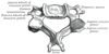
1) Describe lumbar vertebrae?
2) What is the degree of lordosis?
3) what direction do the pedicles orientate as you move caudally?
1) Largest vertebra
- Higher Anteriorly than posteriorly -> lumbar lordosis with aid of discs
- Rectangular non overlapping spinous processes
- Apex of lordosis is L3
- Short laminae and pedicles
Made up anterior vertebral bodyposterior arch(pedicles( postlat projection) andlaminae( post medial project and join in midline)),spinous porcess,transverse process,mammillary process(separate ossification centres-project posteriorly from superior articular facet ,pars interarticularis (bone between sup and inf articulating facets)
2) average lordosis = 60 degrees
3) MEDIALLY
L1 has the smallest diameter of pedicle

Describe the facet joints?
- also called zygapphyseal joints
- Orientation varies throughout the spine in accordance with predominant direction of allowed motion at the level.
- cervical - 45o sagittal to allow flexion- extension, lateral flexion, rotation
- thoracic- allows some rotation, minimal flexion/extension, prevents downward flexion on heart and lungs
- lumbar- 90o sagittal, allows flexion/extension,minimal rotation
- Superior tip of inferior articulating process is a major offending structure in lumbar foraminal stenosis

Describe the intervertebral disc complex?
- Intervertebral disc and vertrebral bodeis support more than 80% of axial load transmitted through the spine
- Disc essential to the functional spinal unit which comprises the vertebra above and below the disk and associated paired facet joints at that level
- consists of outer fibrous annulus fibrosis - obliquely orientated collagen type 1 moelcules
- inner core- nucleus pulosus - type 2 - cushions force

What is the ligamentum flavum?
How can it cause nerve compression?
- A yellow elastic ligament connecting the laminae
- Runs anterior surface of SUPERIOR laminae to POSTERIOR surface of INFERIOR LAMINA
- usually CONSTANT in TENSION
- By HYPERTROPHY

Describe the ligaments in the spine?
1) Supraspinous ligament- lies dorsal to the spinal processes
* Begins at C7 and incontunity with ligamentum nuchae ( C7 to occiput)
2) Interspinous ligament- between the spinous processes

Describe Denis Model of Instability?
- Divided spine into 3 columns
- Anterior= ALL, anterior 2/3rds of ANNULUS & vertebral body
- Middle= Post 1/3rd of body and ANNULUS, PLL
- Posterior= Pedicles, facets, facet capsule, spinous processes, PLL than includes- interspinous adn supraspinous ligmants, ligamentum flavum

What is the sacrum formed from?
What do the foramina transmit?
- 5 fused spinal elements
- 4 sacral foramina thru which S1-S4 nerve roots runs
- S5 root runs inferiorly thru sacral hiatus
- The anterior and posterior foramina transmit ventral and dorsal branches of upper sacral nerves
- sacrum promontory project into pelvis
- sacral canal opens up into sacral hiatus

Describe the coccyx? What is attached to it?
- fusion of 4 lowest spinal elements
- dorsally - gluteus maximus
- external anal sphincter
- coccygeal muscles

1) What is spondylolysis?
2) what are the articulations of the lumbar spine?
3) what is the blood supply of the lumbar spine?
1) A defect in the PARS INTERARTICULARIS
* most common cause of back pain in children and adolescents
2) i) Intervertebral disc
ii) Facet joint articulations- superior and inferior facets project from junction of pedicle and lamina
3) Segmental arteries
What atlanto-dens interval would worry you?
What would this mean in elective surgery?
- ADI Normal <3mm adults
- interval More than 7-10mm worrying
or Posterior Space
- Increase in space in flexion sugggests less space posteriorly for spinal cord
- Stabilisaton should be sort first

What would an increase in ADI mean?
- DDX
-
Transverse ligament injury (ADI 3-5 mm)
- if >5mm implies injury to transverse and alar ligaments
- Jefferson Fracture
- Atlanto axial rotatory fixation
-
atlanto axial subluxation
- RA
- downs syndrome
a) What makes up the CNS?
b) Where does the spinal cord end?
a) The BRAIN and SPINAL CORD
b) At L1 level
What does the PNS consist of?
Cranial Nerves and Peripheral Nerves
What does the autonomic nervous system consist of?
- The Sympathetic and Parasympathetic nervous system
a) What does the sympathetic nervous system consist of?
b) Which ganglia is most at risk? What is observed with injury to this ganglia?
a) 22 ganglia - 3 cervical. 11 thoracic, 4 lumbar , 4 sacral
* Cervical ganglia include the middle, stellate and superior
b) Middle ganglion- at level of C6- close to medial border of the LONGUS COLI muscles
* Horner’s syndrome
What is the parasympathetic nervous system?
- Hypogastric plexus formed from S2-S4 parasympathetic fibres and lumbar sympathetic fibred- splanchnic nerves
a) Where does the spinal cord extend from and to?
b) What is the termination of the spinal cord?
a) Brain stem to L1
b) Conus Medullaris

1) What is the name of the fragment of spinal cord that extends from the conus medularis to the sacrum?
2) what is the name of the nerve roots and filum terminale surrounded by dura that extend from the spinal cord ?
1) Filum terminale
2) Cauda Equina
1) What forms the spinal cord in development?
2) What happens when this fails to fuse?
1) primitive streak deepens-> primitive midsagittal groove deepens within ectoderm and folds in on itself -> Neural Tube
- Neural tube-> spinal cord
- As closes neural crest forms DORSAL to neural tube and notochord remains ventral
- spinal cord changes position with growth
- Conus Level L3 at birth
- skeletal maturity L1-2
2) cranial- > hydrocephalus
* caudal->spinda bifida occulta, meningocele, myelomeningocele

a) What forms the peripheral nervous system?
b) Where does this form in relation to the neural tube?
c) What else does this structure form?
a) Neutral CREST
b) DORSAL
c) PIA MATER
SPINAL GANGLIA
SYMPATHETIC TRUNK

a) What forms the vertebral bodies in the embrology of the spine?
b) What other structures does it make up?
c) Where does it form in relation to the neural tube?
a) NOTOCORD and Somites
b) Interverebral DISCs- nucleus pulposus
c) VENTRAL

How many ossification centres does each vertebra have?
Name them?
- 3
- The centrum- anterior vertebral body
- Neural arch- post elements, pedicles, small portion of anterior vertebra
- costal elements- anterior portion of lateral mass, transverse process or ribs
Can you name the layers of the spinal cord?
- DURA MATER- outside
- ARACHNOID
- PIA MATER- Inside

1) Can you name, describe the location and function of the ascending tracts?
1) All SENSORY
- DORSAL COLUMNS- Deep touch, vibration and proprioception = DVP
- LATERAL SPINOTHALAMIC- Pain and Temperature
- VENTRAL** SPINOTHALAMIC**- LIGHT Touch

Can you describe the location, functions of the descending spinal tracts?
- MOTOR Descending re: cortex down- a motor (car) runs down a hill
-
LATERAL CORTICOSPINAL
- Main Voluntary motor
- UPPER LIMB pathways more MEDIAL (central) = why a central cord injury effects upper limb > lower limb
- VENTRAL CORTICOSPINAL- Voluntary motor

Can you name the blood suply to the spinal cord and what specificially they supply?
1) Anterior spinal artery= ant 2/3rds to lateral corticospinal and ventral corticospinal tracts
2) Posterior spinal artery- primary blood supply to dorsal sensory columns
3) Artery of Adamkiewicz - the largest segemental sartery

Describe where the artery of Adamkiewicz arises?
Does variation of origin occur?
1) Typically from the Left posterior intercostal artery , which branches the aorta, supplies the lower 2/3rds of the spinal cord via the anterior spinal artery
2) Yes, 75% arise on left between T8-L1 vertebral segments
* If injuried can lead to cord infraction

1) What is CSF?
2) where is the subarachnoid space?
3) what is CSF’s Function?
1) Cerebral spinal fluid is an ULTRAFILTRATE of BLOOD PLASMA- colourless fluid
* occupies the SUBARACHNOID SPACE- surrounding the brain, spinal cord, ventricular system
2) Between the _ Arachnoid mater and Pia mater_
3) Provides Mechanical and Immunological Protection for the brain, spinal cord and thecal sac.
1) Where is CSF made?
2) How much CSF exsists within the subacrachnoid space?
3) How much CSF is formed per day and what is its turnover?
1) Produced by the CHOROID PLEXUS in the 3RD, 4TH and LATERAL VENTRICLES of the brain
* An ultrafiltrate of BLOOD PLASMA thru the permeable capillaries of the choroid plexus
2) approx 150 mls
3) approx 500 mls, turnover 3-4 x per day
1) Where do the nerve roots exit in the cervical spine?
2) Where do the nerve roots exit in the thoracic spine?
2) Where do the nerve roots exit in the lumbar spine?
1) ABOVE CORRESPONDING PEDICLES e.g C5 nerve exits above C5 pedicle
- They travel HORIZONTALLY to exit
- There is an extra C8 n root without a corresponding vertebra
2) BELOW CORESSPONDING PEDICLE- T1 exits below T1 pedicle, T2 exits below T2 pedicle
3) nerve roots descend VERTICALLY before exiting
Travel BELOW CORRESPONDING PEDICLE- L1 exits below L1 and L5 below L5

Name the spinal muscles in the thoracic spine?
types of ERECTOR SPINAE
1) SPINALIS- most meidal - spinous process to spinous process
2 ) LONGISSIMUS- intermediate- transverse process to transverse process
3) ILIOCOSTALIS- most LATERAL- origin ribs and ilium- inserts ribs and transverse process

Describe the plaement for a lumbar pedicle screw?
- startinf point is intersection of 2 lines:
- Midpoint of the TRANSVERSE PROCESS used to identify MIDPOINTof PEDICLE in medial to lateral dimenstion
- Lateral border of PARS INTERACRTICULARIS used to identify midpoint in superior to inferior dimension
