Degenerative thoracolumbar spine Flashcards
Discogenic back pan herniated thoracic disc herniated lumbar disc synovial facet cyst Lumbar stenosis
What is discogenic back pain?
- Back pain associated with disc degeneration
- controversy over acceptance of cause of isolated back pain
What are the signs and symptoms of discogenic back pain?
- Axial loading back pain without Radicular symptoms
-
Pain excerbated by
- Bending
- sitting
- axial loading
Signs
- Straight leg raising negative
What investigations are useful in dx discogenic back pain?
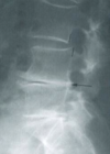
Xrays
MRI
- degenerative disc without significant stenosis/herniation
Provocative Discography
- studies shosn can lead to accelerated disc degeneration and herniation, loss of height and endplate changes

What is the tx of discogenic back pain?
non operative
- NSAIDS, physical therapy , lifestyle modifications
- tx of choice in majority without neurology
Operative
-
Lumbar Discectomy w fusion
- controversial
-
Lumbar total disc replacement
- single level disease with disease free facet
What is the epidemiology of thoracic disc herniation?
- Uncommon
- makes up to 1% of herniated nucleus pulposa
- most seen 40-60 years
- As disc desiccates less likely to actually herniate
- location
- usually involves middle- lower levels
- T11-T12 most common
- 75% disc occur T8-T12
What are the risk factors for thoracic disc herniation?
- Scheuermann’s disease
Describe the types of herinated thoracic disc?
BY herniation
-
Bulging nucleus
- annulus intact
-
Extruded disc
- thru annulus by confined to Post LL
-
Sequestrated
- Disc material free in canal
By Location
- Central
- Posterolateral
- Lateral
What are the symptoms thoracic disc herniation?
Symptoms
-
Pain
- axial back or chest pain- most common
-
thoracic radicular pain
- band pain around course of intercostal n
- arm pain
-
Neurology
- Numbness, parathesia, sensory changes
- Myelopathy
- Paraparesis
- Bowel/ bladder changes- 15-20%
- sexual dysfunction
What are the signs of thoracic disc herniation?
- localised thoracic tenderness
- root symptoms
- dermatomal sensory changes- parathesia/dysesthesia
- cord compression & findings of Myelopathy
- weakness / mild paraparesis
- abnormal rectal tome
-
UMN signs
- Spascitity
- Hyperreflexia
- sustained clonus
- positive Babinski sign
-
Gait changes
- wide based
-
Horner’s syndrome
- seen with HNP T2-T5
What investigations are useful in thoracic disc herniation?
xrays
- lateral radiographs
- disc narrowing
- calcifications (ostephytes)
- MRI
- most useful and dx
- disadv high false positive rate at asymptomatic individuals

What are the tx for thoracic disc herniation?
Non operative
-
Activity modification, physical therapy, symptomatic tx
- majority of cases
- immobilisation & short term rest
- analgesic
- progressive activity restoration
- injections for radiculopathy
- majority improve non op
Surgery
-
Discectomy with possible hemicorpectomy or fusion
- minority of pt
- myelopathic findings, progressive
- persistent and intolerable pain
- debate regarding transthoracic /costotranvserectomy approach
What are the surgical techniques for disectomy of thoracic spine?
-
Transthoracic discectomy +/- fusion
- best approach fo rcentral disc
- complx- intercostal neuralgia
- ca be done video assisted surgery
-
Costotransversectomy +/- fusion
- lateral herniated discs
- extruded or sequestrated discs
- some studie suggest anterior or lateral costotransversectomy is better
What is the epidemiology of lumbar disc herniation?
- 95% involve L4/5 or L5/S1
- most common L5/S1
- peak incidence 40-50 years
- only 5% become SYMPTOMATIC
- male 3: 1 female
What is the pathoanatomy of lumbar disc herniation?
- Recurrent Torsional strain leads to tears in OUTER ANNULUS
- leads to herniation of NUCLEUS PULPOSIS
What is the prognosis of lumbar disc herniation?
- 90% of pts will have improvements of symptom within 3 months with non op care
-
Size of herniation decreases over time ( reabsorbed)
- Sequestered disc herniation- greatest degree of spontaneous reabsorption
- Macrophage phagocytosis is mechanism of reabsorption
Can you describe/draw the anatomy of the interverbral disc?
-
Annulus fibrosis
- type 1 collagen, water, proteogylcans
- extensibility & tensile strength
- high collagen/ low proteogylcan ratio
-
Nucleus Pulposus
- Composed type 2 collagen,water, proteoglycans
-
Compressibility
- low collagen/high proteoglycan
- Proteogylcan interact w H20 & resist compression

Can you describe the nerve root anatomy?
key difference between cervical and lumbar spine is
-
Pedicle/ nerve root mismatch
- C spine C6 n root travels Under C5 pedicle ( mismatch
- L spine L5 n root travels under L5 pedicle ( match)
- Xra C8 nerve root ( no C8 pedicle) allows transition
-
Horizontal (cervical) vs Vertical ( lumbar) anatomy of n
- vertical lumbar root a paracentral & formainal disc will affect different n roots
- Horzontal cervical root a central & foraminal will affect same n

Can you classify the herniation of the lumbar disc?
By location
-
Central prolapse
- assoc back pain only
- can cause Cauda equina
-
Posterolateral ( paracentral)
- most common 90-95%
- PLL is weakest here
- affects transversing n root
- at L4/5 affects L5
-
Foraminal ( far lateral)
- less common 5-10%
- affects exiting n root
- at L4/5 affects L4
-
Axillary
- Can effect exiting and descending roots
BY anatomy
-
Protrusion
- Eccentric bulding annulus fibrosis intact
-
Extrusion
- Tear in annulus, disc herniated thru but continous with disc space
-
Sequestered
- disc material thu annulus & no longer continuous with disc space

What are the symptoms of lumbar disc herniation?
-
Axial back pain
- discogenic/mechanical
-
Radicular pain
- worse with sitting coughing, improves with standing
-
Cauda equina syndrome 1-10%
- bilateral leg pain
- saddle anaesthesia
- LE weakness
- bowel/bladder dysfunction
What are the signs of lumbar disc herniation?
- Motor exam
- Dorsiflexion weakness- L4/5
- EHL weakness L5
- Hip abduction weakness- L5
- Ankle plantar flexion weakness S1
- provocation tests
- Straight leg weakness
- Lesegue sign- SLR aggrevated by forced ankle dorsiflexion
- Gait analysis
- Trendelenberg gait
- gluteus medius weakness - L5
- Trendelenberg gait

What imaging is useful in dx in lumbar disc degeneration?
Xrays
- may show lordosis, loss of height, spondylosis
MRI
- without gadolinium
- highly specific and sensitive
- dx from synovial facet cysts
- high rate of abnormal findings in normal people
- pt with pain >1 month not responding non op tx
-
red flags
- infection- iv du, fever, chills
- tumour- hx cancer
- trauma
- cauda equina- bowel/bladder changes
- MRI With gadolinium for revision surgery
- distinguish post surgical fibrosis ( enhances) vs recurrent herniated disc (doesn’t enhance)
Describe the tx of lumbar disc herniation?
non operative
-
rest. PT, anti-inflammatory
- 1st line
- 90% improve within 3 month
- bed rest then progressive activity
- extension exercises, pilates
- nsaids, ,muscle relaxants, oral steriod taper
-
Selective root corticosteriod injections
- 2nd line in medication fails
- epidural vs selective nerve block
- Long lasting improvement in 50%( surgery90%)
- Best in pts with extruded discs
Surgery
-
Laminotomy and discectomy ( microdiscectomy)
- for persistent disabling pain after 6wks non op
- progressive & significant weakness
- cauda equina syndrome
-
Far lateral microdiscectomy
- for far-lateral disc
- utilises paraspinal approach of wiltse
- avoids injury to lamina or facet joints
- complx- injury to dorsal root ganglia->dysesthesias.- abnormal sensation

What are the outcomes of surgery cf non op tx?
- 70% improvement in back pain
-
neurological recovery less predictable
- 50% motor/sensory recovery
- 25% reflex recovery
good outcome
- if leg pain chief complaint
- positive straight leg raise
- weakness correlates with n root impingment seen on MRI
- married status
- no workers compensation
Bad outcome
- workers compensation
- less relief from symptoms & less improvement in qulaity of life
What are the complications of lumbar spine surgery?
-
Dural tear- 1%
- if have at time of surgery preform water tight repair
-
Recurrent Herniated nucleus pulposus
- can tx non op
- outcomes for revision discectomy = primary
- Discitis- 1%
-
Vascular catastrophe
- break thru ant annulus- injury aorta/vena cava
What is synovial facet cyst?
- A rare cause of spinal stenosis in lumbar spine
What is the epidemiology of synovial facet cyst?
- Rare
- location
- usually Lumbar spine
- 60-90% at L4-L5 level
What is the pathophysiology of synovial facet cyst?
- Trauma
-
Microinstability of the facet leading to
- extruded synovium thru joint capsule
- myxoid degeneration of collagen tissue
- proliferation of fibroblasts with increased hyaluronic acid production
What are the associated conditions of synovial facet cyst?
- Degenerative Spondylolisthesis
What are the signs and symptoms of synovial facet cyst?
Symptoms
- Mechanical back pain
- Radicular symptoms- leg pain
- neurogenic claudication - buttocks/leg pain on walking
Signs
- May see nerve root deficit at associated spinal levels
What imaging is helpful in dx of synovial facet cyst?
- Radiographs
- Ap, lateral , flexion, extension of spine
- usually normal
- look foe segmental instability
- MRI
- significiant leg pain
- best seen on T2 axial/sagittal images

What is the tx for synovial facet cyst?
Non operative
- NSAIDs, rest immobilisation
- mild symptoms
- CT guided aspiration- technically challening and not usually effective
Surgery
-
Laminectomy with decompression
- for symptomatic intraspinal synovial cysts
- high incidence of back pain & cyst formation at 2 years
-
Facetectomy and instrumental fusion
- significant symp failed non op mx
- first line of surgical tx
- lowest risk of persistent back pain & reoccurrance of cyst formation
What are the complications of synovial facet cyst?
-
Cyst reocurrance
- high incidence with resection alone
- facetectomy and fusion
What is lumbar spine stenosis?
- **Reduction in dimensions of central or lateral lumbar spine canal **
- caused by
-
bony structures
- Facet Osteophytes
- Uncinate spur ( posterior vertebral body ostephytes)
- Spondylolithesis
-
Soft tissue structures
- __Herniated/bulging discs
- hypertrophy/ buckling of ligmentum flavum
- synovial facet cysts
-
bony structures
What is the classification of lumbar spine stenosis?
By aetiology
-
Acquired
- Degenerative/Spondylotic change- most common
- Post surgical
- Traumatic - vertebral fractures
- Inflammatory- ankylosing spondylitis
-
Congenital
- short pedicles with medial placed facets= achondroplasia
What is the anatomical classification of lumbar spine stenosis?
-
Central stenosis
- cross sectional area <100mm2 or <10mm AP diameter on axial CT
- thecal sac compressed
- presents with non specific root compression or symptoms of lower nerve root (at L4/5- L5 effected)
-
Lateral recess stenosis
- assoc with facet joint arthropathy adn osteophyte formation
- overgrowth of superior articular facet
- sympt of lower nerve root (L5 at L4/5)
- assoc with facet joint arthropathy adn osteophyte formation
-
Foraminal stenosis
- secondary to disc protrusion, ostephytes, disc collapse
- presents w higher n root (L4 at L4/5)
What are the sign and symptoms of lumbar spine stenosis?
Symptoms
- Back pain
- referred buttock pain
- Claudication
- pain worse with extension- walking/standing
- pain RELIEVED with FLEXION- sitting/leaning
- Leg pain - unilateral
- weakness
- bladder disturbance
- recurrent UTI- 10% due to autonomic sphincter dysfunction
Signs
-
Kemp sign
- Unilateral radicular pain from foraminal stenosis made worse by extension of back
- Straight leg raises- usually negative
-
Valsalva test
- radicular pain not worsened by valsalva- unlike herniated disc
Can you describe the differences between neurogenic claudication and vascular claudication?
-
Neurogenic
- postural changes- yes
- walking upright- causes symptoms
- standing stationary- causes symptoms
- sitting- relieves symptoms
- stair climbing - up easier- back flexed
- stationary bicycle- relieves symp= back flexed
- pulses- normal
-
Vascular
- postural changes- No
- walking upright- causes symptoms
- Standing stationary- relieves
- sitting- relieves
- stairs- down easier- back extended
- stationary bicycle- causes symptoms
- pulses- abnormal
What investigations aid in dx of lumbar spine stenosis?
Xrays
- Standing ap and lateral
- non specific degenerative change- disc space narrowing, osteophyte formation
- degenerative scoliosis
- degenerative spondylolithesis
MRI
- central stenosis with thecal sac <100mm2
- obliteration of perineural fat adn compression of lateral recess foramen
- facet/ligamentum hypertrophy
Ct Myelogram
- more invasive than MRI
- may see
- central and lateral neural element compression
- bony anomalies
- bony facet hypertrophy

What is the tx of lumbar spine stenosis?
Non operative
-
oral medication, PT, corticosteriod injections
- first line
- steriod injections- epidural/transforaminal effective and may obviate need for surgery
Operative
-
Wide pedicle to pedicle decompression
- for persistent pain for 3-6/12 w non op
- progressive neurological deficit
- improved pain and function at 4yrs cf non op
- failure= recurrent disease above/below decompression level
-
Wide pedicle to pedicle decompression with instrumental fusion
- presence of segmental instability-isthmic spondylolithesis,degen spondylolothesis, degenerative scoliosis
- instablilty = complete laminectomy +/- removal of >50% facets

What are the complications of lumbar spine stenosis?
- Increase with age, blood loss and levels fused
- Major Complx
-
Wound infection- 10%
- tx with surgical debridement & irrigation
- Pneumonia
- Renal Failure
- neurologic deficits
-
Wound infection- 10%
- Minor Complx
- UTI 34%
- Anaemia requiring transfusion
- Confusion
- Dural tear
- assoc csf leak
- tx with percutananeous fibrin glue, epidural blood patch or re op if no improvement
- Failure for symptoms to improve
What is the post important pre op prognostic factor in a pt with lumbar spine stenosis who is going to undergo decompression?
- Comorbid medical conditions
What are the outcomes of surgery for lumbar spine stenosis?
- The Spine Patient Outcomes Research Trial (SPORT) is a multi-center randomized controlled trial (RCT) comparing surgical and non-surgical treatment for patients with lumbar disc herniations, lumbar spinal stenosis, and degenerative spondylolisthesis. Results of the randomized cohort were disrupted by a high cross-over rate between the surgical and nonsurgical groups. Therefore, they conducted “as-treated” statistical analysis as a prospective cohort study (non-randomized). Based on the as-treated analysis, two year and four year results for spinal stenosis show improved clinical outcomes in pain and function cf no op.
- Weinstein et al spine 2010
What is degenerative Spondylolithesis?
- A slippage of one lumbar vertebra on another but not due to a defect in the pars
- an absent in pars defect differentiated this from Adult isthmic spondylolithesis

What is the epidemiology of degenerative spondylolithesis?
- More common in
- Africans
- Diabetics
- women >40 yrs
-
x8 more common in women
- increase in ligamentous laxity related to hormonal changes
location
- x5 more common at L4/5 ( cf isthmic L5/S1)
What are the risk factors for developing degenerative spondylolithesis?
- Sacralization of L5 ( transitional L5 vertebra)
- Sagitally orientated facet joints
Describe the pathoanatomy of degenerative spondylolithesis?
-
Forward subluxation (intersegmental instability) of vertebral body is allowed by
- Facet joint degeneration
- Facet joint sagittal orientation
- Intervertebral disc degeneration
- Ligamentous laxity ( hormone changes)
- Degenerative cascade involves
- disc degeneration -> facet capsule degeneration and instability
- microinstabilit-> further degeneration-> macroinstability & anterolithesis
- instability worsens with sagittally orientated facets that allow forward subluxation
What are the neurological symptoms caused by?
-
Central and lateral recess stenosis
- degeneration slip at L4/5 will affect L5 n root in lateral recess
- caused by slippage, hypertrophy of ligamentum flavum, encrochment of spinal canal of osteophytes from facet arthrodesis
-
Foraminal Stenosis
- degenerative slip at L4/5 affect l4 n root
- vertical foraminal stenosis
- loss of disc height
- osteophytes from poastlat corner pushing nerve root up against inferior surface of pedicle
- anteriopost foraminal stenosis- loss of post/anter ior area
- degenerative changes of superior articular facet and post vertebral body

Name the classification of degenerative Spondylolithesis?
Myerding
- Grade 1 = <25%
- Grade 2 =25-50%
- Grade 3= 50 to 75%
- Grade 4 = 75-100%
- Grade 5= spondyloptosis- all the way off
Grade 3 and above are rare in degenerative spondylolithesis

What are the signs and symptoms of degenerative spondylolithesis?
Symptoms
-
mechanical back pain
- most common pc
- relieved with rest and sitting
-
Neurgenic claudication & leg pain
- buttock & leg pain
- relief by sitting
- not relieved by standing in 1 place
- unilat/bilateral
-
Cauda equina
- rare
Signs
-
L4 root involvement - foramen L4/5
- weak quads ( sit to stand manover)
- weak ankle DF ( heel- walk exam)
- decrease patellar reflex
-
L5 root involvement
- weakness ankle dorsiflexion ( heel-walk exam)
- weakness EHL
-
Provocation
- PT walks until symptoms start then stands
- if resolves = vascular
- if sits and resolves = neurogenic
What imaging is useful in dx of degenerative spondylolithesis?
Xrays
- Weight beaing ap, lateral neutral, lateral flexion, lateral extension
- slip evident on lateral xray
- flexion/extension view
- instability= 4mm translation or 10o angulation of motion cf adj motion segement
MRI
- pt with perisitent pain, failed non op tx
- best for inv of impingment of neural elements -t2 weighted sagittal best
Ct
- Identify any bony pathology
- useful when MRI CI- pacemaker!

What is the tx of degenerative spondylolithesis?
Non operative
-
PT & NSAIDS
- most pt tx non operatively
- Epidural steriod injections
Surgery
-
Lumbar wide decompression with Posterolateral instrumented fusion
- laminectomy/wide decompression + foraminotomy
- worse outcome in smokers
- better with pedicle screws and athrodesis
- 79% satisfactory outcome
- cauda equina syndrome
- progressive motor deficit
- in persistent and incapacitating pain failed 6 mo non op
-
Posterior lumbar decompression alone
- only in medically frail pt who won’t tolerate time for fusion
- 69% satisfied, 31% poregressive instability
-
Anterior lumbar interbody fusion (ALIF)
- for revision cases with peudoarthrosis
- Reduction of lithesis is limited in adults
What are the complications of surgical decompression ands fusion on degenerative spondylolithesis?
- Dural tear
- Pseudoarthrosis ( CT better thna MRI to detect failed arthrodesis)
-
Surgical infection
- irrigation & debridment-usually metal work retained as fusion aids stability, increasing and aiding arthrodesis
-
Adjacent segmental disease - 2.5%
- fusions up to L1-3 > risk cf fusion L4 & L5
Summarise what degenerative spondylolithesis is?
- Degenerative spondylolisthesis is the combination of spinal stenosis with intersegmental instability of the vertebrae.
- It most commonly affects L4/5 disc space, causing neurogenic claudication and rarely, cauda equina syndrome.
- Initial treatment is non-operative and includes physical therapy, pain control and injections.
- If non-operative measures fail, surgical management includes posterior decompression with fusion of the unstable segments with or without instrumentation.
What is the evidence for surgical tx in degenerative spondylolithesis cf non op?
- Weinstein et al (SPORT) NEJM 2007 showed patients with degenerative spondylolisthesis treated with surgery had greater improvement in pain and function through 4 years compared to those treated nonoperatively.
- Herkowitz JBJS am 1991et al found patients who had had a concomitant spinal arthrodesis, compared to those who had decompression alone, had improved outcomes with respect to relief of pain in the back and lower limbs.
- Fischgrund et al spine 1997 shows that in patients undergoing single-level posterolateral fusion for degenerative spondylolisthesis with spinal stenosis, the use of pedicle screws may lead to a higher fusion rate.
Describe adult isthmic spondylolithesis?
- Spondylolithesis in an adult caused by a defect in the pars interarticularis (spondylolysis)

What is the epidemiology of adult isthmic spondylolithesis?
- Spondylolysis seen in 4-6% of population
- Increased prevalence in sports that involve Repetitive Hyperextension - gymnasts, weightlifters
location
-
82% occur L5/S1
- due to forces at lumbar spine being greatest at these levels adn the facet more coronal
- only 11% L4/5
What is the pathopysiology of adult ischmic spondylolithesis?
-
Foraminal stenosis
- At L5/S1 ->L5 nerve root impingment
- hypertrophy fibrous repair from pars defect
- uncinate spur formation of post L5 body
- Bulging L5/S1 disc
-
Lateral recess stenosis
- facet arthrosis
- hypertrophic ligamentum flavum
-
Central stenosis
- rare as slips normally grade 1/2
What is the prognosis of adult ischmic spondylolithesis?
- Relatively few patients 5% with spondylolysis will develop spondylolithesis
- Slip progression > in Females
- Slip progression usually occurs in adolscece post puberty
What is the classification system of adult ischmic spondylolithesis?
- Wiltse and Newman
- Type 1 - dysplastic- a congential defect in pars
- Type 2
- A-Isthmic- pars fatigue fx
- B- Isthmic- pars elongation ( multiple healed fx)
- C- Isthmic- acute pars fx
- Type 3
- Degenerative, facet instability wout pars fx
- Type 4
- Traumatic, acute post arch fx other than pars
- Type 5
- Neoplastic- pathological destruction of pars
- DON’t forget Myerding for amount of displacement

What are signs and symptoms of adult isthmic spondylolithesis?
Symptoms
-
Axial back pain
- most common, vary intensity
-
leg pain
- L5 radiculopathy
-
neurogenic claudication
- caused by spinal stenosis
- buttock & leg pain worse w walking
- rare
-
Cauda equina
- rare because slip rarely progress beyond grade 2
Signs
-
L5 radiculopathy
- weakness ankle dorsiflexion/ EHL
What imaging is useful?
Radiographs
- AP, lateral , oblique and flexion-extension
- lateral -see spondylolithesis and pars defect
- flex-exten : instability 4mm translation/ 10o angulation of motion cf adjacent motion segment
What is pelvic index- how can it be measured?
- Pelvic tilt + sacral slope
- a line is drawn from the center of the S1 endplate to the center of the femoral head
- a second line is drawn perpendicular to a line drawn along the S1 endplate, intersecting the point in the center of the S1 endplate
- the angle between these two lines is the pelvic incidence (see angle X in figure above)
- correlates with severity of disease
pelvic incidence has direct correlation with the Meyerding–Newman grade - Child 470, adult 570
- A low PI= low shear forces at lumbrosacral junction and less lumbar lordosis
-
PI greater in pts with spondylolithesis
*

What is pelvic tilt? how can this be measured?
- pelvic tilt = pelvic incidence - sacral slope
- a line is drawn from the center of the S1 endplate to the center of the femoral head
- a second vertical line (parallel with side margin of radiograph) line is drawn intersecting the center of the femoral head
- the angle between these two lines is the pelvic tilt (see angle Z in figure above)

What is sacral slope? How is it measured?
- Sacral slope= pelvic incidence- pelvic tilt
- a line is drawn parallel to the S1 enplate
- a second horizontal line (parallel to the inferior margin of the radiograph) is drawn
- the angle between these two lines is the sacral slope (see angle Y in the figure above)

What is the tx of adult isthmic spondylolithesis?
non operative
- oral medications, lifetsyle modifications, PT
- most pt tx non op
- bracing maybe benefit in acute phase
Operative
-
L5-S1 Decompression & instrumented Posterolateral fusion +/- reduction
- in low grade L4-5 spondylolithesis with persistent and incapacitating pain w failed consx
- progression of slip/neurological deficit
- cauda equina
- improve sagittal balance with reduction
- risk of stretch injury L5 nerve root
- PLIF- common, inserts device medial to facets
- Anterior Lumbar interbody fusion ALIF
- success to tx low grade isthmic spondylolithesis when radicular symptoms
- not used in high grade due to translation & angular deformity
- gd- excellent results at 2years 87-94%
- thru trans-retropertioneal approach
-
Posterior decompression+ insitu PLIF L4-S1 & posterior strut support
- high grade spondylolithesis
- partial reduction better to preserve nerve root L5 than full reduction
What are the complications of surgery for adult isthmis spondylolithesis?
- Pseudoarthrosis
- Dural tear
- Nerve injury- L5 from reduction-weak EHL, ankle dorsiflexion, therefore aim for partial rather than full reduction
What is sacroilitis common in?
-
Ankylosing spondylitis
- assoc HLA- B27
- 1-2% pts will have HLA- B27
-
Reiter’s Syndrome
- oligoarthritis, conjuctivitis, urethritis
- Joint Arthritis
- common in teen- middle aged
- Males > females

What is the pathophysiology of sacroilitis?
- Trauma
- infection
- pregnancy
- Often from Chronic inflammation of SI joint-> fibrosis and ossification
what is the prognosis of sacroilitis?
- depends on cause but most patients will eventually resolve the epidoes and continue without issue
What are the signs and symptoms of sacroilitis?
Symptoms
- pain with prolonged standing
- difficulty climbing stairs
- generalised back pain
- weakness from hip musculature on affected side
- morning stiffness
Signs
-
FABER TEST
- Pain with FLEXION, ABDUCTION & EXT ROTATION
- Ankylosing spondylitis
- spinal flexion deformities
- starting in T and L spine
investigations useful in sacroilitis?
- xrays
- erosive changes but non specific
- calcification/sclerosis at SI joint
- MRI
- gadolinium
- T2 show fluid/inflammation at Si joint ?abscess
- check
- WBC- usually normal/ elevated
- ESR?CRP- elevated
- Blood cultures- posiitve in 50%
- HLA- B27
- Rheumatoid factor- neg in ank spon
How is sacroilitis tx?
Depends on cause
infection
- IV antibiotics: iv then oral. monitor crp
- surgery- if abscess large, crp climbing
Trauma/overuse
- Rest, activity modifications, nsaids, steriod injections
- most resolve
Pregnancy
- observe
- resolves post partum
If ankl spond
- agressive PT, nsaids, tnf inhibitors