Radiology Flashcards

1
Q

Turn me over!

A
2
Q
A
3
Q
A
4
Q
A
5
Q
A
6
Q

Point to them!

A
7
Q
A
8
Q
A
9
Q
A
10
Q
A
11
Q
A
12
Q
A
13
Q
A
14
Q
A
15
Q
A
16
Q
A
17
Q
A
18
Q
A
19
Q
A
20
Q

whatis the abnormality?

A
21
Q

What is the pathology here?

A

Both SBO and LBO

22
Q

Name this pathology

A

Calcified Gall stone

23
Q

What can you see here? What is the likely cause?

A

Cholecystectomy clips - and air in the Biliary tree. The most likely cause is recent laprop surgery.

24
Q

what does this image show?

A

Dilated Colon - Sigmoid Volvulus

This occurs in cases of long-standing chronic constipation where patients develop a large, elongated, relatively atonic colon, particularly in the sigmoid segment. It is often referred to as acquired or idiopathic megacolon. In sigmoid volvulus, a large sigmoid loop full of faeces and distended with gas twists on its mesenteric pedicle to create a closed-loop obstruction. If uncorrected, venous infarction leads to perforation and faecal peritonitis.

25
Q

What does this image show? Can you make any further comments?

A

Imbibed foreign bodies. Denser than bone, likely metal objects.

26
Q

Name this Ivx

A

IV Pyelogram

An intravenous pyelogram (IVP) is a special x-ray examination of the kidneys, bladder, and ureters (the tubes that carry urine from the kidneys to the bladder).

27
Q

Name this pathology

A

Large Bowel Obstruction

28
Q

What is the obvious pathology? Can you see the cause?

A

LBo due to colonic cancer.

29
Q

what is the pathology?

A

LBO

30
Q

Name the pathology

A

LBO

31
Q

Describe the pathology on this image, including its location.

A

LBO - splenic flexure.

32
Q

Name the abnormality on this image.

A

NO LEFT LEG!

33
Q

What does this image show?

A

Neonate with perforated bowell - pneumoperitoneum

34
Q

what is the pathology?

A

Pneumoperitoneum

35
Q

what does this image show?

A

A NORMAL ABDO radiograph!

36
Q

what does this image show?

A

Pneumoperitoneum

37
Q

what does this image show?

A

pneumoperitoneum

38
Q

What does this image show?

A

Pneumoperitoneum

39
Q

What clinical sign is seen here?

A

rigler’s - Air both sides of the bowel.

40
Q

What pathology does this radiograph show?

A

R Pneumoperitoneum

41
Q

Name the pathology?

A

SBO

42
Q

SBO or LBO?

A

SBO

43
Q

Name the pathology

A

SBO

44
Q

Name the pathology

A

SBO

45
Q

Say what you see.

A

small bowel dilatation

46
Q

Name the pathology

A

stagohorn calculi

47
Q

Name the pathology

A

Ureteric calculus.

48
Q
A
49
Q

What pathology is seen here?

A

Acute Extradural haematoma

50
Q

What is the pathology here? Is it chronic or acute?

A

Acute Cerebral HAematoma.

51
Q

What pathology is seen here?

A

Acute on chronic subdural haematoma

52
Q

What pathology is seen here?

A

Acute Subarrachnoid Haemorrhage

53
Q

What pathology is seen here?

A

Acute subdural haematoma

54
Q

What pathology is seen here?

A

Basal ganglia calcification

55
Q

What pathology is seen here?

A

Cerebral HAemorrhage on CT

56
Q

What pathology is seen here?

A

Cerebral Infarction

57
Q

What pathology is seen here?

A

Frontal view of sjull fracture

58
Q

What pathology is seen here?

A

Intra and extra dural Haemmorhage

59
Q

What pathology is seen here?

A

Intracerebral abnormality

60
Q

What pathology is seen here?

A

Intracerebral mass - highlighted with contrast

61
Q

What pathology is seen here?

A

Acute intraventricular haemorrhage

62
Q

What pathology is seen here?

A

Intraventricular Haemorrhage on CT

63
Q

What pathology is seen here?

A

Skull fracture (lateral view)

64
Q

What pathology is seen here?

A

Left MCA infarct

65
Q

What pathology is seen here?

A

Middle cerebral artery thrombosis on CT

66
Q

What pathology is seen here?

A

Nothing! This is a NORMAL Skull (lateral view)

67
Q

What pathology is seen here?

A

Nothing! This is a normal (CHILD) Skull!

68
Q

What pathology is seen here?

A

Nothing! Normal headf ct (albeit old)

69
Q

What view is this?

A

PA

70
Q

Which view is this?

A

Normal Townes view

71
Q

What pathology is seen here?

A

Occipital bone fracture

72
Q

What pathology is seen here?

A

R MCA Infarct

73
Q

What pathology is seen here?

A

R MCA Infarct

74
Q
A
75
Q

What condition is this?

A

Frontal Mucocele

76
Q

What condition is this?

A

Frontal Mucocele

77
Q

What can you see here?

A

Inverted Papiloma

78
Q

What can you see here?

A

Inverted Papilloma

79
Q

Inverted Papilloma

A

What can you see here?

80
Q

What can you see here?

A

Paradoxical curvature

81
Q

What is this?

A

Wegners Granulomatosis

82
Q

What can you see here?

A

Septal Deviation

83
Q

What can you see here?

A

Silent Sinusitis

84
Q

What condition can you see here?

A

Post Nasal Rhabdomyocarcoma

85
Q

What obvious abnormality can you see here?

A

TUMOUR!

86
Q

What can you see here?

A

Septal Floor Squamous Cell Carcinoma

87
Q

What can you see here?

A

Septal Spur

88
Q

What can you see here?

A

Tumour. Horrible, horrible TUMOUR.

89
Q
A
90
Q

What can you see here?

A

Emphysema on ct scan.

91
Q

What can you see here?

A

Pleural Effusion in the sagittal plane view.

92
Q

What can you see here?

A

Pleural effusion in the sagittal plane CT.

93
Q

What is the pathology here?

A

Left lung pleural effusion.

94
Q

What can you see here?

A

Pleural effusion, coronal view CT.

95
Q

What can you see here?

A

PLeural effusion.

96
Q

What can you see here?

A

Uppe right lung Bullae.

97
Q
A
98
Q
A
99
Q

What can you see?

A

This is an apple core cancer.

100
Q

What can you see?

A

This is Caecal cancer.

101
Q

What can you see?

A

Caecal Cancer

102
Q

What can you see?

A

Caecal gas - imminent perforation

103
Q

What can you see?

A

Cancer of the rectum.

104
Q

What can you see?

A

Colitis

105
Q

What can you see?

A

Distal sigmoid carcinoma.

106
Q

What can you see?

A

Epiploic Appendagitis.

107
Q

What can you see?

A

Liver mets

108
Q

What can you see?

A

Large rectal polyp

109
Q

What can you see?

A

Large Polypoid rectal tumour.

110
Q

What can you see?

A

Large bowel obstruction

111
Q

What can you see?

A

Pneumoperitoneum

112
Q

What can you see?

A

Pulmonary mets

113
Q

What can you see?

A

Transverse colon polyp

114
Q

What can you see?

A

Villous Adenoma - Rectum

115
Q

What can you see?

A

Villous Adenoma - Sigmoid

116
Q
A
117
Q

Week 222

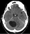
What does this image show?

A

This is an infarction of the posterior inferior cerebellar artery (PICA)

118
Q

Week 222

What does this image show?

A

Posterior cerebellar artery infarction.

119
Q

Week 222

What does this image show?

A
120
Q

Week 222

what does this image show?

A

Subarrachnoid haemorrhage

121
Q

Week 222

what does this image show?

A

Haemangioblastoma

122
Q

Week 222

What does this image show?

A

This is lymphoma.

123
Q

Week 222

What does this image show?

A

Progressive multifocal encephalopathy

124
Q

Week 222

What is Fahrs disease?

A

This is idiopathic basal ganglia calcification.

125
Q
A