PATHO - Reproductive System Flashcards
Female gametes are known as ____________.
Male gemetes are known as __________.
Each gamete contains _____ chromosomes. Female x male gametes unite together to form a ____ chromosome zygote.
Female gamete: ovum
Male gamete: spermatozoon (sperm cell)
Each gamete: 23 chromosomes
Joins together to form a 46 chromosome zygote
Classification of sex hormones
steroid hormones
all made from cholesterol
Function of dehydroepiandrosterone (DHEA) in females and males
- from adrenal gland, ovary, other tissues
- Females & Males : converted to androstenedione ⇒ estrogens, testosterone, both
Function of estrogens in females and males
- estrone, estradiol, estriol - works on estrogen receptors 𝝰 & 𝛃 (in ovary and placenta, small amounts in other tissues)
-
Females: stimulates development of female sex traits:
- breast, uterus, and vaginal maturation
- proliferation of endometrium during menstrual cycle
- mammary gland development during pregnancy
- promotes fetal adrenal gland function
- uteroplacental blood flow
- Males: growth at puberty, fusion of bone growht plate, preventing apoptosis of germ cells
Source and function of testosterone in females and males
- Source: adrenal glands (from DHEA), ovaries
- Females: libido, learning, sleep, protein anabolism, growth of muscle and bone, pubic and axillary hair, activation of sebaceous glands, contributes to acne
- Males: stimulates spermatogenesis, development of 1’ and 2’ sex tracts, bone and muscle growth, pubic and axillary hair, activates sebaceous glands, contributes to acne, maintains libido
Source and function of GnrH in females and males
- Source: neuroendocrine cells in hypothalamus
- Females & males: stimulates secretion of gonadotropins (FSH an dLH) from anterior pituitary
Source and function of FSH in females and males
- Source: gonadotroph cells from anterior pituitary
- Females: promotes development of ovarian follicles, stimulates estrogen secretion
- Males: promotes development of testes, stimulates spermatogenesis by Sertoli cells
Source and function of LH in females and males
- Source: gonadotroph cells in anterior pituitary
- Females: triggers ovulation; rpomotes development of corpus luteum
- Males: stimulates testosterone production by Leydig cells of testes
Source and Function of Inhibin in females and males
- Source: ovary and testes
-
Females & Males: inhibits FSH production in anterior pituitary
- in females, Inhibin B is primarily secreted in follicular phase but spikes when ovulation occurs
- Inhibin A is secreted during luteal phase and further supresses FSH
- also restrains prolactin and GH release, inteferes with GnRH receptors, and promotes breakdown of itnracellular gonadotropins
- activity boosted by follistatin
Source and Function of Human Chorionic Gonadotropin (hCG) in females
- Source: placenta
- Females: supports corpus luteum, which secretes estrogen and progesterone during first 7 weeks of pregnancy
Source and function of Activin in females
- Source: ovary (granulosa cells)
- Females: stimulates secretion of FSH and pituitary response to GnRH and FSH binding in dominant granulosa cells
- inhibited by follistatin
Source and Function of progesterone in females
- Source: ovary and placenta
- Females:
- promotes secretory changes in endometrium during luteal phase of menstrual cycle
- quiets uterine muscle activity
- prevents lactogenesis during pregnancy
Source and function of relaxin
Source: corpus luteum, myometrium (uterine muscle), placenta
Function: inhibits uterine contractions during pregnancy and soften pelvic joints and cervix to facilitate childbirth
In early embryonic development, the reproductive structures of male and female embryos are ____________.
They consist of a pair of primary sex organs known as ________ and two pairs of ducts which are _____________ and _____________.
In early embryonic development, the reproductive structures of male and female embryos are homologous (the same)/undifferentiated.
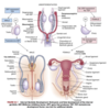
They consist of a pair of primary sex organs known as gonads and two pairs of ducts which are mesonephric (wolffian) and paramesonephric (müllerian) ducts.
What are the mullerian ducts?
How may these ducts change in females vs males?
- precursors of the internal female sex organs (oviducts, uterus, cervix, upper vagina)
- initially formed regardles sof genotypic sex (no SRY signaling needed for development)
- SRY signaling is needed in males for these ducts to regress and prevents deelopment of female reproductive tract
What are wolffian ducts? What is their function?
precursor of male internal sex organs
secretes testosterone and promotes development of male sex organs
The first sign of development of reproductive organs (male/female) occurs during _____ week of gestation.
5th
What kind of effect does the SRY gene have on embryonic differentiation?
- sex-determining region of the Y chromosome
Where is Muwllerian inhibitory hormone (MIF) secreted from?
What is the function of Muwllerian inhibitory hormone (MIF) in reproductive development?
Source: secreted by Sertoli cells in the testes
Function: promotes degeneration of the müllerian ducts
- No MIF, the müllerian ducts would develop and wolffian ducts would degenerate with loss of male sex organ development
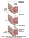
At what age would the male gonads (testes) descend into the scotum?
At what age would the testes produce sperm?
Descend into the scrotum: 9 months
Sperm production: after puberty
How does female gonadal development occur?
- development occurs due to absence of SRY expression
- estrogen is present while testosterone and MIF are absent which lead to loss of wolffian system; also promotes development of external genitalia
- 6-8 weeks’ gestation: two female gonads develop into ovaries
- mesonephric ducts deteriorate and upper ends of paramesonephric ducts decome fallopian tubes
Where do external reproductive structures stem from and how do they develop from these precursor structures?
Source: homologous embyronic tissues (i.e. undifferentiated)
Development: at 7-8 weeks gestation, the male & female embryos develop genital tubercle (elevated structure). The tubercle relies on testosterone for differentiation into external male genitalia or else female genitalia will develop in the absence of ovaries
Describe when and what occurs during development of the endocrine system in utero that contributes to sexual differentiation.
4/5 weeks gestation: anterior pituitary development begins
10 weeks: GnRH produced in hypothalamus - contraols production of LH and FSH in anterior pituitary
12 weeks: vascular connection between hypothalamus and pituitary is established
28 weeks: in female fetus, high levels of FSH and LH are excreted (up until 28 weeks) and stimulate production of estrogen and progesterone by the ovary. At ~28 weeks, etrogen and progesterone levels in ovaries and placenta are high enough so gonadotropin production decreases
Females:
Production of ova occurs (during fetal life/at puberty).
How many female gametes mature per menstrual cycle?
Males:
Production of sperm occurs (during fetal life/at puberty).
How many sperm are produced daily?
Females:
ova production occurs during fetal life
one female gamete matures per menstrual cycle
Males:
sperm production occurs at puberty
Millions are produced daily, for life
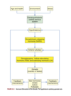
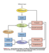
What negative feedback system develops to regulate reproductive hormones?
- present at term pregnancy
- includes gonadostat (gonadotropin-releasing hormone pulse generator) that responds to high levels of placental estrogens by releasing low levels of GnRH
- After birth, steroid hormone level drops (due to loss of maternal placental hormones)
- GnRH is secreted from hypothalamus and LH and FSH are released
- in boys: peaks at 3-6 mos and then falls
- in girls: peaks at 12-18 mos and then falls
- gonadotropins are suppressed until the onset of puberty
Puberty vs Adolescence
Puberty: onset of sexual maturation (different from adolescence) - marked by development of secondary sex characteristics, rapid growth, and ability to reproduce
- Girls: begins ~8-9 y.o. with breast development (therlarche), ends with first ovulatory menstrual period in females (which can take up to 1-2 years after menarche)
- Boys: begins ~11 y.o. but may occur ealier with increased weight and BMI, begins with voice changes and ends with first ejaculation with mature sperm
- Complete when an individual is capable of reproduction
Adolescence: developmental stage between childhood and adulthood
Factors that influence puberty
- genetics
- environment
- ethnicity
- general health
- earlier onset in obese girls
- delayed with low body fat and reduced weight/intense exercise
- nutrition
What physiological/hormonal changes occur with reproductive maturation in girls (i.e. puberty)?
- ~ 1 year before puberty, there are nocturnal pulses of LH and FSH and increased response to GnRH which stimulate gonadal maturation (gonadarche) with estradiol secretion
- estradiol causes breast development (thelarche), maturation of sex organs (vagine, uterus, ovaries), and fat deposition in the hips
- estrogen and increased groth factor production causes rapid skeletal growth
- ovaries start to release matura ova
Adrenarche
- increased production of androgens (from adrenal gland)
- dehydroepiandrosterone
- androstenedione
- these are converted to testosterone and estrogen before puberty
- S/S: hair growth in armpits and pubic area, sweat and sebaceous gland activation
What physiological/hormonals changes occur during reproductive maturation in males (i.e. during puberty)?
- ~ 1 year before puberty, there are nocturnal pulses of LH and FSH and increased response to GnRH which stimulate gonadal maturation (gonadarche) with testosterone secretion
- estrogen and increased growth factor production causes rapid skeletal growth
- Testosterone causes growth of testes, scrotum, penis
- +ve feedback loop of gonadotropins stimulating gonads to produce more sex hormones
- with males, testes begin producing more mature sperm (that can fertilize ovum)
Function of the female reproductive system?
Function of external genitalia?
Overall function: to produce mature ova. provide protection and nourishment to fetus until birth
Function of external genitalia: protect body openings, involved in sexual functioning
What are the major extenal genitalia structures (6) in females?
known collectively as the vulva/pudendum
- Mons pubis
- Labia majoraa
- Labia minora
- Clitoris
- Vestibule
- Perineum
Structure and function of the mons pubis
Structure: fatty tissue layer over pubis symphysis. Gets covered in pubic hair has more active sebaceous/sweat glands during puberty
- moundlike shape due to fat deposition under the skin (caused by estrogen)
Function: protects the pubic symphysis during sexual intercourse
Structure and function of Labia Majora/majus
Structure: two folds of skin extending from mons pubis to fourchette (see diagram) to form a cleft
- During puberty: increased fatty tissue, pubic hair on lateral surfaces, and secretion of lubricants from sebaceous glands on medial surface
- ++sensitive to temperature, touch, pressure, and pain
- homologous to the male scrotum
Function: protects inner structures of vulva
Structure and Function of labia minora/minus
Structure: two smaller, thinner, asymmetric folds of skin within labia majora; forms the clitoral hood (prepuce) and frenulum. Splits to enclose the vestibule and then converge near anus to form fourchette
- hairless, pink, and moist
- well supplied by nerves, blood vessels, and sebaceous glands
Function: sebaceous glands secrete bactericidal fluid that lubricates and waterproofs vulva skin. Labia swell with blood during sexual arousal
Structure and Function of clitoris
Structure: the erectile organ betweeen the labia minora. Small and cylindrical with a visible glans and shaft that lies beneath the skin
- homologous to the penis
Function: major site for sex stimulation and orgasm. Slightly enlarges when it is filled with blood during sexual arousal
Structure and Function of vestibule & associated glands in that area
Structure: area that is protected by labia minora, contains the external opening of the vagina (introitus/vaginal orifice) and urethral opening (urinary meatus). Introitus may also be covered by hymen (thin perforated membrane)
- openings are lubricated by two pairs of glands - Skene glands, Bartholin glands
- Skene glands: aka lesser vestibular/paraurethral glands - has ducts that open on both sides of the urinary meatus. Lubricates urinary meatus
- Bartholin glands: aka greater vestibular/vulvovaginal glands - has ducts that open on either side of introitus. Secrete mucus to lubrivate inner labial surfaces and enhance viability and motility of sperm
Function: tissue underneath vestibule fills with blood and becomes engorged during sexual excitement. Secretions from the ^ glands facilitate sex
Structure of perineum
Structure: area between vaginal orifice and anus. Has less hair, skin, and subQ tissue (so skin is clsoe to underlying muscles).
- Covers the muscular perineal body - a fibrous structure that consists of elastic fibers and connective tissue & is the common attachment point for bulbocavernosus, external anal sphincter, and levator ani muscles
- varies in length (2-5cm +), has elastic properties
- length and elasticity of perineal body influences tissue resistance and injury during childbirth
What are the major internal reproductive organs in females?
ovaries
fallopian tubes
uterus
vagina
What is the structure of the vagina? What are the four layers it consists of?
Structure: an elastric & fibromuscular canal 9-10cm long. Extends up and backwards from the introitus to the lower portion of the uterus
- upper part surrounds the cervix
- Fornix: space around the cervix that is further divided into posterior (deeper) and anterior fornix - occurs due to cervix meeting the vaginal canal at a 90 deg angle
- lengthens, widens, and anterior 1/3rd becomes congested with blood during sexual excitement
Vaginal wall has 4 layers:
- 1) Mucous membrane lining of squamous epithelial cells - thickens and thins in response to hormones (i.e. estrogen). In those that are of reproductive age, this layer is arranged in transverse wrinkles (rugae_)_ that allows stretching during coitus and childbirth
- 2) Fibrous connective tissue - has lots of blood and lymphatic vessels
- 3) Smooth muscle
- 4) Connective tissue - has rich network of blood vessels
Cul-de-sac
pouch that separates the posterior fornix and rectum
What happens to the vaginal wall during sexual arousal?
- vaginal wall becomes engorged with blood, which pushes som fluid to the surface of the mucosa enhancing lubrication
- vaginal wall doesn’t have mucus-secreting glands so secretions drain into the vagina from the endocervical glands or Bartholin/Skene glands
What two factors help with maintaining self-cleansing action of the vagina to defend from infection?
1) Acid-base balance - discourages proliferation of pathogenic bacteria
- before puberty: vaginal pH ~7
- at puberty: pH 4.0 - 5.0
2) Thickness of vaginal epithelium
- before puberty: thin epithelium
- at puberty: thickens
Protection is greatest during sexually active years and when estrogen levels are high and there is a normal population of Lactobacillus acidophilus. These changes are maintained until menopause (pH then rises again to alkaline levels and epithelium thins)
What is the structure of the uterus and how does it develop throughout puberty?
- hollow & pear shaped organ with its lower end opening into the vagina
- loosely held in position by ligaments, peritoneal tissue folds, and pressure of adjacent organs (bladder, sigmoid colon, rectum)
- In most women: uterus is tipped forward (anteverted) which makes it rest on the bladder but can exist in other positions
- Innervation: by motor and sensory fibers of ANS
- During puberty: attains its adult size and descends from abdomen to the lower pelvis (between bladder and rectum)
-
In a mature, non preggo female:
- 7-9 cm long, 6.5 cm wide with muscular walls 3.5cm thick
-
After preggo:
- enlarges ~1cm in all dimensions
What are the two major parts of the uterus?
1) Corpus: body of uterus
- Fundus: top of corpus (above where the fallopian tubes insert). Widest part of the uterus
- Isthmus: area just above the cervix. Narrowest part of the uterus
2) Cervix:“neck” of uterus - extends from isthmus to vagina
The endocervical canal is the passageway between upper opening (internal os) and lower opening (external os) of the cervix
What are the three layers that make up the uterine wall?
1) Perimetrium (parietal peritoneum) - outer serous membrane that covers uterus
2) Myometrium - thick muscular middle layer
3) Endometrium - aka uterine lining
- made of a functional layer (superficial compact layer and spongy middle layer) and basal layer
- Functional layer: responds to estrogen and progesterone; proliferates and sheds monthly between puberty and menopause
- Basal layer: attached to the myometrium and regenerates the functional layer after shedding (menstruation)
Which structure in the cervix does not have an endometrial layer?
What type(s) of cells are found here?
endocervical canal
lined with columnar epithelial cells which is continuous with the lining of the outer cervix & vagina (aquamous epithelial cells)
The point where the columnar and squamous epithelial cells meet is known as the ________________. What is this area particularly susceptible to?
transformation zone/squamous-columnar junction
vulnerable to HPV which can lead to cervical dysplasia or carcinoma. These are the cells that are removed for examination during a pap smear
Functions of the uterus
- anchors and protects a fertilized ovum, provides optimal environment while ovum develops, and pushes the fetus out at birth
- involved in sexual response and conception
- during sexual arousal, cervix dilates slightly while uterus increases in size and moves up and backward (like a tent) that results in the cervix sitting in semen
- during orgasm, rhythmic contractions facilitate sperm movement through cervical opening
Function of the cervix
- acts as a mechanical barrier to infectious microorganisms from the vagina
- external cervical os has a mucous plug (during luteal phase of menstrual cycle and pregnancy)
- During ovulation, mucus changes under estrogen influence and forms watery strands (spinnbarkeit mucus) that facilitates sperm transport into the uterus
- downward flow of cervical secretions also flushes microorganisms away from cervix and uterus
- pH of secretions are also inhospitable to bacteria, and there are enzyms and antibiotices (IgA) that are involved with immune system
Structure of fallopian tubes
Structure: aka oviducts/uterine tubes. Enter the uterus bilaterally just beneath the fundus. Curve up and over the two ovaries. Each tube ~8-12 cm long and 1 cm in diameter. At the ends, it is fringed (fimbriae) and called the infundibulum
Function: direct the ova from the spaces around the ovaries into the uterus (the fimbriae move and create a current that draws the ovum into the infundibulum and then into the fallopian tube. One in the tube, cilia and peristalsis push it towards the uterus
Where does fertilization occur? What happens when the egg is/is not fertilized?
- Fertilization occurs in the ampulla (distal third) of fallopian tube
- Sperm released into the vagina can travel upward through endocarical canal and uterine cavity to get to the fallopian tubes
- If an ovum is present in either tube, fertilization can happen
- ovum becomes a blastocyst and then implants itself in the endometrium of the uterine wall
- If not fertilized, ovum fragments and leaves uterus with menstrual fluids
Describe the structure and function of the ovaries.
Structure: primary female reproductive organs. Almond shaped and on both sides of the uterus. Suspended by mesovarium portions of the broad ligament, ovarian ligaments, and suspensory ligaments
- homologous to testes
- If at reproductive age: 3-5cm long, 2.5cm wide, 2cm thick, weighs 4-8 g
Function: secrete female sex hormones & development/release of female gametes
Describe the changes/process that occur during the ovarian cycle
- Follicular maturation, ovulation, corpus luteum development, corpus luteum degeneration
- At birth there are 1-2 mil ova within the primordial (immature) ovarian follicles
- By puberty, drops to 300k - 500k and some follicles and ova begin to mature (so there are follicles in different stages)
- Every menstrual cycle, one of the follicles that reaches maturation and releases its ovum (the remaining ones fail to develop or degenerate - these are atretic follicles)
- After ovum release, follicle develops into the corpus luteum
- if fertilization occurs, corpus luteum enlarges and secretes hormoens that maintain and support pregnancy
- if fertilization does not occur, it releases these hormones for 14 days and then degenerates which triggers the maturation of another follicle
- cycle occurs from puberty to menopause (except pregnancy or hormonal contraceptive use)
- At menopause: process stops and ovaries atrophy
Theca cells
granulosa cells
two types of cells in the ovarian follicle that secrete sex hormones
Theca cells: produce androgens that migrate to granulosa cells
Granulosa cells: convert androgens to estradiol
Female sex hormones include:
estrogen
androgens
progesterone
What is estrogen and what functions does it have?
generic term for 3 similar hormones derived from cholesterol: estradiol, estrone, estriol
Estradiol (E2) - most potent and plentiful. 95% of it produced by the ovaries (very little from the adrenal cortex and placenta)
Estrone: converted from androgen in ovarian and adipose tissue
Estriol: metabolite of estrone and E2
Functions:
- maturation of reproductive organs
- development of secondary sex characteristics, growth, and maintenance of pregnancy
- closure of long bones after growth spurt in puberty
- bone and skin maintenance
- systemic organ function
What are androgens and its function?
- primarily sex hormones produced in testes but occurs in small amounts (from adrenal cortex) in men and women, and from the ovaries
- some (dehydroepiandrosterone and androstenedione) are precursors of estrogens (estrone, estradiol)
Functions: skeletal growth & pubic/axillary hair growth during puberty. Activate sebaceous glands (acne). Involved in libido
What is progesterone and its function?
What is it: the 2nd major female sex hormone, sometimes known as the hormone of pregnancy. Secreted by the corpus luteum when stimulated by anterior pituitary (which is stimulated by LH).
Function: Works with estrogen to control ovarian menstrual cycle
- LH surges 24-36 hr before ovulation and promotes luteinization of granulosa in dominant follicle
- this results in progesterone production and the development of blood vessels and connective tissue
- thickens endometrium to prep it for implantation of blastocyte
Function/Effects during pregnancy:
- 1) maintaining thickened endometrium
- 2) relaxing smooth muscle in myometrium which prevents premature concentractions and helps uterus expand
- 3) thickening of myometrium to prepare for labour
- 4) promoting growth of lobules and alveoli in the breast to prep for lactation BUT preventing lactation until fetus is born
- 5) preventing additional maturation of ova by supressing FSH and LH (i.e. stops menstrual cycle)
- 6) providing immune modulation (tolerating against fetal antigens so that mom’s immune system does not attack fetus)
Menses (menstrual bleeding) starts with what and ends with what?
What factor(s) affect the onset of menses?
Starts with menarche (first menstruation)
Ends with menopause (cessation of menstrual flow for 1 year)
body weight (% of body fat may change metabolic rate and lead to hormone changes associated with early menarche - 11 years or younger)
How long do menstrual cycles last?
What factors affect menstrual cyclicity and regular ovulation?
- at first, cycles are anovulatory and vary between 10-60 days +
- regular patterns start once adolescence hits, cycles last 21-45 days
- through adulthood, most common the cycle length is 28 days (25-30 days)
- ~2-8 years before menopause, cycles begin to lengthen again
Factors:
- 1) activity of GnRH
- 2) initial pituitary secretion of FSH
- 3) estrogen (estradiol) positive feedback mechanism for FSH/LH surge prior to ovulation, oocyte maturation, corpus luteum formation, and progesterone production
Phases of the menstrual cycle
Two phases:
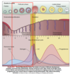
1) follicular/proliferative phase (postmenstrual)
2) luteal/secretory phase (premenstrual)
What happens during the follicular/proliferative phase of the menstrual cycle?
- occurs after menses (functional layer of endometrium sheds)
- Two simultaneous processes occur during this phase:
- 1) maturation of ovarian follicle
- 2) proliferation of endometrium
- GnRH and balanced activin & inhibin levels from granulosa cells contribute to increase in FSH levels from anterior pituitary - this stimulates follicles and rescues a dominant ovarian follicle from apoptosis (days 5-7 of cycle)
- Estrogen and FSH together increase # of FSH receptors in primary follicle which make them more sensitive to FSH. This also induces LH stimulation
- LH and FSH stimulation continue to encourage estrogen secretion and increased levels
- As estrogen levels increase, FSH levels drop due to increase in inhibit-B secreted by granulosa cells in the dominant follicle → this drop decreases the growth of less developed follicles
- estrogen causes endometrial cells to proliferate and stimulate LH production
- surge in FSH and LH is required for final follicular growth and ovulation
What marks the onset of the luteal/secretory phase?
What occurs during this phase?
Onset: ovulation (release of ovum from mature follicle)
What occurs during this phase:
- ovarian follicle begins to transform into corpus luteum
- LH is secreted from anterior pituitary which stimualtes corpus luteum to secrete progesterone which then initiates secretory phase of endometrial development
- Glands and blood vessels in the endometrium branch through the functional layer, glands secret a thin glycogen-containing fluid (which prepares for implantation because it’s full of nutrients)
- Human chorionic gonadotropin (hCG) is secreted 3 days after fertilization by blastocytes and maintains corpus luteum once implantation occurs (day 6 or 7)
- production of estrogen and progesterone will continue until placenta can adequarelt maintain hormonal production
- If no conception/implantation occurs, corpus luteum degenerates and stops its progesteron and estrogen production → this results in endometrium entering an ischemic/menstrual phase causing it to shed
How long do menstrual blood flows typically last? How much blood is lost?
What is menstrual discharge composed of?
- blood flow typically lasts 3-7 days (but can be irregular)
- blood loss can be 30-80ml with most of it being during the first 3 days of menses
- menstrual discharge consits of blood, mucus, and desquamated endometrial tissue, does not clot under normal circumstances
- usually dark and has musty odor on oxidation
What is the ovarian cycle and how is it regulated via hormones?
The process of follicular maturation, ovulation, corpus luteum development, and corpus luteum degeneration
- Decline in luteal phase estrogen, progesterone, and inhibin secretion allows FSH levels to rise (and slight LH increase)
- FSH stimulates granulosa cell growth and initates estrogen production
- A group of ovarian follicles then get recruited and begin to mature → eventually one becomes dominant and the others atrophy
- Dominant follicle begins to secrete progressively larger amounts of estrogen which increases pituitary sensitivity to GnRH (creating a +ve feedback look with FSH and LH surge)
- Ovulation occurs 1-2 hr before final progesterone surge or 12-36 hr after onset of FSH and LH surge
- Progesterone, proteolytic enzymes, and prostaglandins trigger mechanisms that control release of ovum
- FSH and LH surge also transform granulosa cells of follicle into corpus luteum, and corpus luteum secretes progesterone and estrogen
- progesterone acts to suppress new follicular growth during early/midluteal phases
- if pregnancy doesn’t occur, the corpus luteum persists 11-14 days and then regresses and disappears
What are the uterine phases of the menstrual cycle?
Follicular/proliferative phase, luteal/secretory phase, and menstruation
Midfollicular/proliferative phase: increased levels of estrogen contribute to endometrial repair and proliferation leading to endometrial thickness (luteal phase). Cervical mucus is thin and watery
Ovulation: peak estrogen occurs right before which maximally stimulates cervical glands to produce mucus (more abundant and elastic/spinnbarkeit). Tiny channels also develop which allow access for sperm into interior of uterus
Secretory phase: occurs after ovulation (where progesterone levels increase) and endometrial tissu develops secretory characteristics
Menstruation: Endometrial tissue breaks down when implantation does not take place (~11 days after oculation) and then shedding of tissue starts ~14 days after ovulation
What is the vaginal response to cyclic hormonal changes of the menstrual cycle?
- vaginal epithelial cells grow maximally during follicular/proliferative phase (influenced by estrogen)
- Cornification: keratinized cells overgrowing basal epithelium - occurs after ovulation
- Decornification: when leukocytes invade vaginal epithelium and remove the outer layers (near end of luteal phase)
How does body temperature change with menstrual cycle?
- Basal body temperature (BBT) has biphasic changes
- Follicular phase: BBT ~37C
- Luteal phase: BBT avg temp increases by 0.2 to 0.5 C
- Declines again to follicular phase levels at the end of luteal phase, 1-3 days before onset of menstruation