PATHO - Digestive System Flashcards

1
Q

The digestive system includes what components?

A
  • GI tract
  • Accessory organs of digestion
    • salivary glands
    • liver
    • gall bladder
    • exocrine pancreas
2
Q

What movements of the digestive system are controlled by hormones and autonomic NS?

a) chewing
b) swallowing
c) defecation of solid wastes
d) peristalsis

A

d) peristalsis

3
Q

Describe the general pathway of food from ingestion to elimination.

A

1) food breakdown starts in the mouth with chewing and continues in the stomach where it’s churned and mixed with acid, mucus, enzymes and other secretions
2) In stomach, fluid and partially digested food pass into small intestine where more biochemical agents and enzymes secreted by the intestinal cells, gallbladder, and exocrine pancrease break things down even more into components that can be absorbed (proteins, carbs, fats)
3) Nutrients pass through the walls of small intestine into blood vessels and lymphatics, they’re off for storage or further processing
4) Things that are not absorbed in small intestine pass into large intestine where fluid continues to be absorbed. Fluid wastes travel to kidneys for elimination via urine. solid wastes through rectum for defecation.

4
Q

What components does the GI tract consist of?

A
  • mouth
  • esophagus
  • stomach
  • small intestine
  • large intestine
  • rectum
  • anus
5
Q

What digestive processes are carried out by the GI tract?

A
  • Ingestion of food
  • Propulsion of food and wastes from the mouth to the anus
  • Secretion of mucus, water, and enzymes
  • Mechanical & chemical digestion of food particles
  • Absorption of digested food
  • Elimination of waste products by defecation
  • Immune and microbial protection against infection
6
Q

Histology

A

study of microscope structure of tissues

7
Q

What are the four layers of the GI tract, starting from inside out?

A

mucosa

submucosa

muscularis

serosa/adventitia

8
Q

What is the enteric/intramural plexus and why is it important?

A

The enteric plexus is located within different layers of the GI walls and it a network of nerves that control mobility, secretion, sensation and blood flow within the GI tract.

This is all coordinated through through local and autonomic nervous system

9
Q

How many permanent teeth are in an adult mouth? What is the importance of having teeht?

A

32

needed for speech and mastication

10
Q

Function of mouth/tongue

A
  • acts as a reservoir for chewing and mixing food with saliva
  • food particles gets smaller and move around in the mouth where taste and buds and olfactory nerves are continuously stimulated to add to satisfaction of eating
  • Tongue’s surfaces has thousands of chemoreceptors that can distinguish between salty, sour, bitter, sweet, and savoury (umami) tastes and these (along with food odors) help with initiating salivation and secretion of gastric juice in stomach
11
Q

Describe the structures & substances involved with salivation and what functions saliva may have.

A

Structures involved: 3 pairs of salivary glands - submandibular, sublingual, parotid glands that collectively secrete ~1L of saliva/day

  • Innervated by sympathetic and parasympathetic divisions to control salivation; not regulated by hormones (nervous system only)
  • Gland stimulation: cholinergic parasympathetic fibers, β-Adrenergic stimulation from sympathetic fibers
  • Gland inhibition: atropine (anticholinergic), makes mouth dry

Composition of Saliva: mostly water with mucus, sodium, bicarbonate, chloride, potassium, salivary α- amylase (ptyalin), an enzyme that initiates carbohydrate digestion in the mouth and stomach. Composition depends on rate of secretion.

Functions:

  • pH ~7.4 to neutralize bacterial acids and prevent tooth decay
  • also contains mucin, IgA and other antimicrobial substances to help prevent infection
  • Mucin provides lubrication
  • Exogenous fluoride (i.e. from drinking water) also secreted in saliva as additional protection against tooth decay
12
Q

What structural components are involved with swallowing. Describe this process as if you were following a piece of food down to the stomach.

A
  • once food passes the mouth, it enters the esophagus (hollow muscular tube ~25cm long that conducts food from oropharynx into stomach) and moves via peristalsis
    • each end has a sphincter
    • upper esophageal sphincter: keeps air from entering esophagus during respiration
    • Lower esophageal sphincter (Cardiac sphincter): prevents regurgitation from stomach and caustic injury to esophagus. Normally constricted serving as a barrier between stomach and esophagus; relaxes with swallowing
  • pharynx and upper 1/3 of the esophagus is striated muscle (voluntary) that is directly innervated by skeletal motor nueorns that control swallowing
  • Lower 2/3s contain smooth muscle (involuntary) that is innervated by preganglionic cholinergic fibers from vague nerve
    • fibers activated and coordinated by swallowing center in the medulla
13
Q

What is peristalsis and when is it stimulated?

A

Definition: coordinated sequential contraction and relaxation of outer longitudinal and inner circular layers of muscles to move food throguh GI tract

How it works: stimualted when afferent fibers along the length of the esophagus sense changes in wall tension caused by food passing by and stretching the walls. The greater the tension, the greater the intensity of contraction. Intense contractions can cause pain similar to “heartburn” or angina

14
Q

Swallowing is coordinated primarily by the swallowing center in the medulla. There are several phases that make up swallowing. Identify what the two phases are and describe what happens during each phase.

A

1) Oropharyngeal (voluntary) phase: takes <1 second

  1. Food is segmented into a bolus by the tongue and forced posteriorly toward the pharynx.
  2. The superior constrictor muscle of the pharynx contracts so the food cannot move into the nasopharynx.
  3. Respiration is inhibited, and the epiglottis slides down to prevent the food from entering the larynx and trachea.

2) Esophageal phase: takes 5-10 seconds, bolus moves 2-6cm/sec

  1. The bolus of food enters the esophagus.
  2. Waves of relaxation travel the esophagus, preparing for the movement of the bolus.
  3. Peristalsis, the sequential waves of muscular contractions that travel down the esophagus, transports the food to the lower esophageal sphincter, which is relaxed at that point.
  4. The bolus enters the stomach, and the sphincter muscles return to their resting tone.
15
Q

Primary vs Secondary Peristalsis

A

primary peristalsis: peristalsis that immediately follows the oropharyngeal phase of swallowing

secondary peristalsis: if a bolus of food becomes stuck in the esophageal lumen, a wave of contraction and relaxation independent of voluntary swallowing occurs. This is a response to stretch receptor stimulation by increased wall tension which activates impulses from the swallowing centre of the brain.

16
Q

Describe the structural components and functions of the stomach.

A

Structure: hollow, muscular organ below the diaphragm

  • starts at the lower esophageal sphincter where food passes through the cardiac orifice at the gastroduodenal junction into the stomach
  • other end: pyloric sphincter which relaxes as food is propelled through the pylorus/gastroduodenal junction into the duodenum
  • innervated by sympathetic and parasympathetic divisions of ANS
  • at rest, contains about 50ml of fluid and has no wall tension (everything is relaxed)

Function: stores food during eating, secretes and mixes food with digestive juices, propels partially digested food (chyme) into duodenum and small intestine

17
Q

The functional areas of the stomach are:

A

fundus (upper portion)

body (middle portion)

antrum (lower portion)

18
Q

What are the three layers of smooth muscle that make up the stomach?

A

1) outer longitudinal layer
2) middle circular layer
3) inner oblique layer (the most prominent)

these layers become progressively thicker in the body and antrum where food is mixed and pushed into duodenum

19
Q

Describe the vasculature of the stomach

A

receives its blood supply from the celiac artery

abundant blood supply that ischemic changes will occur only after majority of the arterial vessels are blocked off

drains blood via small veins that empty into hepatic portal vein

20
Q

What kind of effect does swallowing have on the stomach and what other hormones facilitate this process?

A

swallowing causes the fundus to relax (Receptive relaxation) to receive a bolus of food from the esophagus

relaxation coordinated by vagal fibers and facilitated by gastrin and cholecystokinin

21
Q

Gastrin - describe its source of secretion, when it is stimulated, and what its action is.

A

Source: Stomach mucosa

Stimulus for secretion: presence of partially digested proteins in stomach

Action: stimulates gastric glands to secrete HCl, pepsinogen, histamine; growth of gastric mucosa

22
Q

Cholecystokinin - describe its source of secretion, when it is stimulated, and what its action is.

A

Source: small intestine mucosa

Stimulus for secretion: presence of chyme (acid, partially digested proteins, fats) in duodenum

Action: Stimulates gallbladder to eject bile and pancreas to secrete alkaline fluid; decreases gastric motility; constricts pyloric sphincter; inhibits gastrin

23
Q

What factors increase gastric motility/contractions?

A
  • initiation of perstaltic waves (go from body to antrum) - rate ~3 contractions/min
  • Gastrin, motilin, vagus nerve all increase rate of contraction by lowering threshold potential of muscle fibers
24
Q

What factors decrease or inhibit gastric motility/contractions?

A
  • sympathetic activity
  • secretin
  • both raise threshold potential
25
Q

Motilin - describe its source of secretion, when it is stimulated, and what its action is.

A

Source: small intestine mucosa

Stimulus for secretion: presence of acid and fat in duodenum

Function: increases GI motility

26
Q

Secretin - describe its source of secretion, when it is stimulated, and what its action is.

A

Source: small intestine mucosa

Stimulus for secretion: Presence of chyme (acid, partially digested proteins, fats) in duodenum

Function: Stimulates pancreas to secrete alkaline pancreatic juice and liver to secrete bile; decreases GI motility; inhibits gastrin and gastric acid secretion

27
Q

Describe the process of gastric mixing and emptying - what is happening in the stomach at this point in time?

A
  • gastric contents at this time (chyme) can take several hours to be mixed and emptied out of stomach
  • Mixing occurs as food is propelled towards antrum
  • Peristaltic wave velocity increases as contents is pushed towards the pylorus BUT this actually causes retropulsion (pushing contents back toward body of stomach) to allow for effective mixing of food with digestive juices and breaking down foods
  • With each peristaltic wave, small portion of chyme passes through the pylorus and into duodenum
  • Gastric emptying: gastric conentes moving into duodenum - rate depends on volume, osmotic pressure, chemical composition of chyme
    • larger volumes increase emptying, solids/fats/nonisotonic solutions (like feeding tubes) delay emptying
  • When digesting fat, cholecystokinin is secreted to reduce gastric motility and emptying so that fats aren’t emptied at a faster rate of bile and enzyme secretion
  • Osmoreceptors in the duodenal wall are sensitive to contents coming in. When activated it causes delayed gastric emptying to facilitate formation of an isoosmotic duodenal environment (so rate of emptying is adjusted to duodenum’s ability to neuralize incoming acidity
28
Q

Phases of gastric secretion

A

all phases promote secretion of acid by stomach

1) cephalic phase (stimulated by the thought, smell, and taste of food)

2) gastric phase (stimulated by distention of the stomach)

3) intestinal phase (stimulated by histamine and digested protein)

29
Q

Gatric juices/secretions are secreted from where and made up of what components?

A
  • Primary secretory units: gastric flands in the fundus and body of the stomach
  • mucus, acid, enzymes, hormones, intrinsic factor, and gastroferrin
  • IF: needed for intestinal absorption of vitamin B12
  • Gastroferrin: facilitates small intestinal absorption of iron
30
Q

What factors may affect the composition of gastric juice?

A
  • volume, flow rate
  • varies with time of day - lowest in the morning, highest in afternoon and evening
  • K+ remains relatively constant levels, but secretion increases with decreased Na+ (see chart)
  • Stimulated by: process of eating (gastric distention), gastrin and paracrine pathways (histamine, ghrelin), NT Ach, other chemicals (ethanol, coffeem protein)
  • Inihibited by:
    • ​stomatostatin (inhibits GH)
    • unpleasant odors and tastes
    • rage, fear, pain
    • sympathetic impulses
31
Q

Describe how and where hydrochloric acid is formed, its functions, and how it is regulated.

A

Formation: produced by parietal cells via hydrolysis of water, requires H+ and Cl- transport from these cells to stomach lumen

Function: dissolve food fibers, act as bactericide against swallowed microorganisms, convert pepsinogen to pepsin

Regulation: acid secretion stimulated by vagus nerve (ACh is released with stimulates gastrin secretion which then stimulates histamine release from enterochromaffin cells). Histamine activates H2 receptors on parietal cells where HCl gets secreted.

  • Stimulators: caffeine, calcium
  • Inhibitors: somatostatin, secretin, other intestinal hormones
32
Q

What is pepsin and how is it secreted, activated/inactivated?

A
  • a proteolytic enzyme (breaks down protein and forms polypetides in the stomach)
  • chief cells are stimulated by ACh, gastrin and secretin to release pepsinogen during eating
  • Pepsinogen converted to pepsin in acidic gastri environment (pH 2 for optimal activation)
  • Inactivated once chyme enters the duodenum due to alkaline environment
33
Q

What functions does a mucosal barrier have in the stomach?

A
  • protection from digestive actions of acid and pepsin (in conjunction with intercerllular tight junctions and gastric mucosal blood flow contributing to protection)
  • Prostaglandins protect mucosal barrier by stimulating secretion of mucus and bicarbonate, and inhibit secretion of acid
  • also impermeable to water but can absorb alcohol and aspirin
34
Q

What factors/substances can cause a break in the protective mucosal barrier?

A
  • ischemia
  • H. pylori
  • aspirin
  • NSAIDs (inhibit prostaglandin synthesis)
  • Ethanol
  • Regurgitated bile
  • Breaks cause inflammation and ulceration
35
Q

Where is the small intestine located and how long is it?

A

coiled within peritoneal cavity

about 5-6 m long

36
Q

What are the small intestine’s functional divisions?

A

1) Duodenum: begins at pylorus and ends where it joins jejunum at the Tretiz ligament; lies retroperitoneally and is attached to the posterior abdominal wall

  • absorbs calcium, magnesium, iron

2) Jejunum: suspended in loose folds from posterior abdominal wall by the mesentery membrane

  • Absorbs fat soluble vitamins, amino acids, fats, water (90%), carbohydrates, water soluble vitamins, alcohol (80% of total), sodium, potassium

3) Ileum: no anatomical barrier between end of jejunum and beginning of ileum; ^also suspended as above;

  • absorbs Vitamin B12, bile
37
Q

Function of the mesentery

A

facilitates intestinal motility and supports blood vessels, nerves, and lymphatics.

38
Q

Describe the structural components of the peritoneum.

A
  • a serous membrane surrounding abdominal and pelvic organs
  • visceral peritoneum - lies on surface of organs
  • parietal peritoneum - lines wall of the body cavity
  • In between is the peritoneal cavity and contains juts enough fluid to lubricate the two layers and prevent friction during organ movement
39
Q

Describe the innervation and regulation of the small intestine

A
  • small intestine innervated by enteric nerves from both division of the ANS
  • Parasympathetic stimulation (via vague nerve) - stimulates secretion, motility, pain sensation, and intestinal reflexes
  • Sympathetic activity - inhibits motility and produces vasoconstriction
  • Intrinsic reflexive activity mediated by myenteric (Auerbach) plexus & submucosal (Meissner) plexus
40
Q

How are the smooth muscles in the small intestine arranged and what function does it serve?

A

longitudinal outer layer and thicker inner circular layer

Circular folds of the small intestine slow the passage of food, which allows more time for digestion and absorption; folds most numerous and prominent in jejunum and proximal ileum

41
Q

Where does absorption occur and what structures are involved with it?

A
  • Absorption occurs through villi (functional units of the intestine) which cover the circular folds of muscle
  • Microvilli on the villi create a mucosal surface called brush border = greatly increase SA available for absoprtion
  • Structure of villus: absorptive columnar cells (enterocytes) held together via tight junctions and mucus-secreting goblet cells of mucosal epithelium; contains tiny projections called microvilli
    • contains the lamina propia which is a connective tissue layer of mucous membrane beneath villi epithelial cells; has lymphocytes and plasma cells to produce Immunoglobulins
    • within each villus, central arterioles ascend in and branch into capillary network. Drained via hepatic portal circulation
    • also contains lacteals
  • Function of villus: secretes some digestive enzymes and absorb nutrients
42
Q

Lacteal

A

lymphatic capillary found within each villus that is needed for absoprtion and transport of fat molecules. Contents of the lacteals flow to regional nodes and channels that eventually drain into the thoracic duct

43
Q

Crypts of Liberkühn - Structure & Function

A
  • found between the bases of the villi and extends to submucosal layer
    • Undifferentiated cells arise from stem cells at the base of the crypt and move toward the tip of the villus, maturing to become columnar epithelial secretory cells (water, electrolytes, and enzymes) and goblet cells (mucus)
    • Once at tip of villus, they function for a few days and are then shed into intestinal lumen and digested (endogenous protein)
  • Entire epithelial population replaced every 4-7 days; influenced by starvation, B12 deficiency, cytotoxic drugs or irradiation suppress cell division and shorten villi
44
Q

Briefly describe the process of digestion and absorption beginning at the stomach.

A
  • Digestion is initiated in the stomach via gastric HCl and pepsin
  • Chyme that passes into the duodenum is a liquid with small particles of undigested food
  • Digestion continues in the proximal portion of the small intestine via pancreatic & intestinal enzymes, and bile salts.
  • In proximal small intestine, everything is broken down (carbs to surgars, proteins degraded to AA, fats emulsified to FAs). These nutrients & water, vitamins, and electrolytes, are absorbed across the intestinal mucosa by active transport, diffusion, or facilitated diffusion
  • Products broken down move into villus capillaries and then to the liver through the hepatic portal vein. Digested fats move into the lacteals and eventually reach the liver through the systemic circulation.
  • Intestinal motility exposes nutrients to a large mucosal surface area by mixing chyme and moving it through the lumen.
45
Q

Digestion and absorption of all major nutrients and many drugs occr in which part of the GI tract? How is it stimulated?

A

small intestine - stimulated by chyme leaving the stomach into the duodenum (helps blend secretions from the liver, gallbladder, pancreas, and intestinal glands)

46
Q

Intestional motility is affected by what two movements?

A

1) Haustral segmentation - Localized rhythmic contractions of circular smooth muscles divide and mix the chyme, enabling the chyme to have contact with digestive enzymes and the absorbent mucosal surface, and then propel it toward the large intestine.

2) Persistalsis - Waves of contraction along short segments of longitudinal smooth muscle allow time for digestion and absorption. The intestinal villi move with contractions of the muscularis mucosae, a thin layer of muscle separating the mucosa and submucosa, with absorption promoted by the swaying of the villi in the luminal contents.

47
Q

Motility, digestion, and absoprtion is also facilitated by neural reflexes. What are these reflexes?

A

1) Ileogastric reflex - inhibits gastric motility when ileum becomes distentded to prevent continued movement of chyme into an already distended intestine

2) Intestinointestinal reflex - inhibits intestinal motility when one part of the intestine is overdistended.

3) Gastroileal reflex - activated by an increase in gastric motility and secretion & stimulates increase in ileal motility and relaxation of the ileocecal valve/sphincter - empties the ileum and prepares it to receive more chyme

48
Q

Ileocecal valve/sphincter - Structure, Function, and Regulation

A

Structure: marks the junction between terminal ileum and large intestine. Intrinsically regulated and normally closed.

Function: controls the flow of digested material from the ileum into the large intestine and prevents reflux into the small intestine.

Regulation:

  • Open: Arrival of peristaltic waves fro last few cm of the ileum causes it to open and allow a small amoutn of chyme to pass; gastroileal reflex
  • Close: distention of upper large intestin to prevent further distention or retrograde flow of intestinal contents
49
Q

Descrribe the structure of the large intestine

A
  • ~1.5m long
  • consists of cecum, appendix, colon (ascending, transverse, descending, sigmoid), rectum, anal canal
50
Q

Cecum & Vermiform appendix

A

Cecum: pouch that receives chyme from ileum; where chyme starts to enter the colon

Vermiform appendix: attached to cecum, is an appendage with little to no function

51
Q

What are the two sphincters controlling flow of intestinal contents through the cecum and colon?

A
  • ileocecal valve: admitting chyme from ileum to cecum
  • rectosigmoid (O’Berine) sphinter: controls movement of wastes from sigmoid colon to rectum
52
Q

Internal & External anal sphincter

A

Internal anal sphincter; thick (2.5-3cm) portiom of smooth muscle surrounding the anal canal

External anal sphincter: aka anus. Distal to ^ and is made of striated skeletal muscle

53
Q

Describe the musculature and cellular make up of the cecum and colon (i.e. what type of muscles and cells and their respective functions).

A

Musculature:

  • longitudinal muscle layer with 3 longitudinal bands (teniae coli) and are shorter than the length of the colon to give it a “gathered” appearance
  • circular muscles of colon separate the gathers into outpuchings called haustra

Cellular structure:

  • mucosal surface of the colon has rugae (folds) especially between the haustra and Liberkühn cryps but no villi
  • Columnar epithelial cells and mucus-secreting goblet cells form the mucosa throughout large intestine ; absorbs fluid and electyolytes (columnar) and lubrate mucosa (goblet cells)
54
Q

What innervation does the large intestine have?

Where does it receive its blood supply from?

A

Innervations:

  • extrinsic parasympathetic innervation via vagus nerve, extends from cecum up to first part of transverse colon; increases rhythmic contraction of proximal colon
  • extrinsic parasympathetic innervation reaches the distal colon through sacral parasympathetic splanchnic nerves
  • Myenteric plexus provides the major innervation of the internal anal sphincter, but responds to sympathetic stimulation to maintain contraction and parasympathetic stimulation (via celiac and superior mesenteric ganglia and the sphincter nerve) that facilitates relaxation when the rectum is full
  • External anal sphincter innervated by pudendal nerve arising from sacral levels of spinal cord

Blood supply: branches of the superior and inferior mesenteric arteries; drains via inferior mesenteric vein

55
Q

What kind of movements (i.e. peristaltic, propulsive, etc.) occurs in the colon, and what benefits do these types of movements have in this region of the GI tract?

A
  • Segmental: primary type of movement in colon. Circular muscles contract and relax at different sites which help shuttle contents bakc and forth between the haustra (most commonly during fasting); massages the contents (fecal mass at this point), to facilitate water absorption
  • Propulsive: occurs with proximal-to-distal contraction of several haustral units; initiated by gastrocolic reflex
  • Peristaltic: promote emptying of the colon
56
Q

Gastrocolic reflex

A
  • reflex that initiates propulsion in the entire colon (usually during or immediately after eating) when chyme enters from ileum
  • causes fecal mass to pass radpily into sigmoid colon and rectum - stimulating defecation
  • gastrin may also contribute to this reflex
  • epinephrine inhibits contractily activity
57
Q

Where does absorption occur in the large intestine and what substances are reabsorbed vs what substances remain in the colon to be eliminated?

A
  • ~500-700 ml of chyme flows from ileum to cecum daily
  • H2O: most of the water is absorbed in colon via diffusion and active transport
  • Sodium: aldosterone also increases membrane permeability to Na (so Na uptake into the cell via diffusion and interstitial fluid via active transport)
  • Macros: some FAs, but no monosaccharides and AA
  • absorption occurs in cecum, ascending/transverse./descending colon; it becomes feces when it gets to the sigmoid colon (food residue, unabsorbed GI secretions, shed epithelial cells, and bacteria)
58
Q

Defecation/Rectosphincteric reflex

A
  • stimulated by movement of feces into the sigmoid colon and rectum
  • rectal wall stretches and internal anal sphincter (smooth muscle with ANS control) relaxes which creates the urge to defecate
  • can be overridden voluntarily by contraction of the external anal sphincter and muscles of the pelvic floor
  • rectal wall gradually relaxes which reduces tension and urge to defecate passes
  • reflex can be inhibited with pain or fear of pain (i.e. with hemorrhoids)
59
Q

What techniques help with defecation?

A
  • sitting/squatting as these positions straighten the angle between rectum and anal canal (increases efficiency of straining via increasing intra-abdominal pressure)
  • Valsalva maneuver: increases intra-abdominal pressure (and intrathoracic) by inhaling and forcing the diaphragm & chest muscles against the closed glottis (pressure is transmitted to the rectum)
60
Q

What kinds of immune defences is the GI tract equipped with?

A
  • mucosa of intestine produces secretions that make antibodies (esp. IgA) and enzymes that provide defenses against microorganisms
  • Paneth cells: found near base of crypts of Leiberkühn - produce defensins and antimicrobial peptides and lysozymes needed for mucosal immunity
  • Peyer patches: lymph nodules with lymphocytes, plasma cells and macrophages - found mostly in the ileum and produce antimicrobial peptides and IgA as a component of gut-associated lymph tissue in small intestine; important for antigen processing and immune defence
61
Q

What role doe the intestinal microbiome play, and what factors may change its composition?

A

Function:

  • increasing number of bacteria from proximal to distal GI tract, highest number in colon
  • no major digestive/absorptive functions but involved with metabolism of bile salts, estrogens, androgens, lipids, carbs, nitrogenous substances, and drugs
  • produce antimicrobial peptides, hormones, NTs, anti-inflammatory metabolites, and vitamins
  • destroys toxins & prevents pathogen colonization
  • alerts immun system to protect against infection

Factors:

  • genetics, diet, environmental pollution, personal hygiene, vaccination, antibiotics/drugs
  • intestinal tract is sterile at birth, becomes colonized within a few hours
  • Wtihin 3-4 weeks after birth, normal flora is establisehd
  • # and diversity of bacteria decrease with aging
62
Q

How much bacteria can be found in the GI tract?

A
  • sparse in the stomach due to acid secretion that kills ingested pathogens or inhibits bacterial growth (wiht exception of H. pylori)
  • Bile acid secretion, intestinal motility, and antibody production suppress bacterial growth in the duodenum
  • Low aerobe concentration in duodenum and jejunum: primarily streptococci, lactobacilli, staphylocci, other enteric bacteria
  • Anaerobes only found distal to ileocecal valve (make up ~95% of fecal flora and contribute to 1/3 of the solid bulk of feces)
  • Bacteroides & Firmicutes are most common intestinal bacteria
63
Q

What is the importance of splanchnic blood flow?

A
  • provides blood to esophagus, stomach, small and large intestines, liver, gallbladder, pancreas and spleen
  • blood flow regulated by cardiac ouput and blood volume, ANS, hormones, and local autoregulatory mechanisms
  • serves as an important reservoir of blood volume to maintain circulation to heart and lungs when needed
64
Q

How do the accesory organs of the GI system contribute to digestion?

A

1) Liver: produces bile which has salts in it that are necessary for fat digestion and absorption. Also receives nutrients absorbed by small intestine & metabolizes them into forms that can be absorbed by body’s cells (releases it into the blood stream or stores it for later use)

2) Gall bladder: Between meals, bile is stored in the gall bladder

2) Exocrine pancreas: produces 1) enzymes needed for complete digestion of carbs, proteins, and fats & 2) alkaline fluid that neutralizes the chyme and makes the pH of the duodenum appropriate for enzymatic action

Alls ecretions are delivered to duodenum through sphincter of Oddi at the major duodenal papilla (of Vater)

65
Q

External structures of liver

A
  • weighs 1200-1600g
  • located under R diaphragm & divided into R and L lobes
    • Larger R lobe further divided into caudate & quadrate lobes
  • Falciform ligament: separates the R and L lobes and attaches the liver to the anterior abdominal wall
  • Round ligament (ligamentum teres): extends along free edge of falciform (from umbilicus to inferior surface of liver)
  • Coronary ligament: branches from falciform and extends over superior surface of R and L lobes (binds liver to inferior surface of the diaphragm)
  • covered by the Glisson capsule - contains blood vessels, lymphatics, and nerves; becomes distended and causes pain when liver is diseased
66
Q

Internal structures of the liver

A
  • Liver lobules: further smaller units within the liver lobes, formed of cords/plates of hepatocytes
  • hepatocytes: functional cells of the liver, can regenerate
  • Sinusoids: small capillaries that are located between the plates of hepatocytes & receive blood from venous and arterial blood from branches of the hepatic artery and portal vein
    • blood from here drains to a central vein in the middle of each liver lobule which then drains into hepatic vein (then into IVC)
    • sinusoids have highly permeable endothelium to allow nutrient transport from sinusoids into hepatocytes where they are metabolized
  • Bile canniculi: small channels that conduct bile (produced by hepatocytes) outwards to bile ducts, eventually drains into common bile duct
    • Common bile duct then empties bile into ampulla of Vater, then into duodenum through major duodenal papillar (Sphincter of Oddi)
67
Q

Blood supply of the liver

A
  • receives blood from both arterial and venous sources
  • Hepatic artery: branched from celiac artery, provides oxygenated blood ~400-500ml/min (~25% of cardiac output)
  • Hepatic portal vein: receives deoxygenated blood from mesenteric veins, splenic veins, gastric and esophageal veins, and delivers 1000-1500 ml/min to liver
    • carries 70% of blood supply to the liver & is nutrient rich that is absorbed from intesitnal tract
68
Q

What cells/structures within the liver are involved with immune functions?

A
  • Kupffer cells (tissue macrophages): part of mononuclear phagocye system (MPS); important for healing of liver injury, are bactericidal, & important for bilirubin production and lipid metabolism
  • Stellates cells: contain retinoids (vit A), are contractile in liver injury, regulate sinusoidal blood flow, may proliferate into myofibroblasts, participate in liver fibrosis, produce EPO, act as APCs, trap bacteria, and remove foreign substances from blood
  • NK cells (pit cells): found in sinusoidal lumen; produce interferon-γ and are important in tumor defense
  • Disse space: between endothelial lining of sinusoid and hepatocytes & drains interstitial fluid into hepatic lymph system
69
Q

What is bile and how much is secreted daily?

A
  • an alkaline, bitter-tasting, yellow-green fluid that contains bile salts (conjugated bile acids), cholesterol, bilirubin, electrolytes, and water
  • formed by hepatocytes and secreted into the bile cannuculi
  • Bile salts: needed for intestinal emulsification and absoprtion of fats; become actively absorbed into the terminal ileum when emulsifying fat and gets returned to the liver via portal circulation to be resecreted (pathway known as enterohepatic circulation)
  • 700-1200ml of bile daily
70
Q

Describe the enterohepatic circulation of bile salts

A
  • bile has two fractional components: acid-dependent and acid-independent fraction
    • hepatocytes secrete acid-dependent fraction - bile acids, cholesterol, lecithin (phospholipid), bilirubin
    • Acid-independent: secreted by hepatocytes and epithelial cells of bile canaliculi (bicarb-rich aq fluid that contributes to alkaline pH of bile)
  • Acids are synthesized into primary bile acids from cholesterol in hepatocytes & then conjugated by two AA (taurine and glycine) into bile salts
  • bile salts either get stored in gallbladder or go to small intestine to emulsify fats
  • Bile salts are then excreted via rectum (15-35%) or transported across intenstinal lume and deconjugated by bacteria into secondary bile acids
  • Secondary bile acids diffuse passively into portal blood (65-85% of bile salts) and are transported back to hepatocytes in liver
71
Q

What is a cholerectic agent?

A
  • a substance that stimulates bile secretion (choleresis)
  • Stimulants:
    • high [bile salts]
    • cholecystokinin
    • vagal stimulation
    • secretin
72
Q

Describe the steps in metabolizing bilirubin

A
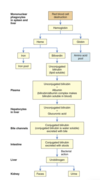
  • Bilirubin - a byproduct of aged RBC destruction & creates that greenish black colour in bile and the yellow tinge in jaundice
  • When RBCs get degraded, they create heme and globin components
    • heme is converted to biliverdin by cleaving iron
    • iron is transported back to liver/bone marrow for new RBCs
    • biliverdin is converted to bilirubin in Kupffer cells and then released into plasma to bind with albumin (aka unconjugated/free bilirubin) which is lipid soluble (May have a role as an antioxidant and provide cytoprotection)
  • In liver, unconjugated bilirubin moves from plasma in sinusoids to hepatocytes
  • In hepatocytes: unconjugated bilirubin joins with glucuronic acid to form conjugated bilirubin (water soluble and secreted in the bile)
  • Once it reaches distal ileum and colon, it is deconjugated by bacteria and converted into urobilinogen (then reabsorbed in intestine and excreted in the urine as urobilin)
  • Small amount excreted in feces (stercobilin) which contributes to poop’s brown colour
73
Q

What vascular and hematologic functions does the liver have?

A
  • has extensive vascular network and can store a large volume of blood (amount stored depends on pressures in arteries and veins)
  • can also release blood to maintain circulatory volume if needed (i.e. hemorrhage)
  • Hemostatic function:
    • synthesizes most clotting factors
    • the bile that is secreted by liver helps with vitamin K absorption which is neede for synthesis of clotting factors
74
Q

How does the liver contribute to metabolism of fats, proteins and carbohydrates?

A

Fats: absorbed by lacteals in intestinal villi then goes into liver via lymphatics as triglycerides. Hydrolyzed and then used to produce ATP or released into bloodstream as lipoproteins (to go to adipose cells for storage). Also makes phospholipids and cholesterol that are used in bile salt production, steroid hormones, etc.

Proteins: In hepatocytes, AA converted to keto acids via deamination (removing NH3). NH3 converted to urea by liver and passes into blood for excretion via kidneys. Liver synthesizes plasma proteins (albumins, globulins) that contribute to maintaining plasma oncotic pressure.

Carbohydrates: liver contributes to stability of BGL by glucose release during hypoglycemia and absorbing glucose during hyperglycemia. Stores it as glycogen or converts it to fat. IF all glycogen stores are used, can also convert AA and glycerol to glucose (gluconeogenesis).

75
Q

How does the liver accomplish metabolic detoxification?

A
  • aka biotransformation
  • alters exogenous/endogenous chemicals like drugs, foreign molecules,s and hormones to make them less toxic or less biologically active
  • Diminiahses the intestinal or renal tubular reabsorption of potentially toxic substances and facilitates their intestinal and renal excretion
  • helps with preventing excessive accumulation and adverse effects from things like EtoH, barbituates, amphetamines, steroids and hormones
  • detoxification is usually protective, but the end products sometimes become toxins or active metabolites.
76
Q

What kinds of minerals and vitamins are stored in the liver

A
  • Minerals: iron and copper (in times of excessive intake)
    • iron stored as ferritin (released as needed for RBC production)
  • Vitamins: B12 and D (several months); vitamin A (several years), E and K
77
Q

Structure and Function of gallbladder

A

Structure: saclike organ on inferior surface of liver

Function: store and concentrate bile between meals

  • between digestive periods, bile flows from liver (via R or L hepatic duct) into common hepatic duct and meets resistance the closed sphincter of Oddi (duodenal papilla) which controls flow into duodenum and prevents backflow
  • Bile then flows through cystic duct into gall bladder for concentration and storage
  • Gallbladder mucosa readily absorbs water and electrolytes which leaves a high concentration of bile salts, pigments, and cholesterol
  • holds ~90ml of bile
78
Q

How does bile get released from the gallbladder?

A
  • Within 30 min after eating, gallbladder begins to contract which forces stored bile through the cystic duct and into common bile duct
  • Sphincter of Oddi relaxes and allows bile to flow into duodenum via major duodenal papilla
  • Gallbladder contraction mediated by vagus nerve & cholecystokinin
  • Relaxation stimulated by vasoactive intestinal peptide, pancreatic polypeptide, SNS stimulation
79
Q

Structure of the pancreas

A
  • ~20cm long, head tucked into curve of duodenum and tail touching the spleen
  • lies deep in the abdomen behind the stomach
  • has endocrine and exocrine functions
  • receives blood via celiac and superior mesenteric arteries, and drains into portal vein (body and tail drains through splenic vein
  • innervation from parasympathetic neurons of the vagus nerve (which stimulate enzymatic and hormone secretion); sympathetic innervation causes vasoconstriction and inhibits pancreatic secretion
    *
80
Q

Structure and Function of exocrine pancreas

A
  • composed of acinar cells that secrete enzymes and networks of ducts that secrete alkaline fluid (both for digestion)
  • Acinar cells are organized in lobes around small secretory ducts where secretions drain into the pancreatic duct (Wirsung duct) which empties into common bile duct at the ampulla of vater then into the duodenum
  • Aq solution secreted: isotonic and contains K, Na, HCO3-, Cl (neutralizes acidic chyme when entering the duodenum to allow for enzymes to digest and fat absorption)
    *
81
Q

What enzymes are released from the pancreas?

A
  • hydrolyzes proteins, fats, and carbs
  • trypsin, chymotrypsin, carboxypeptidase, elastase (but all secreted in their inactive forms to protect the pancreas from its digestive effects)
  • Once in duodenum, they are activated by enterokinase (secreted from duodenal mucosa)
    • trypsinogen is activated first which stimulates conversion of chymotrypsinogen and procarboxypeptidase
  • Once the enzymes are activated in the small intestine, they inhibit the release of more cholecystokinin (that stimulated their release in the first place) to inhibit secretion of any more pancreative exocrine secretion
82
Q

Geriatric changes to the GI system

A
  • Mouth: teeth decay, everything decreases in terms of senses and function
  • Stomach: everything slows down (motility, blood flow, gastric emptying) & mucosal barriers decrease
  • Intestines: change in microbiome, decreased immunity, decline in overall health nutritional status, absorption declines, fecal making changes
  • Liver: decrease in regrowth, detox abilities, bloow flow (thus drug metabolism)
  • Pancreas and Gallbladder: fibrosis, fatty deposits; decreased digestive enzyme secretion; increased risk of gall stones and inflammation
83
Q

Anorexia

A

Definition: lack of desire to eat despite physiological stimuli that would normalyl produce hunger

  • often associated with nausea, abdo pain, diarrhea, stress
  • may also be from side effects of drugs or disorders (cancer, heart/renal disease)
84
Q

Vomiting - Definition and Types

A

Vomiting (emesis): forceful emptying of stomach and intestinal constents via mouth. Vomiting center in medulla oblongata and stimulated with:

  • severe pain; stomach/duodenum distension; presence of ipecac/copper salts in duodenum
  • motion sickness (CN 8 stimulation of vestibular system)
  • drugs
  • trauma to genitourinary structures
  • activaiton of chemoreceptor trigger zone (CTZ) in medulla

Nausea: usually precedes vomiting. Is a subjective experience. Often associated with hypersalivation and tachycardia; can be brought on by things like abnormal pain and spinning movements

Retching: muscular event of vomiting without anything coming out

Projectile Vomiting: spontaneous vomiting not prceded by nausea or retching. Caused by direct stimulation of vomiting center by neuro lesions (IICP, tumors, aneurysms) involving brainstem or a symptom of GI obstruction (pyloric stenosis)

85
Q

What physiological changes/mechanisms occur during the process of vomiting?

A
  • begins with deep inspiration; hlottis closes, intrathoracic pressure falls, and esophagus becomes distended. Simultaneously abdominal muscles contract creating a pressure gradient from abdomen to thorax
  • Lower esophageal sphincter (LES) and body of the stomach relax, but the duodenum and antrum of the stomach spasm
  • The reverse peristalsis and pressure gradient force chyme from the stomach and duodenum up into the esophagus but upper esophageal sphincter is closed so chyme does not enter the mouth
  • As the abdominal muscles relax, esophageal contents drop back into the stomach & process may be repeated several times before vomiting occurs
  • when vomiting does occur, diaphragm is forced high into the thoracic cavity by strong contractions of the abdominal muscles and causes the UES to open and chyme expelled from the mouth
  • Then the stomach relaxes and the upper part of the esophagus contracts, forcing the remaining chyme back into the stomach. The LES then closes & cycle repeats if more vomiting needs to occur
  • A diffuse sympathetic discharge causes the tachycardia, tachypnea, and diaphoresis that accompany retching and vomiting. Parasympathetic system mediates copious salivation, increased gastric motility, and relaxation of UES and LES
86
Q

Constipation

Definition, Pathophysiology, Clinical Manifestations, Diagnosis, Treatment

A

Definition: difficult or infrequent defecation. Common among elderly. (normal bowel habits can be 1-3/day to 1/week)

Pathophysiology: primary or secondary condition

Primary:

  • Normal transit (functional) constipation: normal rate of stool passage but difficulty with evacuation
  • Functional constipation: associated with sedentary lifestyle, low-residue diet (highly refined foods) or low fluid intake
  • Slow-transit constipation: involves impaired colon motor activity with infrequent bowel movements, straining to defecate, mild abdominal distention, palpable stool in sigmoid colon
  • Pelvic floor/outlet dysfunction: inability/difficulty expelling stooling due to dysfunction of pelvic floor muscles/anal sphincter

Secondary: can be caused by diet, meds, neurogenic disorders (CVA, Parkinson’s SCI, MS) where neural pathways or NTs are altered and colon transit time is delayed.

  • Drugs: opiates, antacids with CaCO or AlOH, anticholinergics, iron, bismuth inhibit bowerl motility
  • Endocrine disorders (hypothyroidism, DM, hypokalemia, hypercalcemia)
  • Abdo: pelvic hiatal hernia (herniation of bowel through floor of pelvis), diverticuli, IBS, pregnancy
  • Aging

Clinical Manifestations: 2 of the following for at least 3 months

  • (1) straining with defecation at least 25% of the time
  • (2) lumpy or hard stools at least 25% of the time
  • (3) sensation of incomplete emptying at least 25% of the time;
  • (4) manual maneuvers to facilitate stool evacuation for at least 25% of defecations;
  • (5) fewer than three bowel movements per week

Look for changes in bowel evacuation patterns (less pooping, smaller stool volume, hard stools, straining, feeling bowel fullness and discomfort). Fecal impaction (hard dry stool retained in rectum) leads to rectal bleeding, abdominal cramps, N/C, weight loss, diarrhea. Passage of hard stools may cause hemorrhoidal disease and painful anal fissures.

Diagnosis: History, meds, physical exam, stool diaries

Treatment: Manage underlying cause or disease. Bowel retraining, moderate exercise, increasing fluids and fiber intake. Laxatives/stool softeners for some. Last resort would be surgery.

87
Q

Diarrhea

Definition, Pathophysiology, Clinical Manifestations, Diagnosis, Treatment

A

Definition: presence of loose watery stools. Acute diarrhea is >3 loose stools within 24 h and <14 days. Persistent diarrhea lasts longer than 14-30 days. Chronic diarrhea lasts longer than 4 weeks.

  • high rates of morbidity/mortality in children < 5 y.o. (esp in developing countries) and in elderly
  • Stool volume in normal adult: ~200g/day; infant may be up to 100g/day
  • <150ml of water excreted daily in the stool

Pathophysiology: Large volume diarrhea (feces volume increased; generally caused by ++amounts of water or secretions in intestines). Small volume diarrhea (feces volume not increased; usually from excessive intestinal motility). 3 major mechanisms of diarrhea:

  • (1) Osmotic diarrhea: when there is a non-absorbable substance in the intestine it draws excess water into intestine and increases stool weight and volume, producing large volume diarrhea. Causes: lactase/pancreatic enzyme deficiency, excessive ingestion of synthetic surgars, tube feeding formulas,
  • (2) Secretory diarrhea: Excess mucosal secretion of fluid and electrolytes, makes large volume diarrhea. Causes: viruses (rotavirus), endotoxins (E. coli), exotoxins from overgrowth of C. diff after antibiotics, small bowel bacterial overgrowth
  • (3) Motility diarrhea: Excess motility decreases transit time and opportunity for fluid absoprtion which leads to diarrhea. Causes: resection of the small intestine (short bowel syndrome), surgical bypass of an area of intestine or fistula formation between intestinal loops, IBS, diabetic neuroparhty, hyperthyroidism, laxative abuse.

Clinical Manifestations: dehydration, electrolyte imbalance (hyponatremia, hypokalemia), weight loss. If due to bacterial/viral infection, then fever +/- vomiting or cramping pain. Most infectious diarrhea last <2 weeks. Inflammatory bowel disease: fever, cramping pain, bloody stools, chronic diarrhea. Steateorrhea: fat in stools; common in malabsorption syndromes. Irritation in anal and perineal skin.

Diagnosis: Hx - frequency, volume, duration, presence of blood. Physical exam.

Treatment: restoration of fluid and electrolyte balance, administration of antimotility (loperamide) and water absorbent medications. Treat causes.

88
Q

Abdominal Pain - potential causes and types of abdominal pain

A
  • mechanical, inflammatory, or ischemic causes
  • Abdominal organs are not sensitive to mechanical stimuli (cutting, tearing, etc.) but to stretching and distension as they active nerve endings in structures
  • usually associated with tissue injury and inflammation - mediators like histamine, bradykinin, and 5HT stimulate nerve endings that produce abdo pain; edema and vascular congestion from inflammation also cause painful stretching
  • Parietal (somatic) pain: from parietal peritoneum; more localized and intense than visceral pain (which comes from organs themselves)
  • Visceral pain: arises from a stimulus (distension, inflammation, ischemia) acting on an abdominal organ. Inflammatory mediators can cause pain hypersensitivity. Pain usually poorly localized, diffuse or vague with radiating pattern due to nerve endings in abdominal organs being sparse and multisegmented.
  • Referred pain: visceral pain felt at some distance from diseased or affected organ. Usually well localized and felt in the skin dermatomes or deeper tissues that share a central afferent pathway with the affected organ.
89
Q

Upper GI bleeding

A

Definition: bleeding in esophagus, stomach, or duodenum; characterized by frank, bright red bleeding or dark, grainy digested bloow (coffee grounds) that has been affected by stomach acids.

  • commonly caused by bleeding varices (varicose veins) in esophague, peptic ulcers
  • AVMs (abnormal tangle of blood vessels connecting arteries and veins, which disrupts normal blood flow and oxygen circulation. These can happen anywhere in the body, but most common in the brain and spine)
  • Mallory-Weiss tear at esophageal-gastric junction caused by severe retching (condition marked by a tear in the mucous membrane, or inner lining, where the esophagus meets the stomach. Most tears heal within 7 to 10 days without treatment, but Mallory-Weiss tears can cause significant bleeding)
90
Q

Lower GI bleeding

A
  • bleeding from the jejunum, ileum, colon, or rectum
  • caused by polyps, diverticulitis, inflammatory disease, cancer or hemorrhoids
91
Q

Occult bleeding

A

caused by slow, chronic blood loss that is not obvious and results in iron deficiency anemia (as iron stores in bone marrow are slowly depleted)

92
Q

What physiological responses occur with GI bleeding?

A
  • changes in BP and HR
  • Early stages: peripheral arteries and arterioles constrict to shunt blood to vital organs (brain)
  • Signs of large volume blood loss: postural hypotension, lightheadedness, loss of vision, tachycardia as compensatory
  • eventually hypovolemic shock
  • Decreased UP and potentially oliguria (due to decreased blood flow to kidneys), tubular necrosis, and renal failure
93
Q

Clinical manifesations of GI bleeding

A
  • accumulation of blood in GI tract becomes irritating and increases peristalsis which causes vomiting and diarrhea
  • Hematochezia: bright red stools - usually bleeding from lower GI tract
  • Melena: black or tarry stools that are sticky and have a foul odor - usually bleeding from upper GI tract because blood proteins have been digested
94
Q

Dysphagia

Definition, Pathophysiology, Clinical Manifestations, Diagnosis, Treatment

A

Definition: difficulty swallowing

Pathophysiology: can be from mechanical obstruction of esophague or functional disorder impairing esophageal motility.

Mechanical: Intrinsic obstructions originate in the esophageal lumen wall (tumours, strictures, diverticular herniations/outpouchings). Extrinsic obstructions come from outside the lumen and narrow the esophagus by pressing inwards (most common for these is tumor).

Functional dysphagia: caused by neural or muscular disorders that interfere with voluntary swallowing of peristalsis. Causes: dermatomyositis (muscle disease), neurological impairments 2’ to CVA, Parkinsons, MS, muscular dystrophy, or achalasia

Clinical Manifestations: Distention and spasm of esophageal muscles during eating or drinking, leading to stabbing pain at the level of obstruction. Discomfort 2-4 weeks post swallowing is associated with upper esophageal obstruction. Discomfort occurring 10 to 15 seconds after swallowing is more common in lower esophgeal obstruction. If caused by tumor, pt will have progressive difficulty with swallowing from solids to liquids. Regurgitation of food, unpleasant taste sensation, vomiting, aspiration, weight loss. Aspiration may lead to cough and pneumonia.

Diagnosis: Hx and clinical presentation. Imaging.

Treatment: management of Sx - eating small meals slowly, taking fluids with meals, sleeping with head elevated to prevent regurgitation and aspiration. Formulated food and meds so they can be swallowed. Anticholinergics (like botox) may provide Sx relief.

95
Q

Achalasia

Definition, Pathophysiology, Clinical Manifestations, Treatment

A

Definition: rare form of dysphagia related to loss of inhibitory neurons in the myenteric plexus & smooth muscle atrophy in the middle and lower portions of esophagus.

Pathophysiology; Myenteric neurons attacked by cell-mediated and anti-body mediated immune response against an unknown antigen. Leads to altered esophageal peristalsis and failure of LES to relax causing functional obstruction of the lower esophagus with varying severity. Food accumulates above the obstruction, distends the esophagus, and causes dysphagia. As hydrostatic pressure increases, food is slowly forced past obstruction and into the stomach.

Clinical Manifestations: Coughing, aspiration. Similar to dysphagia symptoms (pain when eating/drinking, pain after swallowing, vomiting, weight loss, etc.)

Treatment: Mechanical dilation or surgical myotomy (cutting muscular layer) of LES

96
Q

Gastroesophageal Reflux Disease (GERD)

A

Deifnition: reflux of acid and pepsin or bile salts from stomach into the esophague that causes esophagitis.

  • Physiological reflux: GE reflux that does not cause symptoms
  • Nonerosive reflux disease (NERD): pt has reflux disease symptoms but no visible esophageal mucosal injury

Risk factors: older age, obesity, hiatal hernia (weakens LES), drugs or chemicals that relax LES (anticholinergics, nitrates, CCBs, nicotine). GERD may trigger astham or chronic cough.

Pathophysiology: Abnormal LES function, esophgeal motility, and gastric motility or epmtying can cause GERD. Resting tone of LES tends to be lower than normal (transient relaxation or weakness of sphincter). Vomiting, coughing, lifting, bending, obesity, or pregnancy increases abdominal pressure - causes reflux esophagitis.

  • Delayed gastric emptying can contribute to reflux esophagitis by: 1) lengthening the period during which reflux is possible and 2) increasing gastric acid content

Reflux esophagitis severity depends on composition of gastric contents and exposure time to esophageal mucosa. Severe if it’s higly acidic or contains bil salts/pancreatic or intestinal enzymes. Weak esophageal peristalsis also allows refluxed chyme to be exposed to esophageal mucosa longer than normal. Reflux causes inflammation with hyperemia, increased capillary permeability, edema, tissue fragility, erosion. Potentiall fibrosis and thickening.

Clinical Manifestations: heartburn (pyrosis), acid regurgitation, dysphagia, chronic cough, astham attacks, laryngitis, upper abdo pain within 1 hour of eating. Sx worsen with lying down or if intra-abdominal pressure increases (coughing, vomiiting, straining to poop). Potentially dysphagia with weight loss (if edema, strictures). Alcohol or acid foods cause discomfort during swallowing.

Diagnosis: history and clinical presentation. Esophageal endoscopy (evidence of hyperemia, edema, erosion, and strictures). Tissue biopsy

Treatment: Proton pump inhibits, H2 receptor antagonists, antacids. Weight reduction, smoking cessation, head elevation, avoiding tight clothing for Sx relief. Most common surgical intervention: larpascopic fundoplication.

97
Q

Eosinophilic esophagitis

Definition, Clinical Manifestations, Diagnosis, Treatment

A

Definition: idiopathic inflammatory disease of esophagus where there is infiltration of eosinphils associated with atopic disease (caused by an allergy) - including asthma and food allergies.

Clinical Manifestations: dysphagia, food impaction (stuck in esophagus), comiting, weight loss

Diagnosis: endoscopy with biopsy

Treatment: elimination diets and steroids

98
Q

Hiatal Hernia

Definition, Pathophysiology, Clinical Manifestations, Diagnosis, Treatment

A

Definition: type of diaphragmatic hernia. Protusion of upper part of stomach through the diaphragm and into thorax

Pathophysiology: several types:

Sliding hiatal hernia (Type 1): most common. Proximal portion of stomach moves into thoracic cavity via esophageal hiatus (opening in diaphragm for esophagus and vagus nerves). Causes: congenitally short esophagus, fibrosis or excessive vagal nerve stim., weakening of diaphragmatic muscles at gastroesophageal junction. Often associated with GERD and made worse with coughing, bending, tight clothing, ascites, obesity, pregnancy.

Paraesophageal hiatal hernia (Type 2): herniation of greater curvature of the stomach through a secondary opening in diaphragm alongside esophagus. Causes congestion of mucosal blood flow, leading to gastritis and ulcers. Major complication is strangulation of hernia, presenting with vomiting and epigastric pain.

Mixed hiatal hernia (Type 3): less common; a combination of sliding and paraesophageal hiatal hernias. Tends to occur in conjunction with several other diseases (reflux esophagitis, peptic ulcer, cholecystitis, cholelithiasis, chronic pancreatitis, diverticulosis

Clinical Manifestations: often asymptomatic. Wide variety of Sx developing later in life related to GI disorders. Heartburn, regurgitation, dysphagia, epigastric pain. Ischemia from hernia strangulation causes acute, severe chest or epigastric pain, N/V and GI bleeding.

Diagnosis: radiology (barium swallow), endoscopy, high resolution manometry. Chest X ray

Treatment: conservative. Eat small frequent meals and avoid laying down after eating. Avoid abdominal supports and tight clothing. Weight control. Antacids. Elevation of the head of bed 6 inchs. Surgery (fundoplication) if other Ix fail.

99
Q

Gastroparesis

Definition, Pathophysiology, Clinical Manifestations, Treatment

A

Definition: delayed gastric emptying without mechanical gastric outlet obstruction. Most commonly associated with DM, surgical vagotomy (one or more branches of the vagus nerve are cut to reduce the rate of gastric secretion ie to treat peptic ulcers), fundoplication (to treat GERD). Can be idiopathic

Pathophysiology: not well understood but involves abnormalities of ANS, smooth muscle cells, enteric neurons, and GI hormones. Diabetic gastroparesis represents a form of neuropathy involving the vagus nerve.

Clinical Manifestations: N/V, abdo pain, post-eating fullness or blating

Treatment: diet management, prokinetic drugs (strengthens LES and increases food emptying). Potentially gastric electrical stimnulation or surgery.

100
Q

Pyloric Obstruction

Definition, Pathophysiology, Clinical Manifestations, Diagnosis, Treatment

A

Definition: aka gastric outlet obstruction - the narrowing/blocking of the opening between stomach and duodenum.

Causes/Pathophysiology: can be acquired for congenital. Acquired - caused by peptic ulcer disease or carinoma near pylorus (duodenal ulcers). Ulceration results in obstruction leading to inflammation, edema, spasm, fibrosis, scarrying.

Clinical Manifestations:

  • Early sx: vague epigastric fullness which becomes more distressing after eating and at end of the day. Nausea and epigastric pain occurs as stomach muscles contract to try and force chyme past obstruction (Sx disappear when chyme finally moves into duodenum)
  • Progression of obstruction: anorexia and weight loss
  • Severe obstruction: gastric distension & feeling fullness, atony (lack of muscle tone); Succussion splash: rolling/jarring of abdomen produces a sloshing sound; vomiting is cardinal sign (copious amounts and several hours after eating; vomitus is undigested food with no bile)
  • Prolonged vomiting leads to dehydration - hypokalemia, hypochloremia leading to metabolic alkalosis
  • infrequent and small stools because food doesn’t go into intestine
  • Prolonged obstruction leads to severe malnutrition, dehydration, and extreme debilitation

Diagnosis: clinical presentation, Hx of ulcer disease, examination of residual gastric contents. Endoscopy if there is gastric carcinoma

Treatment: Conservative if obstruction is due to ulcer. NG tube to aspirate stomach contents and relieve distention, decompress stomach, and stores normal motility (2-3 days). PPIs and H2 receptor antagonists to reduce inflammation and edema 2’ to gastric secretions. IV fluids to restore electrolyte fluid balance. IV nutrition for those severely malnourished. Surgery or pyloric stents for gastric carcinoma or persistent obstruction caused by fibrosis and scarring.

101
Q

Intestinal Obstruction and Functional Obstruciton (Paralytic Ileus)

Definition, Causes, Pathophysiology, Clinical Manifestations, Diagnosis, Treatment

A

Definition: any condition that prevents normal flow of chyme through intestinal lumen (small or large intestine but small usually more obstructed due to narrower lumen). Classified as:

  • Simple obstruction: mechanical blockage of lumen by a lesion; most common type
  • Functional obstruction (paralytic ileus): failure of intestinal motility often after intestinal/abdo surgery, acute pancreatitis, or hypokalemia

Causes: hernia, intussusception (intestine telescoping into another), Torson (volvulus; intestinal twisting), diverticulosis (inflamed diverticuli ), tumor, paralytic ileus (loss of peristaltic motor activity), fibrous adhesions (peritoneal irritation from surgery, trauma, Chron disease leading to fibrosis)

  • Small bowel obstruction: adhesions (2’ to abdo surgery, most common), hernia, tumors, mesenteric ischemia, Crohn disease
  • Large bowel obstruction: colon/rectal cancer (most common), volvulus, diverticular disease

Pathophyisiology: Paralytic ileus 2’ to surgery is due to inhibitory neural reflexes associated with inflammatory mediators and opioids that affect entire GI tract. Small bowel obstruction (SBO) caused by post-op adhesions, tumors, Crohn disease, and hernias & leads to distention caused by impaired absoprtion and increased secretion of fluid and gas. Distention decreases the ability to absorb water and electrolytes, on top of that with copious vomiting as well causes severe fluid electrolyte disturbances. ECF and plasma volume decrease (causing dehydration increased Hct, hypotension, and tachycardia; eventually hypovolemic shock).

  • Metabolic alkalosis initially develops due to excess loss of H+ but then metabolic acidosis kicks in due to prolonged obstruction in intestine as HCO3- from pancreatic secretions and bile cannot be reabsorbed
  • Metabolic acidosis also may be accentuated by ketosis, the result of declining carbohydrate stores caused by starvation. Buildup of lactic acid due to lack of circulation also increases acidosis
  • If pressure from the distention is severe enough, it occludes the arterial circulation (causing ischemia, necrosis, perforation, and peritonitis).
  • Fever and leukocytosis are often associated with overgrowth of bacteria, ischemia, and bowel necrosis. Bacterial proliferation and translocation across the mucosa to the systemic circulation cause peritonitis or sepsis. The release of inflammatory mediators into the circulation causes remote organ failure.
  • Large Bowel Obstruction (LBO): less common, often related to cancer
  • Acute colonic pseudo-obstruction (Oglivie syndrome): rare but massive dilation of large bowel related to excessive sympathetic motor input or decreases parasympathetic motor input with absence of mechanical obstrucition. Primarily in critically ill or immobilized older adults

Clinical Manifestations:

  • SBO: colicky pain (from intestinal distention) followed by N/V. Pain intesnsifies for seconds or minutes as a peristaltic wave of muscle contraction meets the obstruction. If ischemia occurs, pain becomes more constant and severe. Hypotension, increased HR, diaphoresis, fever, ++leukocytosis, abdominal distention, rebound tenderness.
  • Obstruction at pylorus: early, profuse vomiting
  • Partial obstruction: diarrhea and constipation
  • Complete obstruction: constipation only; increased bowel sounds, hypovolemia, metabolic acidosis. Distention may push against diaphragm leading to decreased lung volume, atelectasis and pneumonia
  • Large Intestine obstruction: hypogastric pain, abdominal distention. Variable pain. Vomiting occurs later in obstructive process.

Diagnosis: Clinical presentation and imaging studies.

Treatment: early identification of site and type of obstruction. Fluid and electrolyte replacement, decompression of lume with suctioning. Laparoscopic procedures can release adhesions. Immediate surgery for strangulation, complete obstruction, or perforation

102
Q

What is gastritis? Describe the types of gastritis.

A

Gastritis: inflammatory disorder of gastric mucosa. Can be acute or chronic, affect the superficial mucosa of fundus or antrum, or both. Uncommon (1% in US)

Acute gastritis: caused by injury to mucosal barrier - drugs, chemicals, H. pylori. NSAIDs inhibits COX-1 causing gastritis due to inhibiting prostanglandin synthesis (which normalyl stimulates mucus secretion). Contributing factors: alcohol, histamine, digitalis, metabolic disorders. H. pylori causes inflammation, increased gastric secretion, pain, N/V.

  • S/S: vague abdominal discomfort, epigastric tenderness, bleeding
  • healing occurs spontaneously within a few days. Discontinue injurious drugs, use antacids, or H2 receptor antagonists

Chronic gastritis: tends to occur in elderly and cause chronic inflamamtion, mucosal atrophy, and epithelial metaplasia. Classified as type A, immune (fundal), or type B, nonimmune (antral), depending on the pathogenesis and location of the lesions. Type AB (pangastritis - both types occurring). Type C associated with bile and pancreatic secretion reflux into the stomach causing chemical injury

  • Chronic immune (fundal) gastritis: most rare form of gastritis, associated with loss of T-cell tolerance and development of autoantibodies to gastric H-K ATPase. Gastric mucosa degenerates extensively in the body and fundus of the stomach leading to gastric atrophy. Loss of parietal cells diminish acid and IF secretion lead to pernicious anemia. Feedback mechanism to inhibit gastrin secretion is impaired leading to elevated levels. Usually occurs in association with other autoimmune diseases (RA, T1DM).
  • Chronic nonimmune (antral) gastritis: involves antrum only, more common than ^. Caused by H. pylori and use of EtOH, tobacco, NSAIDs. High levels of HCl secretion with increased risk of duodenal ulcers. H pylori can cause worsening of the gastritis turning it into Type AB. Greater risk of gastric cancer.
  • S/S: anorexia, fullness, N/V, epigastric pain, gastric beeding
  • Tx: management of symptoms by eating smaller meals with soft, bland diet; avoid alcohol and ASA. Antibiotics for H. pylori and vitamin B12 to corrrect pernicious anemia
103
Q

Peptic ulcers

What are they, how do they develop, what are the risk factors, and types of ulcers.

A

Definition: break or ulcertaion in the protective mucosal lining of the lower esophagus, stomach, or duodenum. They develop when mucosal protective factors are overcome by erosive factors (commonly caused by NSAIDs and H. pylori infection)

  • can be single, multiple, acute or chronic, and superficial or deep
  • Erosions: superficial ulcerations - erode mucosa but do not penetrate muscularis layer
  • True ulcers: extend through muscularis mucosa and damage blood vessels leading to hemorrhage, or perforate GI wall

Risk factors: psych stress, H. pylori infeciton, chronic use of NSAIDs, alcohol, smoking, age, chronic disease, type O blood

Types: duodenal, gastric, and stress ulcers

104
Q

Zollinger-Ellison syndrome

A

rare syndrome associated with peptic ulcers caused by gastrin-secreting tumor or multiple tumors of the pancreas/duodenum

increased gastrin = ++secretion of gastric acid which results in gastric and duodenal ulcers, gastroesophageal reflux with abdo pain and diarrhea

105
Q

Duodenal Ulcers

Definition, Pathophysiology, Clinical Manifestations, Diagnosis, Treatment

A

Definition: occur more commonly than other types & commonly due to H. pylori infection and NSAID use. Idiopathic ulcers are rare & associated with altered mucosal defenses, rapid gastric emptying, elevated serum gastrin levels, acid production 2’ to smoking.

Pathophysiology: Acid and pepsin in duodenum penetrate the mucosal barrier and cause ulceration. T and B lymphocytes activated in response to H. pylori infection, neutrophils infiltrated. Inflammatory cytokines released and damages gastric epithelium. H. pylroi causes apoptosis of gastric epithelial cells and promotes inflammation

Clinical Manifestations: **chronic intermittent pain in epigastric region**. Pain starts 2-3 hr after eating when stomach is empty. Can also occur at night and disappear in the morning; relieved rapidly by infestion of food or antacids (pain-food-relief pattern). Some may be asymptomatic and then have their first symptom as hemorrhaging or performation (esp with NSAID/anticoagulant use).

  • Complications: bleeding, perforation, obstruction of duodenum or outlet of the stomach. Bleeding most common cause of mortality in elderly.
    • Perforation occurs with destruction of all layers of the duodenal wall, causes sudden severe epigastric pain
    • Obstruction may be from edema (inflammation) or scarring from chronic injury
  • Exacerbations tend to develop in spring and fall

Diagnosis: endoscopies to visualize lesions, biopsy. Urea breath test to detect H. pylori

Treatment: often heal spontaneously but recur within months without tx. Pain relief allows healing. Treatment: relive causes and effects of hyperacidity and preventing complications. Antacids neutralize gastric contents and relieve pain. H2 receptor antagonists to inhibit acid secretion. PPIs to inhibit acid production. Antibiotics for H. pylori. Surgery for bleeding or perforations.

106
Q

Gastric ulcers

Definition, Pathophysiology, Clinical Manifestations, Diagnosis, Treatment

A

Definition: ulcers of the stomach. Occurs equally in M and W usually between 55-65. Tend to develop in the antral region next to the acid-secreting mucosa of the body.

Pathophysiology: primary defect - an abnormality that increases the mucosal’s barrier permeability to H+. Gastric secretion is normal or less than normal, and there may be decreased pass of parietal cells. Contributing factors:

  • Chronic gastritis - due to limiting mucosa’s ability to secrete a protective layer of mucus, which contributes to gastric ulcer formation
  • Decreased mucosal synthesis of prostaglandins
  • Duodenal reflux of bile and pancreative enzymes damaging the mucosal membrane
  • Use of NSAIDs (decreased prostaglandin synthesis)
  • H. pylori infection

Break in mucosal barrier allows H+ to diffuse into the mucosa which disrupts permeability and cell structure. Vicious cycle of: damaged mucosa → liberates histamine →increased acid and pepsinogen production, blood flow, capillary permeability → edema, bye bye plasma proteins → more bleeding and release of more histamine

Clinical Manifestations: similar to duodenal ulcers (upper abdo pain, intermittent, pain-antacid-relief pattern). Pain occurs immediately after eating. Tends to be chronic rather than remission-exacerbation. Causes more anorexia, vomiting, and weight loss than duodenal ulcers

Diagnosis and Treatment: similar to duodenal ulcers. Endoscopy for visualization. Antacids, H2 receptor antagonists, PPIs. Antibiotics for H. pylori. Surgery for bleeding or perforations.

107
Q

Stress-Related Mucosal Disease (Stress ulcer)

Definition, types, clinical manifestations, treatment

A

Stress ulcer: acute form of peptic ulcer that happens with stress of severe illness or major trauma. Usually has multiple sites throughout stomach or duodenum. Classified as ischemic of Cushing ulcers:

  • 1) Ischemic ulcers: develop within hours after event (hemorrhage, multisystem trauma, severe burns, HF, sepsis). Ischemia caused by shock, anoxia, inflammation, sympathetic responses, disrupting mucosal barrier.
    • Curling ulcers: stress ulcer 2’ burn injury
  • 2) Cushing ulcers: stress ulcer associated with severe brain trauma/surgery. Decreased mucosal blood flow and hypersecretion of acid are caused as a result of overstimulation vagal nuclei. Mucosal barrier damaged leading to erosins and ulceration.

Clinical Manifestations: bleeding (but uncommon) that occurs more readily if someone has coagulopathy and more >48 hrs of mechanical ventilation

Treatment: Prophylactics

108
Q

Who would required surgical treatment for ulcers and what are the goals of such treatment?

A
  • for those who have recurrent or uncontolled bleeding and perforation of the stomach or duodenum
  • objectives of surgery are to reduce stimuli for acid secretion, decrease number of acid secreting cells in the stomach and correct complications of ulcer disease
  • May cause chronic complications due to portion of stomach being removed (gastrorectomy)
109
Q

What is postgastrectomy syndrome? What S/S may occur? (7)

A

group of S/S that occur after gastric resection for treating peptic ulcers, gastric carcinomas, or bariatric surgery

1) Dumping syndrome: Chyme moving too quicky from stomach to small intestine, 10-20 min after eating. Due to decreased gastric capacity, loss of emptying control when pylorus is removed, and loss of feedback control by duodenum. Responds to diety management. S/S: cramping pain, N/V/D, weakness, pallor, hypotension

2) Alkaline reflux gastritis: stomach inflammation caused by reflux of bile and pancreatic secretions from the duodenum into the stomach. Disrupts the mucosal barrier and causes inflammation. S/S: Nausea, vomiting of bile, sustained epigastric pain worsening after eating and not relieved by antacids. Surgery needed.

3) Afferent loop obstruction: obstruction of the duodenal stump after surgery. Biliary and pancreatic secretions accumulate in the stump causing distention, intermittent pain and vomiting. Epigastric fullness after eating. Vomiting relieves symptoms. Tx: low fat diet, surgery.

4) Diarrhea: due to rapid gastric emptying and osmotive attraction of water into the gut, especially after high carb intake. Tx: small dry meals and anticholinergic drugs.

5) Weight loss: due to inadequate caloric intake (pt cannot tolerate normal portions or carbs). Stomach less able to mix, churn, break down food.

6) Anemia: iron malabsorption due to decreased acid secretion/lack of duodenum after procedure. Deficiencies in iron, vitamin B12, folate.

7) Bone and mineral disorders: due to altered calcium absoprtion and metabolism with increased risk for fractures and deformity, and malabsoprtion of vitamins and nutrients.

110
Q

Maldigestion vs Malabsorption

A

Maldigestion: failure of chemical processes of digestion to take place in the lumen or brush border of intestine; generally caused by enzyme deficiencies that are needed for digestion or inadequate secretion of bile salts/reabsorption of bile in ileum

Malabsoprtion: failure of intestinal mucosa to absorbed the digested nutrients; the result of mucosal disruption caused by gastric or intestinal resection, vascular disorders, or intestinal disease

these two frequently occur together

111
Q

Malabsorption Syndromes include:

A

1) Pancreatic Exocrine Insufficiency

2) Lactase Deficiency (Lactose Intolerance)

3) Bile Salt Deficiency

112
Q

Pancreatic Exocrine Insufficiency

Definition, causes, Pathophysiology, Clinical Manifestations

A

Definition: deficient production of enzymes (esp lipase; others: amylase, trypsin, chymotrypsin) by the pancreas that is needed for digestion of proteins, carbs, and fats

Causes: chronic pancreatitis, pancreas carcinoma, resection, CF

Pathophysiology: ++damage/loss of pancreatic tissue must occur before enzyme levels decrease sufficiently to cause maldigestion. Fat maldigestion is the chief problem. Absence of pancreatic bicarbonate in duodenum and jejunum causes an acidic pH that worsens the maldigestion by precipitating bile salts and preventing activation of pancreatic enzymes.

Clinical Manifestations: Fatty stools (steatorrhea) most common sign. Deficit of fat-soluble vitamins (A, D, E, K) and weight loss.

113
Q

Lactase Deficiency (Lactose Intolerance)

Definition, Pathophysiology, Clinical Manifestations, Diagnosis, Treatment

A

Definition: lack of lactase (genetic defect) lead to disaccharide deficiency at brush border of small intestine.

Pathophysiology: Lack of lactase inhibits breakdown of lactose into monosaccharides which therefore prevents lactose digestion and absorption. Can also be acquired in diseases like enteritis, bacterial overgrowth, and gluten-sensitive enteropathy. Lactose is undigested & stays in intestine where bacterial fermentation causes formation of gases. Also causes irritation and osmotic diarrhea (drawing fluid from body to dilute stool).

Clinical Manifestations: bloating, cramps, diarrhea, flatulence.

Diagnosis and Treatment: lactose-tolerance test. Tx is avoiding milk products

114
Q

Bile Salt Deficiency

Definition, Causes, Clinical Manifestations, Treatment

A

Definition: Lack of conjugated bile salts does not allow for micelle formation with fat molecules to make them more soluble and pass through brush border. Bile salts are usually conjugated in the bile secreted from the liver

Causes: Advanced liver disease (decreased bile salt production), obstruction of common bile duct (decreases flow of bile into duodenum - cholestasis), intestinal stasis (lack of motility which allowed bacterial overgrowth that deconjugates bile salts), diseases of ileum (prevents reabsorption and recycling of bile salts aka enterohepatic circulation).

Clinical Manifestations: related to poor intestinal absorption of fat and fat-soluble vitmains (A D E K)

  • Steatorrhea (fatty stools) leads to diarrhea and decrease levels of plasma proteins
  • Vit A deficiency: night blindness
  • Vit D deficiency: decreased calcium absorption with bone demineralization (osteoporosis), bone pain, fractures
  • Vit K deficiency: prolongs prothrombin time, leading to spontaneous purpura and petechiae
  • Vit E deficieny: uncertain, potentially testicular atrophy and neuro deficits in children

Treatment: increase consumption medium chain triglycerides in the diet (ex. using coconut oil to cook). Give vit A, D, and K parenterally. Oral bile salts.

115
Q

What is inflammatory bowel disease (IBD)? Descrine the risk factors and pathophysiology of this condition.

A

Definition: General term used to describe disorders that involve chronic inflammation of the GI tract. Includes chronic relapsing diseases - ulcerative colitis (UC) and Crohn disease (CD)

Risk factors/Pathophysiology: genes x environment, alterations in epithelial cell barrier functions, altered immune response to intestinal microflora

  • Possible loss of discrimination of potentially harmful pathogens from microorganisms in the intestinal mucosa
  • leads to a loss of immune tolerence to normal intestinal antigens, which activates dendritic cells triggering T cells to differentiate.
116
Q

Ulcerative Colitis (UC)

Definition, Pathophysiology, Clinical Manifestations, Diagnosis, Treatment

A

Definition: chronic inflammatory disease that causes ulceration of colon mucosa (most common rectum and sigmoid colon). Susceptible to those 20-40 y.o., less common in people who smoke.

Pathophysiology: Primary lesion begins with inflammation at base of crypt of Lieberkühn in large intestine. Disease begins in the rectum (proctitis) and may extend proximally to entire colon (pancolitis). Mucosa is hyperemic (dark red and velvety) with small erosions forming and turning into ulcers. Abscesses, necrosis, and ragged ulceration occur. Edema and thickening of muscularis mucosae may lead to narrowing of lumen. Mucosal destruction and inflammation causes bleeding, cramps, urge to defecate.

Clinical Manifestations: Intermittent periods of remsission-exacerbation. Mild UC: less bowel movements, bleeding, and pain. Severe UC: abdo pain, fever, increased HR, frequent watery diarrhea (10-20 stools/day) with passing small amounts of blood and purulent mucus. Urgency. cramps/ Dehydration, weight loss, anemia, and fever 2’ to fluid loss, bleeding, and inflammation.

  • Complications: anal fissures, hemorrhoids, perirectal abscess, perforation (rare)
  • Edema, strictures, or fibrosis can obstruct the colon.

Diagnosis: medical hx, clinical manifestations, and lab, serology, radiology, endoscopy, biopsy. Stool culture. Difficult to differentiate from Crohn disease due to similar symptoms.

Treatment: promoting mucosal healing and avoiding surgery. Mild to moderate disease is treated with 5-aminosalicyclate therapy followed by steroids. Thioprine and immunomodulatory agents or vedolizumab are used for serious disease. Surgical resection of colon if other Ix are unsuccessful - results in ileostomy.

117
Q

Crohn Disease (CD)

Definition, Pathophysiology, Clinical Manifestations, Diagnosis, Treatment

A

Definition: aka Granulomatous colitis, ileocolitis, or regional enteritis - an idiopathic inflammatory disorder affecting any part of the GI tract (mouth to anus). Most common affects distal small intestine and proximal large colon. Smoking increases risk of severe disease.

Pathophysiology: Inflammation begins in intestinal submucosa and spreads with discontinuous transmural involvement (skip lesions). Ascending colon and transverse colon most common sites of disease. Involves all layers of intestinal lumen (serosa, muscularis, submucosa, mucosa). Ulcerations can produce fissures that extend inflammation into the lymphoid tissue. One side of the intestinal wall may be affected but not the other. Typical lesion is a granuloma with cobblestone appearance from projections of inflamed tissue surrounded by ulceration. Fistula can form in perianal area between loops of intestine or extend into bladder, rectum, vagina. May develop strictures promoting obstruction.

Clinical Manifestations: No specific symptoms for several years. Sx depends on location but are similar to those for UC. Diarrhea (most common), occasionally rectal bleeding if colon is involved. Weight loss, abdominal pain. If ileum involved, then anemic due to malabsorption of B12, potentially also vitamin D and folic acid. Proteins may also b los leading to hypoalbuminemia.

Diagnosis: Similar to UC but also use imaging of small intestine and endoscopy.

Treatment: Smoking cessation. Surgery may be needed to manage complications (fistula, abscess, obstruction)

  • Similar to US: Mild to moderate disease is treated with 5-aminosalicyclate therapy followed by steroids. Thioprine and immunomodulatory agents or vedolizumab are used for serious disease.
118
Q

Microscopic Colitis

Definition, Cause, Risk Factors, Clinical Manifestations, Treatment

A

Definition: chronic inflammation of the colon, relatively common cause of diarrhea primarily in females and elderly. Two histologic forms:

  • Lymphocytic colitis: shows increase in # of intraepithelial lymphocytes
  • Collagenous colitis: thickened subepithelial collagen layer, changes to vascular mucosal pattern, and mucosal nodularity

Cause: unknown

Risk factors: ≥50 y.o., female, weight loss, absence of abdo pain, use of PPIs or NSAIDs

Sx: frequent, chronic daily water diarrhea & abdominal pain and weight loss.

Treatment: Antidiarrheal and budesonide (Pulmicort; anti-inflammatory steroid)

119
Q

Irritable Bowel Syndrome (IBS)

Definition, Demographics, Pathophysiology, Clinical Manifestations, Diagnosis, Treatment

A

Definition: Symptom-based condition characterized by recurrent abdominal pain with altered bowel habits. Increasing evidence of organic causes of disease

Demographics: more common in women (higher prevalence in youth and middle aged). These individuals also more likely to have anxiety, depression, reduced QoL.

Pathophysiology: unknown cause but potential mechanisms

  • Visceral hypersensitivty/hyperalgesia: potential dysregulation of the brain-gut axis that causes processing of distension in the gut/rectum to be painful.
  • Abnormal gastrointestinal permeability, motility, secretion: rapid colon transit times and increased intestinal permeability leading to diarrhea. Bloating and constipation IBS will have delayed transit times & decreased intestinal permeability.
  • Postinflammatory (non-/infectious) IBS: dx if 2+ occur - fever, vomiting, diarrhea, positive stool culture. Intestinal infection and low grade inflammation altered gut microbiome, immune activaiton in gut tissues, and change intestinal permeability
  • Alteration in gut microbiota (dysbiosis): influences sensory, motor, and immune systems of the gut and interacts with higher brain centers. May contribute to IBS
  • Food allergy/intolerance: food antigens may activate mucosal immune system, alter intestinal flora, or mediate hypersensitivity reactions and IBS symptoms
  • Psychosocial factors (epigenetics): early life trauma, emotional stress contributing to IBS symptoms

Clinical Manifestations: lower abdominal pain/discomfort and bloating. Women - abdominal pain and constipation; men - more diarrhea. Maybe diarrhea-prevalent or constipation-prevalent or both. Gas, bloating, and nausea that is relieved with defecation and do not interfere with sleep.

Diagnosis: S/S, hx, ruling out other structural/biochemical causes of disease. Diagnostics (CT, ultrasound, blood work). Rome III criteria for diagnosis IBS.

Treatment: no cure for IBS. Treating symptoms - laxatives and fiber, antidiarrheals, antispasmodics, prosecretory drugs, low-dose antidepressants, analgesics. Prebiotics and probiotics. Yoga, CBT, dietary interventions, acupuncture

120
Q

Diverticular Disease of the Colon

Definition, Causes/Risk Factors, Pathophysiology, Clinical Manifestations, Diagnosis, Treatment

A

Definitions:

  • Diverticula: herniations/saclike outpouchings of the mucosa and submucosa through the muscle layers (usually sigmoid colon; rarely in small intestine)
  • Diverticulosis: asymptomatic diverticular disease
  • Diverticulitis: inflammatory stage of diverticulosis

Causes/Risk Factors: unknown; associated with increased intracolonic pressure, abnormal neuromuscular function, and alterations in intestinal motility. Predisposing factors: older age, genetic predisposition, obesity, smoking, diet, lack of physical activity, and medication use (ASA, NSAIDs)

Pathophysiology: Can occur anywhere in the GI tract esp. at weak points in the wall where arteries penetrate the tunica muscularis. Most common sites are L sigmoid colon and R colon. Common associated finding is thickening of the circular muscles and shortening o the longitudinal (teniae coli) muscles surrounding the diverticula. Increased collagen and elastin deposited which causes muscle thickening and increased intraluminal pressure + herniation. Pressure can increase so much to the point of rupturing causing inflammation and diverticulitis. Complicated diverticulitis involves abscess, fistula, obstruction, bleeding, perforation.

Clinical Manifestations: If uncomplicated, then vague/absent Sx. Lower abdo cramps with diarrhea, constipation, distention, flatulence. Inflamed diverticula or presence of abscess: fever, leukocytosis, LLQ tenderness.

Diagnosis: often discovered during diagnostics for other problems. Ultrasound, sigmoidoscopy, colonoscopy for direct observation of the lesions.

Treatment: increasing dietary fiber for Sx relief, probiotics & mesalazine. For uncomplicated, bowerl rest and analgesia. If severe, larpascopic resection.

121
Q

Appendicitis

Definition, Pathophysiology, Clinical Manifestations, Diagnosis, Treatment

A

Definition: inflammation of the vermiform appendix. Most common surgical emergency of the abdomen (usually between 10-19 y.o.)

Pathophysiology: Exact mechanism controversial. Obstruction of lumen (stool, tumors, foreign bodies) with consequent bacterial infection is the most common theory. Doesn’t allow drainage of appendix and as mucosal secretion continues, intraluminal pressure increases. Increased pressure decreases mucosal blood flow and appendix becomes hypoxic. Mucosa ulcerates and promotes bacterial or other microbial invasion with further inflammation and edema. Inflammation may involve distal or entire appendix. Gangrene and perforation develop.

Clinical Manifestations: Gastric/periumbilical pain that is vague initially and then gets more severe over 3-4 hours. May subside and then migrate to RLQ indicating extension of inflammation to surrounding tissues. N/V, anorexia, low-grade fever. Diarrhea in some individuals (esp. children) while others may have constipation.

  • Most severe complications: perforation, peritonitis, abscess formation

Diagnosis: Clinical presentation, pain with abdo palpation, rebound tenderness usually referred to RLQ. High WBC. Diagnostics: abdominal ultrasound, CT scanes, MRI

Treatment: Laparoscopic surgery. Antibiotics

122
Q

Mesenteric Vascular Insufficiency

A

Definition: rare condition where the blood vessels supplying the stomach/intestine is inadequate.

  • 3 branches of abdominal aorta supply this area: celiac artery, superior and inferior mesenteric arteries
  • Venous drains: inferior mesenteric vein drains into splenic vein; splenic vein and superior mesenteric vein join portal vein

Risk factors: malignancies, RHF, DVT. Mesenteric venous thrombosis - least common cause.

Types:

Acute mesenteric arterial insufficiency: due to ++reduction in mucosal blood flow to large & small intestines, can be acute or chronic. Predisposing factors: aortic dissections, arterial thrombi, emboli. Superior mesenteric artery has more direct line of flow from aorta so emboli more readily able to enter causing ischemia and necrosis of the small intestine. This alters membrane permeability. Damaged mucosa cannot make enough mucus to protect itself from digestive enzymes so it causes fluids to move from blood vessels into bowel wall and peritoneum. Hypovolemia, shock, fever, bloody diarrhea, leukocytosis.

  • S/S: initially increased motility and need for bowel movement, N/V, severe abdo pain. Then ischemia leads to decreased motility and distention. Eventually gangrene and peritonitis.

Chronic mesenteric ischemia: rare but can develop with atherosclerotic stenosis or occlusion, CHF, acute MI, hemorrhage, thrombus formation, or anything that decreases arterial blood flow. Chronic occlusion often causes formation of collateral circulation which can keep up for the most part but when eating and the intestine needs more blood, the blood supply may be insufficient. Ischemia develops which causes cramping abdominal pain (abdominal angina - cardinal symptom). Continued progressive vascular obstruction eventually causes continuous abdominal pain and necrosis of intestinal tissue.

  • abdominal cramps, weight loss (due to stopping eating for pain relief)

Diagnosis: clinical manifestations, lab findings, imaging. Bruit can often be heard over a partially occluded artery.

Treatment: Aggressive rehydration, antibiotics, anticoagulants, vasodilators, inhibitors of reperfusion injury. Surgery to remove necrotic tissue, repaired narrowed vessels, and revascularize affected tissue.

123
Q

Definition of Obesity and Risk factors

A

Definition: increase in body fat mass. Defined as BMI >30 and generally develops when calories in > calories out. Obesity associated with higher all-cause mortality

Risk factors: 3 leading causes of death associated with obesity - CAD, T2DM, cancer. Major risk factor for morbidity, death, and high healthcare costs.

  • Obesity is a risk factor for: HTN, CVA, DLD, gallstones, GERD, OA, infectious diseases, sleep apnea
124
Q

Pathophysiology of obesity

A
  • Involves interaction of peripheral and central pathways and numerous cytokines, hormones, and neurotransmitters
  • Peripheral: fat cells store triglycerides and increase in size and number. Adipocytes secrete hormones and adipocytokines.
  • Central/visceral white fat accumulation causes dysfunction in the regulation and interaction of these cytokines and hormones
  • Peripheral sources of mediators act on hypothalamus & brainstem to regulate hunger/satiety, which include insulin from the beta cells of the pancreas; ghrelin from the stomach; peptide YY from the intestines; glucagon- like peptide-1 from intestinal endocrine cells; and the adipokines leptin, adiponectin, and resistin
  • Obesity associated with increased levels of leptin, insulin, resistin, and ghrelin; decreased adiponectin and peptide YY
  • Enlarged adipocytes increased lipolysis and secrete proinflammatory adipokines from T lymphocytes and activated macrophages - results in low-grade systemic inflammation
  • Inflammation and accelerated lipolysis contribute to insulin resistance and metabolic syndrome
125
Q

Orexigenic neurons vs anorexigenix neurons

A

neurons that are found in the hypothalamus & interact with peripheral mediators to control food intake and energy expenditure

orexigenic neurons: increased food intake and decreased metabolism

anorexigenic neurons: decrease food intake and increase metabolism

126
Q

Function of leptin in obesity

A
  • acts on hypothalamus to supress appetite and function to regulate body weight
  • levels increase as adipocytes increase however for some reason if it’s too high it leads to leptin resistance where it doesn’t help with decreasing appetite
    • actually promotes overeating and excessive weight gain instead
    • also associated with insulin resistance
127
Q

Function of ghrelin in obesity

A

increased levels of grehlin which stimulate orexigenic neurons (increase foot intake and decreased metabolism)

128
Q

Role of adiponectin and peptide YY in obesity

A
  • adiponectin and peptide YY levels are decreased which decrease the stimulation of anorexigenic neurons (which decrease food intake)
  • Adiponectin: insulin-sensitizing for BGL regulation, promotes glucose uptake, and has anti-inflammatory actions
    • a decrease leads to insulin resistance, CAD, HTN
  • Peptide YY: secreted from intestine and reduces appetite and inhibits gastric motility
129
Q

Obesity - Clinical Manifestations

A

Two different forms of adipose tissue distribution

Visceral obesity: aka intra-abdominal, central, or masculine obesity - occurs when body fat distribution is localized around the abdomen and upper body (apple shape). These people have increased risk for systemic inflammation, metabolic syndrome, OSAS, CV complications, nonalcoholic steatohepatitis (fatty liver) cancer, OA and T2DM.

Peripheral obesity: aka gluteal-femoral, feminine, subQ obesity. Occurs when body fat distribution is extraperitoneal and around the thighs/butt and through the muscle (pear shape) & more common in women. Risk factors are present but less severe than those for visceral obesity.

  • Normal weight obesity (NWO) describes those with normal body weight & BMI with >30% body fat. At risk for metabolic dysregulation, increases in inflammatory cytokines, insulin resistance, CV disease, higher mortality
  • Metabolically healthy obesity (MHO) - people who are obese but no metabolic-obesity associated complications and decreased risk for morbidity and mortality. More prevalent among women and declines with age with adverse LT outcomes
130
Q

Obesity - Diagnosis and Treatment

A

Diagnosis: CT and MRI, measurements and weight for estimating body fat mass. BMI and waist-to-hip ratios. Overweight - BMI > 25kg/m2. Obesity - BMI > 30kg/m2

Treatment: Correcting metabolic abnormalities, diet and exercise programs, psychotherapy, behavioural modification, antiobesity drugs. Weight loss (bariatric) surgery is the most effective tx for decreasing obesity-related morbidity

131
Q

Malnutrition

A

lack of nourish from inadequate amounts of calories, proteins, vitamins, minerals

caused by improper diet, alterations in digestion or absoprtion, chronic disease, or a combination

132
Q

Starvation

Definition and Types

A
  • reduction in energy intake leading to weight loss. Can be ST or LT starvation
  • Therapeutic ST starvation: part of weight reduction programs that cause an initial rapid weight loss that reinforces dieting
  • Therapeutic LT starvation: used in medically controlled environments to facilitate rapid weight loss in morbidly obese individuals
  • Pathologic LT starvation: caused by poverty, chronic disease of CV/pulmonary/hepatic/renal/ digestive systems, malabsorption syndromes, and cancer
133
Q

What is short-term starvation and what occurs in the body while this is happening?

A

Definition: extended fasting where you’re not eating anything for several days.

Physiological changes: once all available energy has been absorbed from intestine, glycogen in liver is converted to glucose (glycogenolysis) which peaks within 4-8 hours. Gluconeognesis also occurs (formation of glucose frrom non-carb molecules: lactate, pyruvate, AA, glycerol portion of fats). Both of these take place in the liever

134
Q

What is long-term starvation and what physiological changes/adaptations occur?

A

Definition: process that starts after several days of dietary abstinence and eventually causes death.

Physiological Characteristics: decreased energy expenditure, decreased dependence on gluconeogenesis, increased ketone bodies use as energy source. Depressed insulin and glucagon levels promote lipolysis in adipose tissue. Lipolysis liberates FA which supply energy (cardiac and muscle cells) and ketone bodies (for brain tissue).

Once all adipose tissue supply is depleted, proteolysis occurs (breakdown of muscle protein). Death occurs due to severe alterations in electrolyte balance and loss of renal, pulmonary, and cardiac function

135
Q

Treatment for starvation

A
  • adequate but slow ingestion of appropriate nutrients
  • in medically induced starvation, body is maintained at a ketotic state until desire amout of adipose tissue has been lysed
  • Enteral or parenteral nutrition for starvation that is 2’ to chronic disease, LT ilness or malabsorption
136
Q

What is the potential complication with ingestion of nutrients after periods of starvation?

A

Refeeding syndrome: condition that causes insulin release & low levels of phosphate, magnesium, and calcium which can be life threatening.

137
Q

Cachexia

A

aka cytokine-induced malnutrition.

Definition: physical wasting with loss of weight and muscle strophy, fatigue, and weakness

Patho: inflammatory cytokines released induce skeletal muscle wasting and blunted response to ghrelin. Adiponectin also suppresses appetite. Things like cancer, AIDS, TB, other chronic progressive diseases contribute to cachexia

138
Q

Of all the digestive accessory organ disorders (re: liver, gallbladder, pancreas), which disease leads to the most significant systemic, life-threatening complications?

A

acute or chronic liver disease

139
Q

Portal Hypertension

Definition, Pathophysiology, Clinical Manifestations, Diagnosis, Treatment

A

Definition: abnormally high BP in portal venous system caused by resistance to blood flow. Normal vs high BP (3 vs 10mmHg)

Pathophysiology: caused by disorders that obstruct blood flow through any part of the portal venous system or vena cava.

  • Intrahepatic causes: vascular remodeling with shunts, clots, inflammation or fibrosis of the sinusoids (ex. liver or biliary cirrhosis, viral hepatitis, schistosomiasis (parasitic infection).
  • Posthepatic causes: occur from hepatic vein thrombosis or cardiac disorders that impair pumping ability of the R side of heart. Causes blood to collect and increases pressure in the veins of the portal system
  • Most common cause: fibrosis and obstruction caused by liver cirrhosis
  • LT portal hypertension can lead to: varices, splenomegaly, ascites, hepatic encephalopathy, hepatopulmonary syndrome

Clinical Manifestations: hematemesis from bleeding esophageal varices is the most common Sx with portal HTN. Bleeding is usually from varices that developed slowly over years. Slow chronic bleeding causes melena. Rupture of varices causes hemorrhaging and voluminous vomiting of dark-colored blood, usually painless & is due to erosion by gastric acid and elevated venous pressure. Mortality 30-60% with esophageal varices. Hemorrhoidal varices: hematochezia and copious rectal bleeding. Most die within 1 year.

Diagnosis: Often dx with variceal bleeding at the same time. Upper GI endoscopy and evaluation of portal venous pressure. Pt may have hx of jaundice, hepatitis, alcoholism, cirrhosis.

Treatment: Beta-blocking drugs to reduce portal pressure and variceal bleeding risk. Emergency management of bleeding varices: vasopressors, compression of varices with balloon/inflatable tube, sclerotherapy, variceal ligaiton, portacaval shunt. Liver transplant if liver failure was the cause.

140
Q

What is varices and how it caused by portal hypertension?

A

Definition: distended, tortuous collateral veins.

Pathophysiology: Prolonged elevation of pressure in the portal vein causes collateral veins to open between the portal vein and systemic veins and turn them into varices. Common in lower esophague and stomach, but can also be abdominal wall (Medusa head) and rectum (hemorrhoidal varices). Rupture can be life-threatening hemorrhage.

141
Q

What is splenomegaly and how is it caused by portal hypertension?

A
  • enlargement of the spleen
  • caused by increased pressure in splenic vein which branches from portal vein
  • most common Sx: thrombocytopenia
142
Q

What is hepatopulmonary syndrome?

A

vasodilation, intrapulmonary shunting, & hypoxia

caused by LT portal hypertension

143
Q

What is portopulmonary hypertension?

A

pulmonary arterial hypertension that is associated with portal HTN

presents with pulmonary vasoconstriction and vascular remodeling

144
Q

Ascites

Definition, Cause, Pathophysiology, Clinical Manifestations, Diagnosis, Treatment

A

Definition: fluid accumulation in peritoneal cavity. Fluid is trapped and cannot escape, so there’s less actual fluid available for normal physiological function.

Cause: _Most common cause - c_irrhosis. Others: HF, constructive pericarditis, abdominal malignancies, nephrotic syndrome, malnutrition. If it’s cirrhosis-related, 25% die within 1 year

Pathophysiology: several contributing factors - portal hypertension, decreased synthesis of albumin by the liver, splanchnic arterial vasodilation, & renal sodium and water retention.

  • Portal hypertension & reduced albumin: causes capillary hydrostatic pressure to exceed capillary osmotic pressure which pushes water into the peritoneal cavity. Portal HTN also increased production of hepatic lymph which “weep” into peritoneal cavity.
  • Splanchnic arterial vasodilation: due to increased NO produced by disease liver. Can decrease effective circulating blood colume which activates aldosterone and ADH and promote renal sodium and water retention and further portal HTN.
  • Bacterial translocation and endotoxin release can cause peritonitis and increased capillary permeability and fluid shift into peritoneal cavity

Clinical Manifestations: abdominal distention, increased abdominal girth and weight gain. Fluid displaces the diaphragm and causes dyspnea by decreasing lung capacity. RR increases and person takes Semi-Fowler position to relieve it. Some peripheral edema. 10% of cases develop bacterial peritonitis (fever, chills, abdo pain, decreased bowel sounds, cloudy ascitic fluid).

Diagnosis/Treatment: clinical presentation and identification of liver disease. Dietary salt restriction and use of potassium-sparing diuretics. Albumin administration. Paracentesis to aspirate fluid for culture. Goal of treatment to relieve discomfort & try to restore liver function. Removal of too much fluid via paracentesis relieves pressure on blood vessels and potential risk of hypotension, shock, death. If pt has ascites x portal HTN, best tx is liver transplant

145
Q

Hepatic Encephalopathy

Definition, Pathophysiology, Clinical Manifestations, Diagnosis, Treatment

A

Definition: aka portal-systemic encephalopathy. complex neurologic syndrome of impaired behavioural, cognitive, and motor function 2; to severe liver disease. Condition develops rapidly during fulminant hepatitis or slowly during cirrhosis

Pathophysiology: results from biochemical alterations affecting neurotransmission and brain function. Liver dysfunction and collateral circulation development shunts blood around the liver which permits toxins (particularly ammonia) absorbed from GI tract accumulate and circulate to the brain. This alters cerebral energy metabolism, neurotransmission, and cause edema.

Clinical Manifestations: Initial manifestations - subtle changes in personality, memory loss, irritability, disinhibition, lethargy, and sleep disturbances. Sx progress to confusion, disorientation to time and space, flapping tremor of the hands (asterixis), slow speech, bradykinesia, stupor, convulsions, coma (usually sign of liver failure, ultimately death). Variceal bleeding and ascites may develop concurrently. Often associated with bleeding varices and ascites.

Diagnosis: hx of liver disease, clinical manifestations, psychometric tests, exclusion of other causes of brain dysfunction. Track ammonia levels to assess tx effectivenesss.

Treatment: Correcting fluid-electrolyte imbalances and stopping depressant drugs metabolized by the liver. Dietary protein maintained to prevent malnutrition.

146
Q

Jaundice

Definition, Causes, Pathophysiology, Clinical Manifestations, Diagnosis, Treatment

A

Definition: aka icterus. Yellow/greenish colour of skin caused by hyperbilirubinemia

Causes: 1) extrahepatic (posthepatic) obstruction to bile flow; 2) intrahepatic obstruction (ex. bil canaliculi); 3) prehepatic obstruction of unconjugated bilirubin (excessive hemolysis of RBCs)

Pathophysiology:

Obstructive (posthepatic) Jaundice: due to extra-/intra-hepatic osbtruction. Extrahepatic develops if common bile duct occludes (gallstone, tumor, inflammation). Bilirubin conjugated by hepatocytes cannot flow through obstructed common bile duct into duodenum so it accumulates in the liver and enters bloodstream causing hyperbilirubinemia

Hemolytic (prehepatic) Jaundice: due to excess destruction of RBCs. Increased unconjugated bilirubin formed through metabolism of heme component of destroyed RBCs. Too much to handle by the liver causing bilirubin levels to rise in the blood.

Clinical Manifestations: darkened urine several days before onset of jaundice. Light coloured stools if complete obstruction of bile flow from liver to duodenum. Normal stool colour and bilirubin present in urine for partial obstructions. Fever, chills, pain with jaundice is likely viral or bacterial inflammation. Jaundice may start in sclera of eye and then progress to skin as bilirubin attaches to elastic fibers. Itching often accompanies jaundice due to bilirubin accumulating in the skin.

Diagnosis: bloodwork to look at elevated levels of bilirubin. Hx, physical exam to identiffy underlying disorders.

Treatment: correct underlying cause

147
Q

Hepatorenal Syndrome

A

Definition: functional renal failure that develops as a complication of advanced liver disease. Renal failure not cauted by primary renal disease but rather portal HTN, cardiac impairment, other circulatory changes with advanced liver disease.

Pathophysiology: Two types

  • Type 1: sudden decrease in blood volume 2’ to massive GI or variceal bleeding and hypotension (from bleeding) and peripheral vasodilation associated with failing liver function. Hypotension can also be caused by excessive use of diuretics to treat ascites or decreased CO. Causes decreased renal perfusion, glomerular filtration, and oliguria.
  • Type 2: develops slowly and is related to ascites. Ineffective circulating blood volume causes decreased glomerular filtration and oliguriq.

Clinical Manifestations: Acute or gradual. oliguria, sodium and water retention. Complications of advanced liver disease - jaundice, ascites, peripheral edema, hypotension, GI bleeding. Anorexia, weakness, fatigue.

Diagnosis: Blood urea level increases followed by increased creatinine concentration. Urine osmolality increases and specific gravity.

Treatment: poor prognosis due to failing liver. Liver transplant needed.

148
Q

Acute Liver Failure (Fulminant liver failure)

Definition, Causes, Pathophysiology, Clinical Manifestations, Diagnosis, Treatment

A

Definition: rare condition of severe impairment/necrosis of liver cells without preexisting liver disease/cirrhosis.

Causes: Leading cause - acetaminophen OD; others - concurrent liver disease (acute on chronic liver failure), viral hepatitie (hep B especially), metabolic liver disorders.

Pathophysiology: Edematous hepatocytes and patchy areas of necrosis and inflammatory cell infiltrates disrupt parenchyma. Hepatocytes die with viral/toxic injury.

Clinical Manifestations: usually develops 6-8 weeks after initial sx of viral hepatitis or a metabolic liver disorder, OR within 5 days to 8 weeks of acetaminophen overdose. Initial Sx: Anorexia, vomiting, abdominal pain, and progressive jaundice. Later symptoms: Ascites, GI bleeding. Neuro changes like lethargy and altered motor functions if there is hepatic encephalopathy. Coma if cerebral edema, ischemia, and brainstem herniation.

Diagnosis: Liver function tests with elevates levels of bilirubin. Prolonged prothrombin time.

Treatment: rapid evaluation and critical care. Hepatic necrosis is irreversible and 60-90% of children die. Liver transplant required as lifesaving tx.

149
Q

Cirrhosis

Definition, Causes, Pathophysiology

A

Definition: irreversible inflammatory, fibrotic liver disease. Develops slowly over years, severity and rate of progression depend on cause.

Causes: Common - Hep B and C, ++alcohol, idiopathic. Nonalcoholic fatty liver disease/nonalcoholic steatohepatitis, autoimmune disorders, prolonged expousre to drugs/toxins

Pathophysiology: Cell structure changes due to injury. Fibrosis 2’ to inflammation conributes to obstruct biliaryt channels and blood flow, leading to jaundice and portal HTN. New vessels form shunts that bypass liver and toxins accumulate and circulate to the brain. Regeneration is disrupted by hypoxia, necrosis, atrophy and liver failure. Cobbly appearance liver due to formation of fibrous bands and regenerating nodules changing the structure. Form and hard on palpation.

150
Q

Alcohol Liver Disease

Definition, Pathophysiology, Clinical Manifestations, Diagnosis, Treatment

A

Definition: Coexisting liver disease x alcohol toxic effects. Includes alcoholic fatty liver, alcoholic steatohepatitis, and alcohol cirrhosis

Pathophysiology:

  • Alcholic fatty liver (steatosis): mildest form of alcohol liver disease. Can be casued by small amounts of alcohol and be asymptomatic. Reversible with stopping drinking. Fat deposits in liver is mainly due to increased lipogenesis, cholesterol synthesis, decreased fatty acid oxidation by hepatocytes
  • Alcohol Steatohepatitis (alcoholic hepatitis): precursor of cirrhosis. Increased hepatic fat storage, inflammation, and degeneration and necrosis of hepatocytes with infiltration of neutrophils and lymphocytes. Fibrosis is characteristic of cirrhosis.
  • Alcohol cirrhosis: caused by toxic effects of alcohol metabolism on liver, inflammatory cytokines, oxidative stress, and malnutrition. Alcohol is transformed to acetaldehyde and ++amounts alter hepatocyte function and activate stellate cells that are involved in fibrosis. Alcoholic cirrhosis impair the hepatocytes ability to oxidize fatty acids, synthesize enzymes and proteins, degrade hormones and clear portal blood of ammonia and toxins. The inflammatory response includes excessive collagen formation, fibrosis and scarring, which obstruct bile canaliculi and sinusoids. Bile obstruction causes jaundice. Vascular obstruction causes portal hypertension, shunting and varices.

Clinical Manifestations: Anorexia, nausea, jaundice, and edema develop with advanced fatty infiltration or the onset of alcoholic steatohepatitis. S/S of alcoholic steatopatitis can be mild or severe. Non-specific sx: fatigue, weight loss, anoreaxia. Manifestations of acute illness: nausea, anorexia, fever, abdo pain, jaundice. Cirrhosis is a multi-system disease (enlarged spleen, liver, ascites, portal HTN, GI bleeding, hepatic encephalopathy, esophageal varices).

Diagnosis: enlarged liver and hx of continuous alcohol intake within previous weeks/months. Hx and presentation. Serology - elevates serum enzymes and bilirubin, decreased albumin, prolonged prothrombin time. Liver biopsy

Treatment: No specific tx for alcoholic steatohepatitis or cirrhosis - Rest, vitamin supplements, nutritious diet, corticosteroids, antioxidants, drugs that slow fibrosis, and managing complications will slow disease progression. Cessation of alcohol. Liver transplant.

151
Q

Nonalcoholic Fatty Liver Disease (NAFLD)

A

Definition: infiltration of hepatocytes with fat (primarily triglycerides) but in the absence of alcohol intake. Associated with obesity, high cholesterol and triglyceride levels, metabolic syndrome, T2DM. Most common chronic liver disease in US

some with NAFLD will develop nonalcoholic steatohepatitis (NASH)

Treatment: behavioural modifications, diet counseling, regular exercise

152
Q

Nonalcholic steatohepatitis (NASH)

Definition, Sx, Treatment

A

Definition: advanced form of nonalcoholic fatty liver disease (NAFLD) - there is liver cellul injury, inflammation and fibrosis. Difficult to distinguish from alcohol-induced liver fibrosis.

Sx: usually asymptomatic and remain undetected for years. Most severe forms may progress to cirrhosis and end-stage liver disease.

Treatment: behavioural modifications, diet counseling, regular exercise

153
Q

Biliary Cirrhosis

A

Definition: Liver disease that starts with damage and inflammation in the bile canaliculi and bile ducts rather that hepatocytes (which is for alcoholic cirrhosis). Two types:

Primary biliary cirrhosis: chronic, autoimmune, cholestatic liver disease. Cause by T-lymphocytes and antimitochondrial antibody destruction of bile ducts. Often accompanies other autoimmune diseases. Inflammation, destruction, fibrosis, and obstruction of the intrahepatic bile ducts.

  • S/S: insidious onset - pruritus, hyperbilirubinemia, jaundice, light/clay-coloured stools to cirrhosis, portal HTN, encephalopathy.
  • Life expectancy 5-10 years after onset of Sx if untreated
  • Tx: ursodeoxycholic acid to slow disease progression. Liver transplant

Secondary biliary cirrhosis: caused by prolonged partial or complete obstruction of the common bile duct/branches by gallstones, tumors, fibrotic strictures, or chronic pancreatitis. Necrotic areas develop and lead to proliferation and inflammation of portal ducts = edema, fibrosis, cirrhosis if untreated.

  • Tx: Surgery/endoscopy to relieve obstruction and resolve Sx
154
Q

Viral Hepatitis

Definition, Pathophysiology, Clinical Manifestations, Diagnosis, Treatment

A

Definition: common systemic disease primarily affecting the liver and due to viral infection. Type Hep A to E. Hep A formerly known as infectious hepatitis, hep B as serum hepatitis.

Pathophysiology: All 5 types can cause acute icteric (jaundice) illness. All can cause hepatic cell necrosis, scarring, Kupffer cell hyperplasia and infiltration of liver tissue by phagocytes. Cell injury is promoted by cell-mediated immune mechanisms. These changes obstruct bile flow and impair hepatocyte function, leading to cholestasis and obstructive jaundice.

  • Damage tends to be most severe in Hep B and C. These are also the most common
  • Hep E most common in preggos

Clinical Manifestations: similar Sx between types but can range from no Sx to fulminating hepatitis and rapid onset of liver failure and coma. Acute viral hepatitis causes abnormal liver function test results. 3 (not including incubation) phases within the course of hepatitis

  • Incubation: 15-60 days for E, 60-180 for B
  • Prodromal (preicteric) phase: begins ~2 weeks after exposures and ends with looking jaundiced. Fatigue, anorexia, malaise, N/V, headache, hyperalgia, cough, low grade fever. Infection is HIGHLY transmissible during this time.
  • Icteric phase: the actual phase which starts 1-2 weeks after prodromal phase and lasts 2-6 weeks. Jaundice, dark urine, clay-coloured stools are common. Liver is enlarged, smooth, and tender, pain with palpation. GI and resp sx subside but fatigue and abdominal pain may persist.
  • Recovery phase: begins with resolution of jaundice (6-8 weeks) after exposure. Sx diminish but liver remains enlarged and tender. Liver function returns to normal 2-12 weeks adter onset of jaundice.

Diagnosis: Serology - anti-HAV and anti-HCV antibodies found for dx (respectively). Hep E is self limiting except in underdeveloped countries.

Treatment: Ig & symptomatic support (HAV, HEV), inferon-alpha (HBC, HCV, HDV). Restricted physical activity and low-fat/high carb diet if obstructed bile flow. Prevention with vaccines and hand hygiene.

155
Q

Types of hepatitis

A

Hepatitis A to E

156
Q

Hepatitis A infection

Major routes of transmission

Acute or chronic?

Carrier state?

Age group?

Pathophysiology?

A

Major routes of transmission: fecal-oral, parenteral, sexual

Acute or chronic? acute

Carrier state? No

Age group affected: children and YA

Pathophysiology: hepatocyte injury caused by immue responses (T and NK cells)

Prevention of transmission: hand hygiene, gloves for disposing bedpans/fecal matter. Preexposure vaccine.

157
Q

Hepatitis B infection

Major routes of transmission

Acute or chronic?

Carrier state?

Age group?

Pathophysiology?

A

Major routes of transmission: Parenteral, sexual, across placenta

Acute or chronic? Acute or chronic

Carrier state? Yes

Age group: Any

Pathophysiology: viral replication, co-infection with viral mutation, inflammation, cellular necrosis

Prevention of transmission: hygiene, HBV vaccine, gloves when handling blood/bodily fluids

158
Q

Hepatitis C infection

Major routes of transmission

Acute or chronic?

Carrier state?

A

Major routes of transmission: Parenteral, sexual, across placenta

Acute or chronic? acute or chronic

Carrier state? Yes

Age group: any

Pathophysiology: Hepatocyte injury caused by immune response, inflammation, and fibrosis leading to cirrhosis

159
Q

Hepatitis D infection

Major routes of transmission

Acute or chronic?

Carrier state?

A

Major routes of transmission: Parenteral, fecal-oral, sexual

Acute or chronic? chronic

Carrier state? Yes

Age group: any

Pathophysiology: Co-infection with HBC resulting in severe cell injury, inflammation progressing to cirrhosis

160
Q

Hepatitis E infection

Major routes of transmission

Acute or chronic?

Carrier state?

A

Major routes of transmission: fecal-oral

Acute or chronic? acute or chronic

Carrier state? No

Age group: Children and YA

Pathophysiology: Viral replication, liver is cytotoxic, immune response causes inflammation and cholestasis

161
Q

Chronic active hepatitis

A
  • A result of persistent clinical manifestations and liver inflammation after acute stages of HBV, HBV/HDV coinfection, and HCV infection.
  • Liver tests remain abnormal for 6+ months and hep B surface antigen persists.
  • Chronic active HBV and HCV is a predisposition to cirrhosis and primary hepatocellular carcinoma
  • is also a carrier state (mom to baby)
162
Q

Cholelithiasis (Gallstones)

Definition, Demographics/Risk Factors, Pathophysiology, Clinical Manifestations, Dx, Treatment

A

Definition: gallstone formation (aggregates of bile substances)

Demographics/Risks: prevalent in developed countries. Risk factors: obesity, middle aged, female, use of birth control, rapid weight loss, Native American ancestry, genetics, disease to gallbladder, pancreas, or ileum.

Pathophysiology: Gallstones are formed from impaired metabolism of cholesterol, bilirubin, and bile acids. All contain cholesterol, unconjugated bilirubin, bilirubin calcium salts, FAs, calcium carbonates and phosphates, and mucin glycoproteins. 3 types depending on composition:

  • cholesterol (70% cholesterol and most common); form in bile supersaturated with cholesterol with allows for crystal formation which then aggregate to go from microstones to macrostones. This occurs in gallbladder and stones lie dormant or become lodged in cystic/common duct causing pain when gallbladder contracts & cholecystitis
  • pigmented (<30% cholesterol and black & hard/brown & soft); Browns form from calcium bilirubinate and FA soaps that bind with calcium. Blacks are rare & associated with chronic liver disease and hemolytic disease. Has calcium bilirubinate and mucin glycoproteins.
  • mixed

Clinical Manifestations: Often asymptomatic. Cardinal signs - epigastric & R hypochondriac pain, intolerance to fatty foods. Vague sx: heartburn, flatulence, epigastric discomfort, food intolerances (fats and cabbage esp.). Biliary colic (pain) occurs 30 mins - several hrs after eating a fatty meal. Caused by lodging of gallstones in duct during contraction of organ. Intermittent or steady and usually occurs in RUQ radiating to mid upper back. If jaundice, then indicates that stones are located in common bile duct

Diagnosis: medical hx, physical exam, imaging studies

Treatment: Oral bile acids to prevent/dissolve cholesterol stones but stones may recurr. Dietary changes (reducing fat and caffeine, increasing fiber). Endoscopic removal of gallstones

163
Q

Cholecystitis

Definition, Pathophysiology, Clinical Manifestations, Diagnosis, Treatment

A

Definition: inflammation of the gallbladder or cystic duct. Can be acute or chronic but both forms are almost always caused by a gallstone lodged in the cystic duct.

Pathophysiology: Obstruction causes gallbladder to become distended and inflamed. Pressure againste the distended wall decreases blood flow and may result in ischemia, necrosis and perforation.

Clinical Manifestations: similar pain to gallstones (RUQ). Fever, leukocytosis, rebound tenderness, abdominal muscle guarding.

Diagnosis: serum bilirubin and alkaline phosphatase levels elevated. Imaging studies.

Treatment: narcotics for pain, antibiotics. Cholecystectomy for acute attacks. Note that obstruction may lead to reflux of bile into the pancreatic duct causing acute pancreatitis

164
Q

Pancreatitis -Definition and Risk factors

A

Definition: inflammation of the pancreas. Relatively rare, can be acute or chronic. Cause is unknown in 15-20% of cases

Risk factors: obstructive biliary tract disease (esp. cholelithiasis), alcoholism, obesity, peptic ulders, trauma, HLD, hypercalcemia, smoking, drugs, and genetic factors

165
Q

Acute pancreatitis

Definition, Pathophysiology, Clinical Manifestations, Diagnosis, Treatment

A
166
Q

Acute Pancreatitis

Definition, PAthophysiology, Clinical Manifestations, Diagnosis, Treatment

A

Definition: mild form and resolves spontaneously but 20% of ppl will develop severe acute form requiring hospitaliztion. Develops due to obstruction to the outflow of pancreatic digestive enzymes caused by bile and pancreatic duct obstruction (eg. gallstones). Other causes - cell injury from EtOH, durgs, viral infection.

Pathophysiology: Pancreatic secretion and activation + release of enzymes is backed up within the acinar cells BUT because the enzymes are activated it causes autodigestion of pancreatic cells and tissues which leads to inflammation. Autodigestion also causes vascular damage, coagulation necrosis and fat necrosis. Pseudocysts occur (wall-off clumps of pancreatic secretions). Edema within the pancreatic capsule occurs which leads to ischemia and more necrosis.

With EtOH abuse, acinar cells metabolize ethanol and create toxic metabolis that accumulation and injure the acinar cells causing release od activated enzymes. Chronic alcohol use may also cause formation of protein plugs in the pancreatic ducts ans sphincter of ODdi spasm leading to obstruction (and more release of enzymes, autodigestion, inflammation, pancreatitis).

Systemic effects: proinflammatory cytokines released into bloodstream, injruy to vessel walls, coagulation shit with vasodilation, hypotension, shock. ARDS, HF, renal failure, intra-abdominal HTN, SIRS. Paralytic ileus and GI bleeding. Bacteria can also translocate and cause sepsis. Recurrent inflammation leads to fibrosis, stricutres and further duct obstruction that turns into chronic pancreatitis.

Clinical Manifestations: Cardinal signs - epigastric/midabdominal constant pain (mild to severe; may radiate to the back). Pain caused by edema (causing distension), chemical irritaiton and inflammation of peritoneum, irritation/obstruction of biliary tract, and inflammatoin of nerves.

  • Fever, leukocytosis, N/V (from paralytic ileus), jaundice (from duct obstruction)
  • Abdominal distention with decreased bowel motility & fluid accumulation in peritoneal cavity
  • hypovolemia, hypotension, tachycardia, myocardial insufficiency, shock due to lost plasma volume with inflammatory mediators vasodilating
  • Tachypnea and hypoxemia develop secondary to ascites, pulmonary edema, atelectasis, or pleural effusions
  • Shock; multiple organ failure & SIRS accounr for most deaths with this

Diagnosis: Clinical presentation, lab studies, imaging, identifying associated disorders. Elevated lipase levels in serum is primary diagnostic marker

Treatment: stop process of autodigestion and prevent systemic complication - Narcotics for pain, oral foods and fluids withheld initially and gastric suction to provide relief to pancreas. Feeding after 24-48 h. Parenteral fluids to restore blodo volume and prevent hypotension and shock.

167
Q

Chronic Pancreatitis

Definition, Causes, Patho, Clinical Manifestations, Diagnosis, Treatment

A

Definition: process of progressive fibrotic destruction of the pancreas (from chronic inflammation).

Causes/Risk Factors: Chronic alcohol abuse most common. Risks - gallstones, smoking, genetics

Pathophysiology: toxic metabolites and chronic release of inflammatory cytokines contribute to the destruction of acinar cells and islets of Langerhans in pancreas. Pancreatic tissue is destroyed and replaced with fibrous tissue, strictures, calcification, ductal obstruction, and pancreatic cysts (pseudocysts).

Clinical Manifestations: continuous/intermittent abdominal pain and weight loss. Pain associated with increased intraductal pressure, ischemia, neuritis, intra-abdominal HTN (compartment syndrome), ongoing injury, and peripheral and central pain sensitization. Late stages will have steatorrhea or malabsoprtion syndrome.

Treatment: oral enzyme replacments before/during meals to prevent malabsorption, Diabetes treatment if islet cells are lost causing insulin lost. Alcohol cessation. Endoscope or drainage of cysts. Chronic pancreatitis is a risk factor for pancreatic cancer

168
Q

Cancer of the Esophagus

Definition, Risk Factors, Pathophysiology, Clinical Manifestations, Diagnosis, Treatment

A

Definition: rare type. SCC or adenocarcinoma

Risk Factors: 65+, chronic alcohol use combined with smoking or chewing tobacco, male, hot and irritant (alcohol) drinks, reflux, food containing nitrosamines, sliding hiatal hernia, obesity and achalasia

Pathophysiology: SCC more common in thoracic and cervical areas of esophagus. Adenocarcinomas more common at gastroesophageal junction. Adenocarcinoma more prevalent in males (smoking, obesity, GERD shit) and often 2’ to infiltration by a gastric carcinoma or presence of Barrett esophagus (columnar instead of squamous epithelium in lower esophague) which is dysplasia that can lead to metaplasia.

Clinical manifestations: Asymptomatic at early stages. main ones are chest pain and dysphagia. Most common pain is heartburn, initiated by spicy or highly seasoned foods & recumbent position. Pain on swallowing (odynophagia) cold liquids. Dysphagia usually is presure-like and radiate to the back, progresses rapidly.

Diagnosis: Endoscopic ultrasound and CT

Treatment: radiation and chemotherapy. If treated early and no malignancy to other sites, good prognosis. Otherwise poor prognosis cause that shit metastasizes rapidly

169
Q

Cancer of the Stomach

Definition, Pathophysiology, Clinical Manifestations, Diagnosis, Treatment

A

Definition: SCC or adenocarcinoma type. Fatality is 75%. Higher in males in Asia.

Pathophysiology: associated with atrophic gastritis and H. pylori. Hereditary diffuse adenocarcinoma is rare and occurs at a younger age. Most adrenocarcinomas are sporadic and associated with consuming heavily salted and preserved foods (eg. nitrates in pickled or salted foods such as bacon), low intake of fruits and vegetables, use of taboacco and alcohol. Salt in diet enahnces nitrates being converted into carcinogenic nitrosamines in the stomach → caustic to the stomach, delays gastric emptying, and can cause chronic atrophic gastritis. Insufficient acid secretion occurs which creates an alkaline environment and allows bacterial growth.

Gastric adenocarcinoma usually starts in the glands of distal stomach mucosa. 50% of all gastric cancers are located in the prepyloric antrum. Weight loss, upper abdo pain, vomiting and hematemesis, anemia develop only after the tumor has penetrated the wall of the stomach.

Clinical Manifestations: early stages are asymptomatic or have vague symptoms - loss of appetite (especially for meat), malaise, indigestion. Later Sx - unexplained weight loss, upper abdo pain, vomiting, change in bowel habits, anemia due to persistent occult bleeding. Poor prognosis because sx don’t show up until shit has metastasized.

Diagnosis: Clinical sx and Barium x-ray. Biopsies.

Treatment: Screening and tx for H. pylori infection is the best preventative approach. Improved outcomes with early dx and chemo and surgery in early stages.

170
Q

Colorectal Cancer (CRC)

Definition, Risk factors, Pathophysiology, Clinical Manifestations, Diagnosis, Treatment

A

Definition: THIRD MOST common cause of cancer and cancer death. Tends to occur in those 50+, rare in children.

Risk Factors: advanced age, high fat (esp. egg consumption), red and processed meats, low fiber. Alcohol, smoking, obesity, family hx, low exercise, IBD, T2DM

Pathophysiology: most are sporadic (acquired) or associated with family hx. Caused by gene alterations and environment. Both below are family-linked autosome dominal inheritance traits.

  • Familial adenomatous polyposis (FAP) - mutation of APC gene & most common hereditary cause of colorectal cancer
  • Hereditary nonpolyposis colorectal cancer (HNPCC), or Lynch syndrome, is associated with several DNA mismatch repair

Sporadic ones are due to loss of function or mutation of tumor-suppressing genes. CRC begins with formation of adenoma (“tumor initiation”) and then progresses to carcinoma (“tumor progression”) over the course of 8-10 years.

Colorectal polyps closely associated with cancer development (Polyp - projection from mucosal epithelium). Most polyps are benign but neoplastic ones are premalignant. The larger the polyp, the greater risk for colorectal cancer. These tumors have a long preinvasive phase and when they invade, they tend to grow slowly. Lesions start in epithelial stem cells at the base of the intestinal crypts, creep into the layesr and then to the bloodstream and lymphatics where they metastasize.

Clinical Manifestations: Sx depends on location, size, shape of lesion and are silent in early stages. Liver metastasis is common.

  • Tumors on ascending (proximal) colon are polypoid and extend along one wlal of cecum and ascending colon. Silent tumors that turn into painful, palpable masses in RLQ, anemia, fatigue, dark red blood in stool. These can become larger and bulky with necrosis and ulceration leading to continusous beleding and anemia. More common in women
  • Tumors on descending (distal) colon are small elevated masses that grow circumferentially (encircles entire bowel wall) and eventually ulcerates in the middle, which penetrates blood supply. Obstruction is common but slow. Stools become more narrow and pencil shaped. Progressive abdomina distention, pain, vomiting, constipation, need for laxatives, cramps, bright red blood on stool surface. More common in men

Diagnosis: those with hereditary polyps should begin screening at early age and removal of polayps if found.

Treatment: Vitamind D, calcium, fiber, folate, diet modifications, weight control, exercise decreases risk of colorectal cancer. Tx for all stages is surgery.

171
Q

Rectal carcinomas

A

Definition: tumors occur up to 15cm from anal opening, ~30% of colorectal carcinoma. These can spread through the rectal wall to nearby structures: prostate (men) and vagina (women).

  • penetration occurs more readily in lower third of rectum due to no serosal covering
172
Q

Cancer of the Liver

Definition, Risk Factors, Pathophysiology, Clinical Manifestations, Diagnosis, Treatment

A

Definition: leading cause of cancer death worldwide. Rare before 40s, most common 60+. Usually caused by metastatic spread from a primary site elsewhere.

Risk Factors: Mycotoxin exposures, alcohol abuse, obesity, chronic liver disease (cirrhosis), infection (Hep B, C, D) .

Pathophysiology: primary carcinomas are hepatocellcular or cholangiocellular

  • Hepatocellcular carcinoma (HCC): develops in hepatocytes and can be nodular, massive, or diffuse. Closely associated with chronic hepatitis and cirrhosis. Invades hepatic and portal veins and often spreads to heart and lungs, brain kidney spleen.
  • Cholangiocellular carcinoma: rare; develops in bile ducts. Associated with primary sclerosing cholangitis (autoimmune diseaese associated with UC). Occurs anywhere along the bile duct and extends directly into the liver.

Clinical Manifestations: HCC usually asymptomatic and S/S develop slowly or abruptly with vague abdominal symptoms (N/V, fullness, pressure, dull ache in R hypochondrium). Those with cirrhosis, HCC will cause deepening jaundice or abrupt lack of appetite. Ascites and pain can develop if there is tumor causing obstruction and portal HTN.

Cholangiocellular carcinoma more commonly insidious as pain, loss of appetite, weight loss, and gradual onset of jaundice. Some liver carcinomas may rupture spontaneously causing hemorrhaging.

Diagnosis: clinical manifestations, lab findings, imaging. Liver scans, CTs, no biopsy.

Treatment: Primary prevention with vaccinating against HBV, reducing contamination of food with aflatoxins. Surgical resection effective only if tumor is localized to removable lobe of the liver. Surgery is hazardous if they have cirrhosis. Radiofrequency (thermal) ablation. Chemo, immunotherapy, and radiation. Liver transplant. Typically poor prognosis.

173
Q

Cancer of the Gallbladder

Risk factors, Pathophysiology, Clinical manifestations, Diagnosis, Treatment

A

Risk factors: gallstones, age, female, anomalous pancreaticobiliary duction junction, obesity. Typically between 50-60y.o. in women. Primary carcinoma is rare and associated with larger gallstones. Most gallbladder cancer is due to metastasis.

Pathophysiology: Not clear. Most are adenocarcinomas, rarely SCC. Chronic inflammation may trigger dysplasia and then metaplasia. Gene mutrations involved. Invasion of the liver and lymph nodes occurs early. Infection always follows gallbladder cancer and can cause potential complications of generalized peritonitis, gangrene, perforation, and liver abscesses

Clinical manifestations: early stages asymptomatic, usually presents itself when it’s advanced. Steady RUQ pain for ~2 months, diarrhea, belching, weakness, loss of appetite, weight loss, vomiting. Obstructive jaundice if enlarge tumor presses on extrahepatic ducts.

Diagnosis: early dx is rare and often found by coincidence. Screening for elderly women

Treatment: Complete resection of gallbladder is the only effect tx for early stages of disease. Recurrence is common. Complete removal of tumor tissue and lymph nodes with chemo for more advanced stages. Poor prognosis

174
Q

Cancer of the Pancreas

Definition, Causes/Risk factors, Pathophysiology, Clinical Manifestations, Diagnosis, Treatment

A

Definition: 4th leading cause of cancer deaths in US. Incidence steadily rises with age.

Causes/Risks: unknown cause. Risk factors - smoking, obesity, diet (high fat foods, processed meats), DM, chronic pancreatitis, family hx of pancreatic cancer, Lynch syndrome, gene mutations.

Pathophysiology: can arise from exocrine or endocrine cells. Most pancreatic tumors arise from metaplasia of exocrine cells in the ducts (ductal adenocarcinomas). Chronic pancreatitis and inflammatory cytokines promote tumor growth. Tumors from different parts of the pancreas invade surrounding tissues and blodo vessels to cause metastasis & lymphatic invasion occurs early and rapidly (to the liver, ex.). If tumor implants onto peritneal surface, it obstructs veins and causes ascites.

Clinical Manifestations: early stages asymptomatic. Sx occur when things are malignant. Vague upper abdominal pain that radiates to the back. Jaundice due to bile duct obstruction; enzymes are blocked and can’t go to duodenum so fat and protein malabsorption occurs resulting in weight loss. Metastases are found in cervical lymph nodes, lungs, brain. Most die of hepatic failure, malnutrition, or systemic diseases.

Diagnosis: no specific biomarker, usually dx once tumor has spread. Ultrasounds and CTs.

Treatment: pancreas removal. pain management. Treatment is palliative and mortality is nearly 100% at 5 years.

175
Q

Cleft lift (CL) & Cleft Palate (CP)

Definition, Pathophysiology, Clinical Manifestations, Diagnosis, Treatment

A

Definition: congenital orofacial anaomalies affecting the mouth. Can be unilaterla or bilateral, partial or complete.

  • Syndromic CLP: when CL and CP occurs with other chromosomal, mendelian or teratogenic (substances that cause malformations) syndromes
  • Nonsyndromic/isolated CLP: CL or CP that occurs in isolation; more common in females
  • CL +/- CP is more common in males
  • Prevention: Vit B, folate, folic acid and reduced tobacco and alcohol

Pathophysiology: embryonic developmental anomalies that vary in severity. Epigenetic influences: maternal smoking, alcohol, steroid or statin use, folate or vit B deficiency, disordered metabolism. Causes reduced amount of neural crest mesenchyme (tissue) that migrate into the area that develops into the face eventually.

Cleft lip: caused by incomplete fusion of the nasomedial or intermaxillary process starting at 4th week of embryonic development (rapid development period). Cleft causes structures of the face and mouth to develop without normal restraints of encircling lip muscles. May also affect nose, nasla cartilages, nasal septum, and alveolar processes. Cleft usually just beneath the center of one nostril. Defect may be bilateral, symmetrical or asymmetrical. The greater the cleft lip, the more the teeth will be missing/malformed.

Cleft palate: May affect only the uvula and soft palate or may extend forward to the nostril and involve the hard palate and the maxillary alveolar ridge. Unilateral or bilateral with cleft occupying midline posteriorly and as far forward as alveolar process where it deviates to the involved side. If facial bones are involved then nasal cavity may freely communicate with oral cavity. Teeth in that area may be missing/deformed. Increased risk for middle ear infections.

Clinical Manifestations: easily recognized on facial structure. Feeding difficulty is the most significant clinical manifestation due to oronasal communication and inability to generate -ve pressure needed for normal sucking. Potential swallowing difficulties.

Diagnosis: Prenatal ultrasound, postnatal imaging.

Treatment: Surgery. Feeding specialist if CP and specialized equipment. Education for family and referral to more specific services (SLP, plastic surgeon, etc.).

176
Q

Esophageal Atresia (EA)

Definition, Risk Factors, Pathophysiology, Clinical Manifestations, Dx, Treatment

A

Definition: most common congenital atresia (closed passage) of esophagus where the esophagus ends in a blind pouch. Usually accompanied by a fistula between esophagus and trachea (tracheoesophageal fistula [EA/TEF])

Risk Factors: Maternal exposure to methimazole, exogenous sex hormones, infectious diseases, alcohol, smoking; materal diabetes, advanced maternal age, maternal employment in agriculture.

Pathophysiology: unknown. Defective growth of endodermal cells and impaired enbryonic development of trachea and esophagus.

Clinical Manifestations: Diagnosed by noting increased amount of amniotic fluid (polyhydramnios). Fetus normally swallows amniotic fluid in utero and it is absorbed into the placental circulation. If the fetus can’t swallow, amniotic fluid accumulates in the uterus. EA will be diagnosed at birth on the basis of drooling, inability to swallow secretions or choking with feeding, and respiratory distress. Confirmed by trying to put a gastric tube into the stomach and can’t. If fistula connects trachea with distal esophagus, abdomen fills with air and become distended possibly interferes with breathing. Intermittent cyanosis may occur.

Pulmonary complications occur due to reflux of air and gastric secretions into the tracheobronchial tree via fistula. Severe chemical irritation. Those without fistula will have scaphoid (boat-shaped) gas-less abdomens. Those with fistula but no atresia will have recurrent aspiration, pneumonia, atelectasis.

Diagnosis: as above^. Evaluated with ultrasound, echocardiogram and x-rays.

Treatment: continuous suction to decrease risk of aspiration. Slight elevation of head of bed to assist draining of upper pouch. No PO feeding. Surgical repair. 95% survival rate.

177
Q

Infantile Hypertrophic Pyloric Stenosis (IHPS)

Definition, Pathophysiology, Clinical Manifestations, Diagnosis, Treatment

A

Definition: acquired narrowing and distal obstruction of the pylorus. Common cause of postprandial vomiting. Unclear etiology but probs genes x environment.

Pathophysiology: Individual muscle fibers thicken which cause entire pyloric sphincter to enlarge and become inflexible. Muscle layers of the stomach may become hypertrophied as well due to extra peristaltic effort needed to force gastric contents through narrowed pylorus.

Clinical Manifestations: Between 2-8 weeks post birth. Infant initially fed well and gained weight now vomits immediately after feeding followed by demands to be refed. Constipation occurs due to little food reaching the intestine. If severe/untreated, fluid and electrolyte imbalances occur due to gastric peristalsis and vomiting. Malnutrition, weight loss that is fatal within 4-6 weeks. Irritable little things because of hunger with discomfort in esophagus from vomiting so much. Vomitus may have blood in in due to ruptured vessels.

Diagnosis: history, clinical manifestations, and findings on abdominal ultrasound. Force and timing of vomiting to help distinguish it from GERD (GERD not forceful and more than 10 minutes after eating). Palpable and firm pylorus due to hypertrophy.

Treatment: pyloromyotomy (loosen tight muscle via splitting and separating). Pre-op and post-op management of fluid electrolyte balance.

178
Q

Obstructions of the Duodenum, Jejunum, Ileum (Small intestine)

A

Duodenal obstruction: upper abdo distention, visible peristaltic waves, decrease in size and frequency of meconium stools, progressive weight loss, persistent vomiting, dehydration. Can be congenital causes (malformations like atresis, stenosis, or external pressures). Usually occur new major duodenal papilla.

  • S/S: classic “double bubble” sign on imaging - large proximal bubble is air in dilated stomach. Distal smaller “bubble” is air in dilated proximal duodenum

Jejuneum and Ileum: attributable to atresia, stenosis, meconium ileus (Stickier larger poop), megacolon, intussuception, Meckel diverticulum, intestinal duplication, strangulated hernia. Intestine ends blindly, proximal and distal to an interruption in its continuity +/- gap in mesentery.

  • Stenosis causes: Dilation proximal to obstruction, luminal collapse distally
179
Q

Malrotation

Definition, Patho, Clinical Manifestations, Diagnosis, Treatment

A

Definition: term describing spectrum of abnormalities of embryonic development of the midgut associated with abnormal intestinal rotation or fixation (or both).

Pathophysiology: small intestine lacks a normal posterior attachment. Loops that are mobile twist upon themselves (volvulus) which lead to sx of bowel obstruction. Twisting can partly or completely occlude superior mesenteric artery causing infarction and necrosis of the entire midgut.

Clinical Manifestations: most cases develop during neonatal period (90% cases <1 y.o.). Classic Sc: intermittent or persistent bile-stained vomiting after feedings and epigastric distention. Dehydration and electrolyte imbalance. Fever occurs with pain and scanty stools. If twisting progresses, diarrhea and bloody stools occur due to further infarction. Intermittent or partial volvulus is more common in older children and adults; May be asymptomatic or cause minor discomfort and be discovered during unrelated abdominal surgery.

Diagnoiss: clinical presentation, confirmed with X-rays

Treatment: Lapro or open surgery to reduce volvulus. Resection of necrotic bowel and anastomosis. Potential for enterostomy. Most have good outcome but there complications include adhesion-related bowel osbtruction & short bowel syndrome due to resection of large amounts of bowel.

180
Q

Meckel Diverticulum

Definition, Pathophysiology, Clinical Manifestations, Dx, Tx

A

Definition: true diverticulum (contains all layers of intestinal wall) that is a remnant of the embryonic yolk sac. Most prevalent congenital abnormality of the small bowel (esp ileum).

Pathophysiology: rule of 2’s - occurs in ~2% of the population, typically located within 2 ft of the ileocecal valve, is 2 inches in length avg, and is sx onset occurs before 2 y.o.

Ectopic gastric mucosal cells are contained in the diverticuli may cause peptic ulcer and painless bleeding.

Clinical Manifestations: most are asymptomatic but the most common sx is painless rectal bleeding. Others: intestinal obstruction, intussusception, and volvulus (more common in adults).

Dx: Sx presentation and imaging

Tx: for symptomatic pts - surgical resection

181
Q

What is meconium?

A

substance that fills the entire intestine before birth. A dark greenish mass of desquamted cells, mucus, and bile that accumulates in the fetal bowel

typically discharged during first 12-48 hours after birth

182
Q

Meconium ileus (MI)

A

Definition: intestinal obstruction caused by meconium in neonatal period. Meconium is abnormally thick and sticky which leads to partial or complete obstruction in ileum. Two forms

  • Simple
  • Complex MI - surgical emergency; usually also has other pathologies going on like bowel atresia, necrosis, perforation

Common in cystic fibrosis (20%), due to abnormal mucus production in the intestine or impaired pancreatic enzymes.

183
Q

Meconium Plug Syndrome (MPS)

A

Definition: aka functional immaturity of the colon. A transient condition of the newborn colon where they is delayed passage of meconium (over 24-48 hours) and intestinal dilatation.

  • similar to meconium disease (MD) - meconium plug found in distal ileum/proximal colon which results in obstruction of passage of meconium from rectum
184
Q

Meconium Disease (MD)

A

Definition: condition resulting from conbination of ++Sticky meconium in colon/ileum and poor intestinal motility which results in mechanical bowel obstruction

  • often associated with severe preemie or low birth weight
  • similar to MPS - meconium plug found in distal ileum/proximal colon which results in obstruction of passage of meconium from rectum
185
Q

Distal intestinal obstruction syndrome (DIOS)

aka meconium ileus equivalent

A

condition characterized by complete or incomplete intestinal obstruction of sticky fecal accumulation in the terminal ileum and proximal colon;

seen in those with CF

186
Q

Meconium Syndromes (MI, MPS, DIOS, MD)

Pathophysiology

Clinical Manifestations

Diagnosis and Treatment

A

Pathophysiology: terminal ileum is plugged with thick viscous meconium resulting in the formation of an insoluble, calcium-glycoprotein compound in abnormal mucus. Proximal to the obstruction is distended with liquid contents and hypertrophied walls. Distal to the obstruction is collapsed and filled with small pellets of pale coloured stool. Meconium at the level of the obstruction is thick like glue. Peristalsis cannot propel the obstruction and it becomes impacted.

Clinical Manifestation: abdominal distention develops during first few hours after birth. As air is swallowed, distention worsens and infant begins to vomit bile-stained material. Distended abdomen shows patterns of the dilated loops, feels dough-like on palpation. If the kid has CF, there is pulmonary involvement (tachypnea, intercostal retractions, grunting).

Diagnosis: radiographic exam to confirm meconium in ileum. Sweat test to measure Cl and rule out CF (if CF, Cl- would be increased in sweat).

Treatment: intestinal lavage (irrigation) and oral laxatives. If not, then surgical removal of meconium. If no complications, primarily good outcomes.

187
Q

Idiopathic Intestinal Pseudo-obstruction

Definition, Sx, Tx

A

Definition: disorder of impaired intestinal motility. Caused by nerve or peristaltic muscle dysfunction that affects movement of food, fluid, and air through intestine.

Sx: abdominal swelling or bloating, cramps, N/V/D or constipation.

Dx/Tx: difficult to diagnose. Tx includes intestinal decompression, nutritional support, sx management

188
Q

Hirschsprung Disease

Definition, Pathophysiology, Clinical Manifestations, Diagnosis, Treatment

A

Definition: aka aganglionic megacolon - functional obstruction of the colon. The most common cause of colon obstruction _(_1/3 of all GI obstructions in infants). Mostly in males.

Pathophysiology: characterized by absence of parasympathetic intrinsic ganglion cells in the submucosal & myenteric plexuses & absence of peristaltic movement in bowels. Mostly only affects rectal end of sigmoid colon (80%), rare but can affect entire colon. This abnormal innervation impairs fecal movements which causes proximal colon to become distended (megacolon)

Clinical Manifestations: typically asymptomatic during first 24-72 hr after birth with delayed passage of meconium. Mild-severe constipation with poor feeding, poor weight gain, and progressive abdominal distention. First sign may be diarrhea because only water can travel around impacted feces.

  • Most serious complication: enterocolitis - related to fecal impaction. Bowel dilation stretches and partly occludes vessels which lead to edema, ischemia, infarction, and ++outflow of fluid into the bowel lumen. Copious liquid stools result
  • Infarction and destruction of mucosa allow enteric microorganisms to penetrate bowel wall leading to gram-ve sepsis with fever and vomiting
  • Can cause severe fluid and electrolyte changes that lead to hypovolemia, septic shock or death

Diagnosis: radiocontrast enema. Definitive dx is a rectal biopsy that shows absence of ganglion cells in submucosa of colon

Treatment: Surgery for all cases. May need bowel retraining and have some LT constipation/fecal incontinence

189
Q

Anorectal Malformations (ARMs)

Definition, Diagnosis, Treatment

A

Definition: a spectrum of anomalies of the anus and rectum. Include: anorectal stenosis, imperforate anus, anorectal atresia, and rectal atresia

  • Persistent cloaca is the most severe type of anorectal malformation, occurs exclusively in girls (a defect in which the rectum, vagina and urinary tract are connected into a single common channel)
  • ~40% of infants with ARMs have other developmental anomalies ‘

Diagnosis: most are identified in routine physical exam during neonatal period.

Treatment: depends on type of malformation & child’s health status. Dilations (for anals stenosis) and surgical corrections (for the other malformations)

190
Q

Gastroesophageal Reflux (GERD)

A

Definition: passage of gastric contents into the esophagus independent of swallowing. Normal and nonpathologic in health infants and may be asymptomatic or may be regurgitating/vomiting. Highest frequency in preemies but resolves without treatment (95%) by 12-14 mos.

Not the same as GERD – the cause of troublesome symptoms/complications. Greatest risk of developing GERD - preemies, neurologic impairment, esophageal atresia, obesity, hiatal hernia, achalasia, cystic fibrosis.

Pathophysiology: In newborns reflux is normal because neuromuscular control of the gastroesophageal sphincter is not fully developed. The most common cause is due to transient lower esophageal sphincter relaxations. Factor that maintain lower esophageal sphincter integrity in children: location of gastroesophageal junction in a high-pressure zone within abdomen, mucosal gathering within sphincter, and angle at which esophagus is inserted into the stomach. Reflux persists if ^ are altered. Reflux of acidic gastric contents results in inflammation of the esophageal epithelium (esophagitis) and stimulation of the vomiting reflex.

Clinical Manifestations: ++regurgitation or vomiting; food refusal/anorexia, unexplained crying, choking, or gagging. Sleep disturbance, dysphagia, abdominal or epigastric pain or both. Potential esophageal complications: esophagitis, hemorrhage, stricture, Barrett esophagus (metaplasia) & adenocarcinoma (rare).

Extraesophageal Sx: coughing, wheezing, laryngitis, pharyngitis, dental erosions, sinusitis, recurrent otitis media, Sadifer syndrome (neuro disorder)

Diagnosis: clinical manifestations.

Treatment: Normal physiological GER resolves without treatment. Recommendation of elimination maternal cow milk protein consumption. Prone position (for those older than 1 y.o. to acoid SIDS). Lifestyle changes - weight loss, smoking cessation, avoidance of caffeine, alcohol, spicy foods

191
Q

Intussusception

A

Definition: telescoping of proximal segment of the intestine into a distal segment causing an obstruction. The most common cause of small bowel obstruction in children.

Demographics: most cases occur between 5 and 7 mos. More common in males. Can occur in children with polyps, tumors, CF, Meckel diverticulum, intestinal adhesions, or immediately post-op abdo surgery.

Pathophysiology: The most common scenario is the ileum telescopes into the cecum and part of the ascending colon by collapsing through the ileocecal valve. Also drags is mesentery into the enveloping lumen. Initially, mesentery is constricted which obstructs venous return. Within hours it turns to venous stais, engorgement, edema, exudation, and further vascular compression. The tension of the mesentary on the proximal part that’s telescoped in to arch the bowel in a curve. Edem and compression obstruct blood flow of chyme through intestine. If untreated, ischemia and necrosis occur.

Clinical Manifestations: Classic signs are colicky abdo pain, irritability, fetal position, abdominal mass, vomiting, bloody (currant jelly) stools

Diagnosis: clinical manifestations, onset of symptoms, and imaging studies.

Treatment: Enema reduction to avoid progression to ischemia and perforation (large bowel intussusception). Laparotomy for small bowel intussusception. Untreated is nearly always fatal. Most will recover if treated within 24 hours.

192
Q

Cystic Fibrosis (CF) - GI manifestations

A

Definition: autosomal recessive disease involving multiple organ systems, leads to death at an earlier age.

Pathophysiology: GI presentations of CF are due to dysfunction of the CF transmembrane regulator (CFTR) protein which is located on epithelial membranes and regulates Cl- and Na- ion channels. Found throughout the airways, sweat glands and GI tract.

  • Hallmark triad of CF: obstruction, infection, and inflammation evident throughout GI tract and within the airways
  • Dysfunction of CFTR protein results in altered sodium, chloride, and potassium resoprtion
  • Mucus obstruction of pancreatic ducts block the flow of enzymes which causes maldigestion and malabsorption and degenerative/fibrotic changes to the pancreas and GI tract
  • DM commonly develops from damage to insulin-producing beta cells and insulin resistance

Clinical Manifestations: GI symptoms usually precede pulmonary ones. 85% of those with CF will have pancreatic insufficiency early in life - which causes malabsorption and failure to thrive in children. Steatorrhea and abdominal distention are common.

  • Partial intestinal obstruction with severe cramping pains
  • Pancreas: decreased vit A D E K absoprtion, glucose intolerance, maldigestion and malabsorption
  • Liver: fatty liver
  • Salivary glands - patchy and fibrotic

Diagnosis: newborn screening for CF via blood test to detect immunoreactive trypsinogen. Sweat test and genetic testing.

Treatment: goal is to reduce malabsorption of nutrients and improve growth. Most CF children will take pancreatic enzyme replacement therapy (PERT) life long. Proper diets for malnutrition.

193
Q

Celiac Disease (CD)

A

Definition: autoimmune disease that damages small intestinal villous epithelium when there is ingestion of gluten (protein component in cereal grains - wheat, rye, barley, malt)

  • common multiorgan disease with strong genetic predisposition associated with human leukocyte antigen (HLA) DQ2 and DQ8.

Factors: CD presents more frequently in children with T1DM, autoimmune thyroid/liver disease, Downs, Turner syndrome, Williams syndrome, selective IgA deficiency, Addision disease, and 1st-degree relatives with CD.

Pathophysiology: genetics and immunological factors. Environmental factors: early infections, gut microbiota in infants, feeding patterns, timing and amount of gluten. Major characteristics is T-cell mediated autoimmune injury to small intestinal epithelial cells. Villi atrophy and flatten, crypt hyperplasia in upper small intestine, malabsorption of most nutrients when there is gluten.

Mucosa of duodenum and jejunum also get damaged which exacerbates malabsorption. Intestinal hormone secretion is diminished and thus pancreatic enzymes. Inflammation from the damage occurs, and water & electrolytes are secreted leading to watery diarrhea. K+ loss leads to muscle weakness. Magnesium and calcium malabsorption cause seizures and tentany. Fat malabsorption causes fatty stools. Vitamin deficiencies.

Clinical Manifestations: onset depends on age of infant when gluten was added to diet. 50% has onset that usually occurs by 18 months. Diarrhea, abdo pain and distension, vomiting, anorexia, constipation. Others: fatigue, weight loss, growth failure, delayed puberty.

  • Celiac crisis: unusual complication in infants characterized by severe diarrhea, dehydration, hypoproteinemia due to malabsorption and protein loss.

Diagnosis: Serology autoantibody measurements again IgA. Screening for HLA

Treatment: lifelong adherence to gluten-free diet. Lactose intolerance may also be present due to villi damage so no milk either. Vit D, iron, and folic acid supplements to treat deficiences.

194
Q

Kwashiorkor vs Marasmus

A

collectively known as protein-energy malnutrition (PEM); both are states of LT starvation resulting from widespread nutritional deficiencies among children in developing countries (partically those with HIV infections)

Kwashiorkor: deficiency of dietary protein; usually in 1-4 y.o. who’s weaned from breast milk to high starch, protein-deficient populations. Higher death rate

Marasmus: all forms of inadequate nutrient intake. Can occur at any age but common those <1 y.o. Starvation due to lack of protein and carbs. Risk factor: early weaning of breast-fed infants to overdiluted commercial formulas.

both conditions lead to delays in physical, cognitive, and behavioural development and academic performance.

195
Q

Kwashiorkor

Pathophysiology, Clinical Manifestations, Diagnosis, Treatment

A

Pathophysiology: unclear but includes inadequate dietary protein, leaky gut syndrome (compromised gut barrier), and intestinal inflammation. Potential stunting of gut microbiome maturation which may generate products that compromise energy metabolism. Lack of proteins in plasma results in generalized edema with substantial loss of K. Liver swells with stored fat due to no hepatic proteins and inability to make lipoproteins. Pancreatic atrophy and fibrosis. Also causes malabsorption, reduced bone density, and impaired renal function. Poor prognosis if not reversed.

Clinical Manifestations: appropriate stores of protein and fats that are just not adequately mobilized. Marked generalized edema, dermatosis (skin eruptions), hypopigmented hair, distended abdomen, hepatomegaly, almost normal weight (due to edema).

Evaluation: nutritional history and clinical manifestations.

Treatment: supplement with nutrients that are deficient (resolves in 4-6 weeks). Early Ix is needed for avoid developmental consequences.

196
Q

Marasmus

Pathophysiology, Clinical Manifestations, Diagnosis, Treatment

A

Pathophysiology: body responds with sustained protein and lipid supply during periods of decreased dietary intake. Metabolic processes are preserved but growth is ++retarded (i.e. liver function continues but caloric intake is too low to support protein synthesis for growth or fat storage). Muscle and fat wasting occur and anemia is common and can be severe

Clinical Manifestations: greater wasting of protein and fat stores, muscle wasting, fatty liver and hepatomegaly, diarrhea, dermatosis, low hemoglobin level, and infection. Loss of subQ fat and no edema.

same thing as kwashiokor for dx tx

Evaluation: nutritional history and clinical manifestations.

Treatment: supplement with nutrients that are deficient (resolves in 4-6 weeks). Early Ix is needed for avoid developmental consequences.

197
Q

Failure to Thrive (FTT)

A

Definition: aka growth faltering (GF). physical sign showing that a child is receiving inadequate nutrition for optimal growth and development. Manifests as deceleration in weight gain, low BMI, low weight-height-head circumference ratio. Common problem and can happen any time in childhood. ~80% present before 18 mos.

Pathophysiology: multifactorial (biological, psychosocial, environmental) condition. Could be due to feeding difficulties, inadequate caloric intake or absorption, incorrect preparation of formula, unsuitable feeding habits, behavioural problems that affect eating, and parental-child stress.

Clinical Manifestations: Delayed growth with malnutrition or an underlying disease (or both). Frequent feeding problems, pale dry cracked skin, sparse hair, poor musculature, decreased subQ fat, swollen abdomen from malabsorption, diarrhea, anorexia, signs of vitmain deficiencies like rickets.

Social or emotional manifestations: low energy or response to environment, social isolation, spasticity and rigidity when held or touched, inability to make eye contact or smile, refusal to eat, and rejection of foods. Potential LT effects on cognitive, behavioral, and academic performance

Diagnosis: if child falls below 3rd percentile for weight, or shows delay in length/weight.

Treatment: Treating underlying cause if any, increase calories and frequency of breast feeding, structuring meals, eliminating things like fruit juice and soda to improve appetite and nutrient absorption. Medications to stimulate appetite. Potential NG tube

198
Q

Necrotizing Enterocolitis (NEC)

A

Definition: ischemic, inflammatory condition that causes bowel necrosis and perforation. Not a specific dx, just a bunch of S/S. Most common severe neonatal GI emergency affecting small and most preemies.

Pathophysiology: unclear etiology. Potential contributing factors - infections, abnormal bacterial colonization, intestinal ischemia, immature immune responses, exaggerated inflammatory resopnses, immature intestinal motility and barrier function, perinatal stress, med effects, feeding practices, genetics.

Immature mucosal barrier delays digestion and slows motility which allows for accumulation of noxious substances that damage intestine, increase permeability, and risk for infection. Bacterial translocation also contribute to inflammation. Immature innate immunity and imbalance of normal vs pathological bacteria in intestine also promotes inflammation and release of proinflammatory cytokines. Gas accumulation in intestine can cause pressure that decreases blood flow.

Clinical Manifestations: sudden onset, within weeks of premature birth. Classic S/S - feeding intolerance, abdominal distension, blood stools after 8-10 days of age, septicemia with elevated WBC, falling platelet levels. Nonspecific: Unstable temperature, bradycardia, and apnea. Bowel perforation potentially, peritonitis, even death.

Diagnosis: Clinical manifestations, laboratory results, Abdominal radiographs

Treatment: Prevention - encourage breastmilk feeding, appropriate fluid management to prevent fluid overload, confirmation of patent ductus arteriosus (predisposing factor). Treatment - feeding cessation & use gastric suction to decompress intestines, fluid balance, antibiotics for sepsis. Surgical resection for perforation and peritoneal drainage. High mortality.

199
Q

Diarrhea in children

Why is prolonged diarrhea concerning for children.

A

Definition: increased water content, volume, frequency of stools; 3+ watery/loose stools in 24h. Chronic diarrhea is 4+ weeks. LEADING CAUSE OF DEATH IN YOUNG CHILDREN

  • severe, acute infectious diarrhea occurs 1-3 times during first 3 years of life but most are self-limiting and resolve within 72 hours.

More dangeous in infants and children due to smaller fluid reserves and more rapid peristalsis and metabolism than adults. Dehydration can develop rapidly if anything causes increasde fluid secretion into GI lumen (secretory diarrhea), draws fluid in via osmosis (osmotic diarrhea), reduces intestinal transit time (intestinal dysmotility) or causes inflammation that results in malabsorption and increased gut motility (inflammatory diarrhea)

200
Q

Acute infectious diarrhea - children

A
  • associated with viral or bacterial gastroenteritis (rotavirus, norovirus, adenovirus)
  • Rotavirus: the most common cause in young children, associated with higher death rate in low income countries
    • Rotavirus vaccine is effective in prevention
  • Bacteria or parasites can contaminate water and cause diarrhea
  • C. diff is often associated with previous antibiotic therapy

S/S: rapid onset, watery stools sometimes mixed with blood, abdominal cramping, fever, vomiting, and weight loss. Severe dehydration, acidosis, and shock can occur quickly from diarrhea and vomiting.

Diagnosis: thorough hx to identify cause, stool testing for common pathogens, lab analysis

Treatment: treat underlying cause if identified. Fluid electrolyte replacement, antibiotics if pathogen is found. Antispasmodics to relieve abdominal cramping and probiotics. Prevention includes clean water, environmental sanitation, and good hygiene.

201
Q

Primary lactose intolerance in children

A

Definition: inability to digest milk sugar due to inadequate production/impaired activity of lactase. Common cause of diarrhea.

Pathophysiology: Malabsorption of lactose results in osmotic diarrhea accompanied by abdominal pain, bloating, flatulence. Systemic manifestations: skin disease, rheumatological complaints, chronic fatigue, and FTT

Diagnosis: eliminitation of dietary lactose or implementation of hydrogen lactose breath testing

Treatment: reducing milk consumption or supplementing diet with oral lactase.

202
Q

Neonatal Jaundice

A

Definition: Yellow pigmentation of the skin caused by an increased level of bilirubin in the bloodstream. Hyperbilirubinemia of the newborn is common in otherwise health newborns due to lack of maturity of bilirubin uptake and conjugation. Other contributing factors may be due to poor caloric intake or dehydration (inadequate breast feeding)

  • In older infants and children, most common causes of unconjugated hyperbilirubinemia are hemolytic processes resulting in bilirubin overproduction

Risk Factors (for pathologic jaundice): fetal-maternal blood type incompatibility (ABO and Rh incompatibility, hemolytic disease of the newborn), premature birth, exclusive breast feeding, maternal age ≥25, male gender, delayed meconium passaing, excessive birth trauma

Pathophysiology: a combination of factors that cause:

  • 1) increased bilirubin production (hemolysis)
  • 2) imapired hepatic uptake or excretion of unconjugated bilirubin
  • 3) delayed maturation of liver bilirubin conjugating mechanisms

Most common cause is ABO Rh incompatibility. Unconjugated bilirubin is lipid osluble and bound to albumin in blood. In free form, it can readily cross the BBB in infants.

Clinical Manifestations: Usually becomes clinically apparent when the serum bilirubin concentration is greater than 2 mg/dL. Physiologic jaundice develops during 2nd/3rd day after birth and usually subsides 1-2 weeks in full term infants, 2-4 weeks in preemies. After this, continued increase in bilirubin values and persistent jaundice is pathologic.

  • Yellow skin, dark urine, light coloured stools, weight loss
  • Preemis with resp distres, acidosis, and sepsis are at greater risk for kernicterus and development of CP, speech and hearing impaired

Diagnosis: Monitoring of total and direct (conjugate) bilirubin levels. Ruling out other causes of jaundice.

Treatment: depends on degree of hyperbilirubinemia. Physiologic jaundice is commonly treated with phototherapy. Pathologic jaundice requires exchange transfusion and treating underlying cause.

203
Q

Kernicterus

A

aka chronic bilirubin encephalopathy

caused by deposition of toxic, unconjugated bilirubin in brain cells and usually does not occur in healthy full term infants. Mechanism unclear.

204
Q

Physiologic vs Pathologic Jaundice

A

Physiologic Jaundice:

  • Common in healthy newborns
  • Caused by lack of maturity of bilirubin uptake and conjugation
  • Can be associated with hemolytic disease, metabolic and endocrine disorders, liver abnormalities, and infections

Pathologic Jaundice:

  • Associated with severe illness
  • Risk factors include fetal-maternal blood type incompatibility, premature birth, exclusive breast feeding, maternal age >25 years, male gender, delayed meconium passage, glucose-6-phosphate dehydrogenase deficiency, and excessive birth trauma
205
Q

Biliary Atresia (BA)

A

Definition: rare congenital malformation characterized by absence or obstruction of intrahepatic or extrahepatic bile ducts. Most common cause is neonatal cholestasis.

Pathophysiology: unclear as to etiology of duct injury but potentially embryonic, congenital, or genetic abnormality OR acquired, perinatal viral-induced progressive inflammation with innate autoimmune destruction. The atresia or obstruction of the bile ducts leads to plugging, inflammation, fibrosis of the bile canaliculi, and cholestasis. Progressive obstruction leads to secondary biliary cirrhosis, portal HTN, liver failure.

Clinical Manifestations: primary Sx is jaundice, hepatomegaly and clay colored stools. Lack of bile salts so fat absorption impaired. Abdominal distension caused by hepatomegaly and ascites may cause anorexia and FTT. Manifestations of cirrhosis and liver failure include ascites, hypoalbuminemia, hypercoagulation, pruritus, esophageal varices, and gastrointestinal bleeding that may lead to death.

Diagnosis: Best outcome is dx and treatment in the first 30-45 days of life. Late diagnosis of BA does not respond well to current surgical treatment

Treatment: Liver transplantation is a successful long-term therapy. 80% with BA die before 3 years if not treated.

206
Q

Hepatitis A virus (HAV)

A
  • 30% to 50% of hepatitis A infections occur in children
  • Outbreaks tend to occur in day care centers with kids who are not potty-trained and staff members who practice poor handwashing techniques
  • Fecal-oral; mother-baby tranmission (rare)
  • S/S: usually mild, asymptomatic but may involve N/V/D. Jaundice.
  • Tx: almost all recover without residual liver damage. Reduced incidence with vaccination program
207
Q

Hepatitis B virus (HBV)

A
  • Risk Factors: infants of moms who are chronic HBV surface antigen carriers, children from endemic areas, children who abuse parenteral drugs or engage in uprotected sex
  • 90% of newborns are infected by their mothers
  • 25-50% of children between 1-5 y.o. who are acutely infected will develop chronic infection
  • Chronic infection may develop due to infant’s immune system being immature
  • Infected infants at risk for cirrhosis and hepatocellular carcinoma (HCC); most serious consequence: fulminant hepatitis
208
Q

Hepatitis D virus (HDV)

A
  • depends on active HBV infection
  • Children with HBV-HDV infection have higher risk of HCC, fulminant hepatitis, and increased mortality
  • Preventative vaccines are best
  • Treatment is conservative and antivirals used for chronic disease
209
Q

Hepatitis C virus (HCV) - children

A
  • most commonly from vertical transmission, enhanced with maternal coinfection with HIV
  • Risk factors: internal fetal monitoring, prolonged rupture of membranes, fetal anoxia. Exposure to infected blood or contaminated materials, sex with HCV infected partners.
  • Prognosis: Spontaneous resolution (if from vertical tranmission) is 40%, Otherwise, disease is usually mild in children and cirrhosis is rare
210
Q

Chronic hepatitis

A
  • mainly caused by HBV and HCV
  • S/S: malaise, anorexia, fever, GI bleeding, hepatomegaly, edema, transient joint pain; often no symptoms
  • Serum alanine aminotransferase and bilirubin levels are elevated
  • Treatment: no curative therapy for chronic HBV/HCV. Treated with antiviral drugs and continued monitoring. Potentially liver transplant for chronic hepatitis
  • autoimmune hepatitis (AIH): autoimmune form of chronic hepatitis
    • unknown etiology
    • thought to be loss of tolerance to hepatocyte-specific autoantigens, environemental and genetic in nature
    • These diseases present with elevations in the levels of aminotransferases, autoantibodies, and immunoglobulin G (IgG)
    • more common in female children
    • treated with immunosuppressive therapy
211
Q

Cirrhosis - in children

A

Definition: fibrotic scarring of liver in response to inflammation and tissue damage resulting in obstruction to flow of blood and bile.

  • Chronic liver diseases in children can progress to cirrhosis, but it is infrequent
  • Similar complications as those in adults (portal HTN, opening of collateral vessels between portal and systemic veins, varices)
  • Other S/s:
    • growth failure caused by nutritional deficits
    • developmental delay (particularly in gross motor function due to ascites and weakness)
212
Q

What are the two basic causes of portal hypertension in childen?

A

1) increased resistance to blood flow within portal system
2) increased volume of portal blood flow (rare)

213
Q

Types of portal hypertension

A

1) Extrahepatic portal hypertension: prehepatic

  • portal venous obstruction - causes 50-70% of cases of extrahepatic portal HTN in children (but no specific cause in 2/3 of cases)
  • Obstruction almost always in portal vein, usually thrombosis from abdominal trauma, pancreatitis, abdominal infection, systemic disorder
  • Life-threatening bleeding and coagulation disorders can occur
  • Collateral circulation (bypass) restores normal portal flow and corrects the HTN

2) Intrahepatic portal hypertension:

  • caused primarily by liver fibrosis which can lead to cirrhosis with increased resistance to portal blood flow by constriting and reducing compliance of hepatic sinusoids
214
Q

Portal hypertension in children

Clinical Manifestations, Diagnosis, Treatment

A

Clinical Manifestations:

  • 1) splenomegaly
  • 2) upper GI tract bleeding
  • 3) ascites
  • 4) hepatopulmonary syndrome
  • 5) hepatorenal syndrome
  • 6) hepatic encephalopathy

Diagnosis: objectives are to 1) locate site of venous block and 2) identify what’s responsible for the portal HTN

Treatment: same as adults. Recovery depends on cause. Children with extrahepatic disease are expected to recover with little morbidity. For children with intrahepatic disease, varied prognoses.

215
Q

Metabolic disorder: Galactosemia

A

Definition: autosomal recessive condition affecting carbohydrate metabolism - body’s ability to convert galactose to glucose is affect.

Patho: deficiency of galactose-1-phsophate uridylyltransferase. Leads to toxic accumulation of galactose in body tissues, liver, and brain

Clinical Manifestations: high levels of galactose in blood, vomiting, hypoglycemia (because can’t convert to glucose), may have FTT. Sx of cirrhosis at 2-6 months (jaundice). If untreated, intellectual disabilities and cataracts.

Evaluation: newborn screening, presence of reducing substances in urine when infant is receiving lactose

Treatment: Galactose-free diet

216
Q

Metabolic disorder: Fructosemia

A

Definition: autosomal recessive trait where body cannot metabolic fructose, sucrose, or honey. Occurs when breast milk is replaced with cow’s milk.

Patho: Deficiency of fructose-1-phosphate aldolase. Leads to toxic accumulation of fructose in body tissues.

Clinical Manifestations: high blood fructose levels, vomiting, hypoglycemia, may have FTT. hepatomegaly, jaundice, seizures.

Evaluation: dietary hx, liver/intestinal mucosa biopsy.

Treatment: fructure, sucrose, & honey-free diet. Vit C supplementation

217
Q

Metabolic disorder: Wilson Disease

A

Definiton: autosome recessive defect on chromosome 13 causing defect in copper excretion by liver.

Pathophysiology: impaired copper transport into bile/blood caused by diminished transport protein (ceruloplasmin). Leads to toxic accumulations of copper in liver, brian, kidney, corneas

Clinical Manifestations: intention tremors (during voluntary movement), indistinct speech, dystonia, greenish yellow rings in cornea, hepatomegaly, jaundice, anorexia, renal tubular defects

Evaluation: low plasma ceruloplasmin levels

Treatment: Chelation therapy to remove copper from body. Decreased dietary intake of cooper. Liver transplant.