Patho - Neurological System Flashcards

1
Q

Describe how the nervous system is divided STRUCTURALLY and its components.

A

1) Central Nervous System (CNS)

  • brain (enclosed within cranial vault)
  • spinal cord (enclosed within vertebrae)

2) Peripheral Nervous System (PNS)

  • cranial nerves
  • spinal nerves
  • ^their respective ganglia
  • Peripheraly nerve pathways differentiated into:
    • afferent pathways (ascending pathways): carry sensory impulses towards CNS
    • efferent pathways (descending pathways): carry motor impulses and innervate skeletal muscle/effector organs (away from CNS)
2
Q

Describe how the peripheral nervous system is divided FUNCTIONALLY.

A

1) Somatic NS - has motor and sensory pathways that regulate voluntary motor control (i.e. skeletal muscle)

2) Autonomic NS - motor and sensory pathways involved with regulating internal environment (viscera) via involuntary control or organ systems; located in both CNS and PNS

  • further divided into:
    • sympathetic - responds to stress by mobilizing energy stores, prepares body for defense
    • parasympathetic - conserves energy and body’s resources
    • both systems function at the same time more or less
3
Q

Define: effector organs

A

organs that are innervated by specific components of the NS

4
Q

What are the two types of cells that make up nervous tissue? Describe what they are and their function in the nervous system.

A

Neurons: primary cell of NS; electrically excitable cell and transmits information

Non-neuronal neuroglial cells: provides structural support, protection, nutrition for neurons, facilitate neurotransmission

  • in CNS: astrocytes, microglia, oligodendrocytes
  • in PNS: Schwann cells (neurilemma), satellite cells (a type of Schwann cell)
5
Q

Structure and Function of neurons

A

Structure: varies throughout the nervous system (adapted to perform specialized functions)

  • three components:
    • Soma: cell body (mostly located within CNS and densely packed called nuclei; in PNS, they are in dense groups called ganglia)
    • Dendrites: thin branching fibers of the cell; receptive portion of neuron - carry nerve impulses toward cell body
    • Axons: long conductive projections, carry nerve impulses away from cell body
      • usually one axon per neuron

Function: detect environmental changes, initiate body responses to maintain dynamic steady state

6
Q

Cellular components of a neuron

A

composed of:

  • microtubules: transport substances within cell
  • neurofibrils: very thin supportive fibers extending throughout neuron
  • microfilamnes: transport of cellular produces
  • Nissl substances: ER and ribosomes; protein synthesis
7
Q

Fuel source for neuron

A

predominantly glucose (without need for insulin for glucose uptake)

8
Q

Definition: Axon hillock

A
  • cone-shaped process where axon leaves the cell body (i.e. initial segment of axon)
  • first part of axon hillock has lowest threshold for stimulation (action potentials begin here)
9
Q

Axons are wrapped in what material, and what purpose does this serve?

A

myelin sheath membrane (segmented layer of lipid material called myelin)

insulating substance that speeds impulse propagation (allows action potential to leap between segments rather than flowing along entire axon length = increased velocity)

10
Q

Definition: Nodes of Ranvier

A
  • periodic gaps in the myelin sheath
  • allows for nutrient exchange at these nodes (because you can’t along the myelin sheath) and for axons to branch (forming axon collaterals)
11
Q

Myelin is produced by what?

A

In the CNS: produced by oligodendrocytes

In the PNS: produced by Schwann cells

12
Q

Axons end with what structure?

A

Telodendria - forms the presynaptic vesicles (synaptic knobs) for neurotranmission

13
Q

Divergence vs. Convergence (in the context of neurons)

A

Divergence: ability of branching axons to influence many different neurons

Convergences: branches of various numbers of neurons “converge” on and influence a single neuron

14
Q

Define Saltatory conduction

A

describes the way an electrical impulse (flow of ions) skips from node to node down the full length of an axon allowing for faster conduction of an action potential rather than travelling down entire length of axon

15
Q

Conduction velocity depends on what factors?

A

1) Myelin coating: presence of myelin = increased velocity
2) Axon diameter: larger axons transmit impulses at a faster rate

16
Q

Neurons are structurally classified based on number of processes/projections extending from the cell body. What are the four types of neuron configuration/cell types?

A

1) Unipolar: have one process/projection that branches shortly after leaving cell body (ex. in retina)

2) Pseudounipolar: one axon process and one dendritic process but both fused to each other near cell body; dendritic portion extends away from CNS and axon portion projects into the CNS (ex. sensory neurons in cranial and spinal nerves)

3) Bipolar: have two distinct processes (one axon, one dendrite) arising from cell body (ex. connecting rods and cones in retina)

4) Multipolar: most common; multiple processes with extensive branching (ex. motor neurons)

17
Q

How are neurons functionally classified?

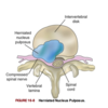
A

1) Sensory: afferent, mostly pseudounipolar - carry impulses from peripheral sensory receptors to CNS

2) Associational (aka interneurons): multipolar - transmits impulses from neuron to neuron (sensory to motor neurons); solely located in the CNS

3) Motor: efferent, multipolar - transmit impulses away from CNS to an effector (skeletal muscle or organ)

18
Q

In skeletal muscle, the end processes of the neuron form a ________________.

A

neuromuscular (myoneural) junction - basically where neuron meets muscle

19
Q

In a synapse, the space between the two neurons is called:

A

synaptic cleft

20
Q

Neuroglia (i.e. nonneuronal cells) comprise ~ _____ the total brain and spinal cord volume, and are ___ times more numerous than neurons.

A

half; 5-10x

21
Q

Function of astrocytes

A

contribute to synaptic function in CNS

  • surround blood vessels & fill spaces between neurons: form specialized contacts between neuronal surfaces and blood vessels
  • contribute to synaptic function in CNS
    • provide rapid transport for nutrients and metabolites
    • essential component of the blood-brain barrier
    • scar forming cells of the CNS (involved with seizures)
    • assist in processing information and memory storage
22
Q

Function of oligodendroglia/oligodendocytes

A

form myelin sheaths within CNS

23
Q

Function of ependymal cells

A

line the CSF-filled cavities (ventricles and choroid plexuses) of the CNS

24
Q

Function of microglia

A

remove debris (phagocytosis) in the CNS

25
Q

Function of Schwann cells

A

form myelin sheath around axons and direct axonal regrowth and functional recovery in the PNS

26
Q

Function of non-myelinating Schwann cells

A

provide metabolic support

27
Q

True or false. Mature nerve cells do not divide therefore injury can cause permanent loss of function

A

True

28
Q

What is Wallerian degeneration? Describe what happens in this process.

A

What it is: degeneration of a nerve fiber in the distal axon after by injury or disease

What happens:

  • 1) swelling in the portion of the axon DISTAL to the cut
  • 2) neurofilaments hypertrophy
  • 3) myelin sheath shrinks and disintegrates
  • 4) axon degenerates and disappears

myelin sheaths re-form into Schwann cells that align in a column between severed part of axon and effector organ

29
Q

Describe what happens during injury to the proximal end of the axon.

How about injury to the cell body?

A
  • in the proximal end of the injury, changes similar to Wallerian degeneration occur but only up to the next node of Ranvier
  • cell body swells and dies via chromatolysis (dispersing Nissl substance) or apoptosis
30
Q

What happens during the repair process of nerve injury ?

A
  • during repair: increased protein synthesis & mitochondrial activity
  • new terminal sprouts project from proximal end and may enter remaining Schwann cell pathway (~7-14 days after injury)
  • this is limited to myelinated fibers and generally occurs only in the PNS
  • regeneration gets harder once more scar formation occurs, and is limited depending on the nature of the myelin formed by the oligodendrocyte, type of injury, inflammation, and location of injury
31
Q

Which of the follow nerve injuries in each set would have greater chances of regeneration?

a) injury closer to cell body vs injury further away from cell body
b) crushing injury vs cut injury
c) peripheral nerves injured near spinal cord vs further away from spinal cord

A

a) injury further from cell body - the closer the injury to the cell body, the more likely nerve cell will die, not regenerate
b) crushing injury - crush injuries sometimes fulley recovery, whereas cut nerves form connective tissue scars that block/slow regeneration of axonal branches
c) peripheral nerves injured further away from spinal cord - if injury to the nerve is close to the spinal cord, it’s slow and poor recovery because super long distance between cell body and where the axon terminates (like in the arms and legs)

32
Q

How are electrical and chemical impulses generated and conducted by neurons?

A

By selectively changing electrical potential of the plasma membrane and influencing the release of neurotransmitters

33
Q

All-or-none response

A

type of response where the neuron either fires or it doesn’t at all (there is no partial response); as long as the stimulus is strong enough, the membrane will be excited if it reaches the threshold potential

otherwise there is no response if it is sub-threshold

34
Q

Presynaptic vs postsynaptic neurons

A

presynaptic neurons: neurons that relay a nerve impulse towards the synapse

post-synaptic neurons: neurons that relay a nerve impulse away from the synapse

35
Q

In a presynaptic neuron, the nerve impulse reaches the vesicles where NTs are stored in what structure?

A

synaptic bouton

36
Q

Neuroplasticity

A

aka synaptic plasticity - the ability for brain synapses to change in strength and number throughout lift (basically the ability of the brain to adapt to new conditions via re-organizing neural pathways and forming new synapses)

allows recovery from injury

37
Q

What are neurotransmitters (NTs) and where are they localized?

A

chemicals synthesized in the neuron and localized in he presynaptic terminal (synaptic bouton)

38
Q

Describe where the following NTs are usually located and whether they are excitatory or inhibitory (or both).

a) ACh
b) Norepinephrine
c) Serotonin
d) Dopamine
e) Histamine

A

a) ACh - throughout brain, spinal cord, NMJ in skeletal muscle, ANS synapses; excitatory OR inhibitory

b) Norepinephrine - throughout brain, spinal cord, in some ANS synapses; Excitatory OR inhibitory

c) Serotonin - many areas of brain and spinal cord; inhibitory

d) Dopamine - some areas of brain and ANS synapses; excitatory

e) Histamine - posterior hypothalamus; excitatory (H1 and H2 receptors) AND inhibitory (H3 receptors)

39
Q

Describe where the following NTs are usually located and whether they are excitatory or inhibitory (or both).

a) GABA
b) Glycine
c) Glutamate and Aspartate
d) Endorphins and Enkephalins
e) Substance P
f) Vasoactive Intestinal peptide

A

a) GABA - most neurons in CNS; post-synaptic inhibition in brain

b) Glycine - spinal cord; post-synaptic inhibition in spinal cord

c) Glutamate and Aspartate - brain and spinal cord; excitatory

d) Endorphins and Enkephalins - throughout CNS and PNS; inhibitory

e) Substance P: spinal cord, brain, pain sensory neurons, GI tract; excitatory

f) Vasoactive Intestinal peptide (VIP) - GI tract; excitatory

40
Q

After a NT binds to the receptors on the post-synaptic neuron, depending on the NTs two things can happen:

excitatory postsynaptic potentials (EPSPs)

inhibitory postsynaptic potentials (IPSPs)

Explain what these terms mean.

A

Excitatory postsynaptic potentials (EPSPs): post-synaptic neuron is depolarized (i.e. excited); If EPSP reaches threshold potential then an action potential (AP) is initiated

Inhibitory postsynaptic potentials (IPSPs): post-synaptic neuron is hyperpolarized (i.e. inhibited); membrane is less likely to reach threshold potential so AP is inhibited

41
Q

Temporal summation vs spatial summation

A

Temporal summation: the effects of successive rapid impulses received from a single neuron at the same synapse

Spatial summation: combined effects of impulses from multiple neurons onto a single neuron at the same time (together, they release enough NTs to exceed threshold potential and an AP occurs)

42
Q

What is facilitation when referring to membrane potentials?

A

refers to the effect of EPSP on membrane potential (i.e. plasma membrane is “facilitated” when summation brings it closer to threshold potential which reduces the amount of stimulus needed for an AP to occur)

43
Q

How much does the brain weigh, and how much of the total cardiac output (in % and in ml) does it receive?

A

3lbs; receives 15-20% of total cardiac output

800-1000ml of blood flow/min

44
Q

What are the three major structural divisions of the brain?

A

1) forebrain (prosencephalon - telencephalon & diencephalon)
2) midbrain (mesencephalon)
3) hindbrain (rhombencephalon - cerebellum, pons, and medulla)

45
Q

What is the brainstem made up of and what structures does the brainstem connect to?

A

midbrain

medulla

pons

connects: brain hemispheres, cerebellum, and spinal cord

46
Q

The largest part of the brain is _______ and contains two types of matter which are ______ and ______ matter.

A

cerebrum; gray & white

47
Q

A collection of nerve cell bodies (nuclei) in the brainstem makes up the ____________. What is the function of this structure?

A

reticular formation

Function:

  • connects brainstem to cortex
  • controls vital reflexes (CV function and resp)
  • essential for maintaining wakefulness and attention
  • some function in balance and posture
48
Q

The reticular activating system (RAS) is comprised of:

A

nuclei in reticular formation

fibers that conduct sensory information to the nuclei

fibers that conduct from nuclei to cerebral cortex

49
Q

The forebrain (pronsencephalon) is split up into two secondary vesicles, _______________ and _____________. What are the structures in each of these secondary vesicles?

A

1) Telencephalon: cerebral hemispheres, cerebral cortex, basal ganglia

2) Diencephalon: epithalamus, thalamus, hypothalamus, subthalamus

50
Q

Structures in the midbrain

A

1) Tegmentum - floor of midbrain - composed of red nucleus, substantia nigra, and basis pedenculi
* cerebral peduncles - tegmentum and basis pedunculi
2) corpora quadrigemina - located on the tectum (celing of midbrain) - composed of two pairs of each superior and inferior colliculi

51
Q

Structures in the hindbrain (rhombencephalon)

A

1) Metencephalon - cerebellum, pons
2) Myelencephalon - medulla oblongata

52
Q

Define the following terms:

Gyri (gyrus)

Sulci (Sulcus)

Fissures

A

Gyri (gyrus): ridges/convolutions on the surface of the cerebral cortex

Sulci (Sulcus): grooves in between gyri

Fissures: deeper grooves in between gyri

53
Q

Composition of gray matter vs white matter

A

Gray matter: neuron cell bodies (outer layer)

White matter: myelinated nerve fibers (lies beneath cerebral cortex)

54
Q

Longitudinal fissure

A

deep groove that separates the two cerebral hemispheres

55
Q

The posterior margin (border) of the frontal lobe is on the ____________, and the inferior margin of the frontal lobe is on the _______________.

A

posterior border: Central Sulcus (Fissure of Rolando)

inferior border: Lateral Sulcus (Sylvian fissure, lateral fissure)

56
Q

Function of prefrontal area

A
  • goal-oriented behaviour (e.g. ability to concentrate)
  • ST or recall memory
  • elaboration of thought
  • inhibition of limbic areas of CNS
57
Q

Function of premotor area (Brodmann area 6)

A

programming motor movements

also contains the cell bodies that make up part of the basal ganglia system

58
Q

The frontal eye fields in the frontal lobe area also known as ____________. They are involved with what function(s)?

A

Brodmann area 8 (lower portion); involved with controlling eye movements

59
Q

The primary motor area is also known as __________ and is located where? Describe its general function(s).

A
  • aka Brodmann area 4
  • located along the precentral gyrus in the frontal lobe
  • forms the primary voluntary motor area (organized into a homunculus) - Function: generate signals to direct the movement of the body
    • means that electrical stimulation over specific areas in this cortex cause specific muscle movements
60
Q

Pyramidal system

A

two-neuron system that are comprised of corticobulbar tract and corticospinal tracts

  • corticobulbar tract: synapses in the brainstem and provides voluntary control of muscles in head and neck
  • corticospinal tracts: originate in precentral gyrus (decussate in medulla oblongata), descend into spinal cord, provide voluntary control of muscles throughout the body
    • has the same somatotopic organization as the body
61
Q

Contralateral control

A

phenomenon where cerebral impulses control function on the opposite side of the body

62
Q

Broca speech area

Brodmann area #, Location and Function

A
  • Brodmann areas 44, 45
  • Location: on the inferior frontal gyrus (FRONTAL LOBE); usually on L hemisphere
  • Function: motor aspects of speech (speech production)
63
Q

Parietal Lobe

Location, function

A
  • Location: see photo - borders: central sulcus, parietoccipital sulcus, & lateral sulcus
  • Function: somatic sensory input (along postcentral gyrus) - storing, analyzing, and interpreting sensory stimuli
64
Q

Occipital Lobe

Location and Function

A
  • Location: behind parietoccipital sulcus, superior to cerebellum (see photo)
  • Function: primary visual cortex (Brodmann area 17)
    • receives visual input from retinas
    • Visual association (Brodmann areas 18, 19)
65
Q

Communication between motor and sensory areas in the cerebral cortex are accomplished through what structures?

A

association fibers

66
Q

Temporal Lobe

Location, Structures, and Function

A
  • Location: inferior to lateral fissure
  • Structure: composed of superior, middle, and inferior temporal gyri
  • Function:
    • primary auditory cortex (Brodmann area 41) and related association area (Brodmann area 42)
    • Wernicke’s area: sensory speech area - receiving and interpreting speech
    • also involved in memory consolidation and smell
67
Q

Insula (Insular lobe)

Location and Function

A
  • Location: hidden in lateral sulci between temporal and fronal lobes of each hemisphere
  • Function: processes sensory and emotional information and routes the info to other areas of the brain
68
Q

The two cerebral hemispheres are connected by a mass of white matter pathways collectively known as _________. What is the importance of this structure?

A

corpus callosum (transverse or commissural fibers)

lies beneath longitudinal fissure; connects the hemispheres through sensory and motor contralateral projections of axons that is needed in coordinating activities between the hemispheres

69
Q

Basal ganglia system - what is this system comprised of and what is the main function of the basal ganglia system?

A

Structures: caudate nucleus, putamen, globus pallidus

  • putamen x globus pallidus - lentiform nucleus (shaped like a lentil)
  • caudate nucleus x putamen - striatum

Other structures: substantia nigra, nucleus accumbens, subthalamic nucleus

Function: involved with voluntary movement, and cognitive and emotional functions (i.e. dopamine and pleasure/reward functions)

70
Q

Substantia nigra synthesizes what neutrotransmitter that contributes to pleasure and reward functions?

A

dopamine

71
Q

What is the internal capsule

A

thick layer of white matter that carries information (sensory and motor pathways) to and from cerebral cortex, and between caudate and lentiform nuclei

72
Q

Extrapyramidal system - what is it comprised of and what is its function(s)?

A

Structures: basal ganglia + interconnections with thalamus, premotor cortex, red nucleus, reticular formation, and spinal cord

Function: motor system network responsible for involuntary control and modulation of muscles (reflexes and movement, stabilizing effect on motor control)

73
Q

Limbic system

Location, Structure and Function

A

Location: between telencephalon and diencephalon, surrounds corpus callosum

Structure: composed of amygdala, hippocampus, fornix, hypothalamus, and related autonomic nuclei

Function: mediates emotions, LT memory, primitive behavioural responses, visceral reaction to emotion, motivation, mood, feeding behaviours, biologic rhythms, and smell

  • just remember 5 F’s: feeding (hunger/satiety); forgetting (memory); fighting (emotional response), family (sexual reproduction/maternal instincts), fornicating
74
Q

Describe the structures that the diencephalon is comprised of and their respective locations and functions.

A

1) Epithalamus: roof of third ventricle; the most superior portion of the diencephalon ⇒ controls vital functions and visceral activities (closely associated with limbic system)

2) Thalamus: borders and surrounds third ventricle ⇒ integrating center for impulses that head to cerebral cortex & relay center for info from basal ganglia and cerebellum to motor areas

3) Hypothalamus: forms base of diencephalon

  • maintains constant internal environment
  • implement behavioural patterns
  • influences body via endocrine system & neural pathways

4) Subthalamus: flanks the hypothalamus laterally ⇒ important for motor activities

75
Q

Functions of superior colliculi and inferior colliculi

A

Superior colliculi: involved with voluntary and involuntary visual motor movements (tracking moving objects)

Inferior colliculi: motor activities affecting auditory system (i.e. positioning head to improve hearing)

76
Q

Input and output of the red nucleus

A

receives ascending sensory info form cerebellum

projects rubrospinal tract (motor pathway) to cervical spinal cord

77
Q

Basis pedunculi composition

A

made up of efferent fibers of cortocospinal, corticobulbar, and corticopontocerebellar tracts

78
Q

What is the cerebral aqueduct (aqeduct of Sylvius)

A

The cerebral aqueduct is a channel that connects the third ventricle with the fourth ventricle and allows cerebrospinal fluid to pass between them.

79
Q

Cerebellum

Structure and Function

A

Structure: gray and white matter; lots of ridges; divided by a central fissure into two lobes (R and L) connected by the vermis

Function:

  • reflexive, involuntary fine-tuning motor control
  • maintaining balance and posture (via connections with medulla and midbrain)
80
Q

Pons

Structure and Function

A

Structure: between the midbrain and medulla

  • houses the nuclei for CN V to CN VIII

Function: acts as a “bridge” to transmit information from cerebellum to brainstem & between the two cerebellar hemispheres

81
Q

Medulla Oblongata

Structure and Function

A

aka myelencephalon

Structure: lowest portion of the brainstem

  • nuclei of CN IX (9) through XII (12) also located here

Function: controls reflex activities - HR, RR, BP, coughing, sneezing, swallowing, and vomiting

  • the medulla is also where a lot of descending motor pathways decussate
82
Q

The term for motor nerves crossing contralaterally in the medulla oblongata is

A

decussate (crossing to the other side)

83
Q

Spinal Cord

Structure and Function

A

Structure: lies within the vertebral canal, protected by vertebral column

  • originates in medulla oblongata, ends at L1 or L2 in adults

Function:

  • connects brain and body
  • conducts somatic and autonomic reflexes
  • provides motor pattern control centers
  • modulates sensory and motor function
84
Q

The cone shaped structure at the end of the spinal cord is termed

A

conus medullaris

85
Q

At the end of the spinal cord, spinal nerves continue downwards and form a nerve bundle known as

A

cauda equina (like a horse’s tail)

86
Q

What filament anchors the conus medullaris to the coccyx?

A

filum terminale

87
Q

The ____________ lies in the center of the butterfly-shaped gray matter in a cross section of a spinal cord. It originates from the __________, extends through the spinal cord and is filled with _________.

A

central canal; fourth ventricle; CSF

88
Q

The gray matter of the spinal cord is divided into three regions, which are what?

Describe their composition.

A

1) Posterior horn/dorsal horn: primarily interneurons and sensory neuron axons (cell bodies in dorsal root ganglion)

  • substantia gelatinosa: a structure located at the tip of the posterior horn that is involved in pain transmission

2) Lateral Horn: contains cell bodies involved with ANS

3) Anterior horn/ventral horn: contains cell bodies for efferent pathways that leave the spinal cord via spinal nerves

89
Q

Spinal tracts exist in (gray/white) matter. How are they named?

A

white matter

named based on where they begin and end (ex. spinothalamic tract = carries nerve impulses from spinal cord to thalamus)

90
Q

Spinal tracts are grouped into _______ according to their location within the white matter. These groups include:

A

Columns

anterior, lateral and posterior (dorsal) columns

91
Q

What are reflex arcs?

A

neural circuits in the spinal cord that control a reflex; responds to stimuli as a protective mechanism

Components:

  • receptor
  • afferent (sensory neuron)
  • efferent (motor neuron)
  • effector muscle/gland

motor effects of reflex arcs generally occur before the event (ex. touching a hot stove) is even registered and perceived in higher centers of the brain (i.e. you even realizing you’re touching a hot stove, you’ve already pulled away)

92
Q

Upper motor neurons (UMNs) vs Lower motor neurons (LMNs)

Location/Structure and Function

A

Upper motor neurons (UMNs): completely contained within CNS

  • primary role: controlling fine motor movement and influencing/modifying spinal reflex arcs and circuits
  • synapse with interneurons

Lower motor neurons (LMNs): cell bodies originate gray matter of brainstem and spinal cord & projections out of CNS into PNS

  • directly influence muscles
  • interneurons synapse with LMNs
93
Q

Composition of a motor unit

A

neuron and the skeletal muscle it stimualtes

94
Q

The region between the axon of a motor neuron and plasma membrane of a muscle call is called the __________________.

A

Neuromuscular (myoneural) junction (NMJ)

95
Q

List the 5 clinically relevant motor pathways

A

In the pyramidal system:

  • corticospinal tract
  • corticobulbar tract

Extrapyramidal system:

  • reticulospinal tract
  • vestibulospinal tract
  • rubrospinal tract
96
Q

Reticulospinal tract

Structure and Function

A

Struction: arises in the reticular formation of medulla or pons; termins within spinal cord

Function: controls body movements by inhibiting and exciting spinal activity; for balance and posture

97
Q

Vestibulospinal tract

Structure and Function

A

Structure: origins from vestibular nucleus in the pons

Functions: causes extensor muscles of the body to rapidly contract (such as seen when someone starts to fall backwards); in charge of maintaining balance while a person is preparing for movement

98
Q

Rubrospinal tract

Structure and Function

A

Structure: originates in red nucleus, decussates and terminates in the cervical spinal cord

Function: involved with muscle movement and fine muscle control in UE

99
Q

What are the three important spinal afferent sensory pathways?

A

posterior column

anterior spinothalamic tract

lateral spinothalamic tract

100
Q

Posterior (dorsal) column

Structure and Function

A

Structure: 3-neuron chain

  • 1st neuron: sensory neuron (afferent) - synapses at medulla oblongtata
  • 2nd neuron: crosses contralaterally at medial lemniscus and synapses in thalamus
  • 3rd neuron: originates in thalamus, continues into cerebral cortex
  • forms two large bundles of nerve fibers: fasciculus gracilis and fasciculus cuneatus

Function: carries fine-touch sensation, two-point discrimination, and propioceptive/epicritic information

101
Q

Anterior spinothalamic tract

Structure and Function

A

Structure: 3-neuron chain

  • primary afferent neuron (1st neuron): synapses in posterior horn of spinal cord
  • 2nd order neuron: crosses contralaterally in the lateral column and ascends to thalamus
  • 3rd neuron: thalamus to cerebral cortex

Function: vague touch sensation (protopathic)

102
Q

Lateral Spinothalamic tract

A

Structure: same as anterior spinothalamic tract - 3-neuron chain

  • primary afferent neuron (1st neuron): synapses in posterior horn of spinal cord
  • 2nd order neuron: crosses contralaterally in the lateral column and ascends to thalamus
  • 3rd neuron: thalamus to cerebral cortex

Function: pain and temp perception (protopathic)

103
Q

The cranium is composed of ____ bones

A

8

104
Q

Galea aponeurotica - what is it?

A

thick fibrous band of tissue that lies over the cranium between frontal and occipital muscles as added protection to the skull

105
Q

How is the cranial floor divided and what brain structures lie in each respective division?

A

divided into three fossae (depressions)

anterior fossa: front lobes

middle fossa: temporal lobes, base of diencephalon

posterior fossa: cerebellum

106
Q

Describe the layers of the meninges.

A

Dura mater: composed of two layers with venous sinuses between them

  • outer layer: periosteum (endosteal layer)
  • inner layer: inner dura (meningeal layer) - forms rigid membranes that support and separate brain structures

Arachnoid mater: spongy, web-like structure that follows the conoturs of cerebral structures

Pia mater: delicate layer that adheres to contours of the brain and spinal cord; supports blood vessels serving brain tissue

Meninges covers the brain and continues done the spinal cord, continue beyond the end of the spinal cord to the sacrum

107
Q

Falx Cerebri

Structure and Function

A

Structure: a fold of the dura mater that dips between the two cerebral hemisphers along the longitudinal fissure

  • anchored to the ethmoid bone

Function: separates the two cerebral hemispheres

108
Q

Tentorium cerebelli

A

fold/membrane of the dura mater that seprates cerebellum and cerebrum

109
Q

Subdural space

A

space between dura and arachnoid mater that contains lots of little bridging veins

110
Q

The structures that arise from the pial membrane and produce CSF are known as _________________.

A

choroid plexuses

111
Q

True or false. The meninges forms potential AND real spaces.

A

True

112
Q

Epidural space

A

aka extradural space

a potential space between dura mater and skull

113
Q

Describe what cerebrospinal fluid (CSF) is, what produces it, and what is the purpose/function of CSF.

A

a clear, colour fluid (similar to blood plasma and IF) that allows intracranial and spinal cord structures to float in and prevent tugging of structures/vessels/roots, therefore serves as protection

produced by choroid plexuses in the lateral, third, and fourth ventricles (a highly-vascularised structure of epithelial cells located in the ventricles of the brain)

114
Q

How much CSF is circulating wtihin the ventricles and subarachnoid space at any given time?

How much CSF is produced daily?

A

125-150ml (circulating at any given time)

600ml produced daily

115
Q

What causes CSF to flow through the CNS?

A
  • CSF flow results from pressure gradient between arterial system and CSF-filled cavities
  • CSF exerts ~5 - 14 mmHg (or 80-180mm of water pressure) on brain and spinal cord when supine, doubles when patient is upright
116
Q

Describe the pathway in which CSF flows through

A

1) CSF formed from the blood and secreted via choroid plexuses
2) begins in lateral ventricles and flows through interventricular foramen (foramen of Monro) into third ventricles
3) then passes through cerebral aqueduct (aqueduct of Sylvius) into fourth ventricle
4) Fourth ventricle to then passing through paired lateral apertures (foramen of Luschka) or median aperture (foramen of Magendie) to subarachnoid spaces
5) reabsorbed into venous circulation through arachnoid villi

117
Q

Function of arachnoid villi

A

reabsorbs CSF and functions as one-way valves directing CSF outflow into the blood but preventing blood flow into the subarachnoid space

118
Q

Vertebral column divisions

A

7 cervicle

12 thoracic

5 lumbar

5 fused sacral

4 fused coccygeal

119
Q

Between each vertebrae (except fused sacral and coccygeal vertebrae) is an _________________

A

intervertebral disk

120
Q

Intervetebral disk

Structure and Function

A

Structure: in between each vertebrae; at the center has a pulpy mass of elastic fibers (nucleus pulposus)

Function: absorbs shocks, prevent damage to vertebrae

121
Q

Why are intervertebral disks one of the most common sources of back problems?

A

because where there is too much stress applied to the vertebral column, the disk contents may rupture and protrude into the spinal canal, which then causes compression of the spinal cord or nerve roots

122
Q

What is the primary regulator for blood flow within the CNS?

A

CO2 - a potent vasodilator and ensures an adequate blood supply

i.e. hypoxia with increased CO2 levels would cause vasodilation to increase blood flow to the brain

123
Q

What are the two systems in which the brain receives its arterial supply from?

A

1) Internal carotid arteries: supply greater blood flow to brain

  • origin: common carotid arteries → enter cranium at base of skull → through cavernous sinus → branch and divide into anterior & middle cerebral arteries

2) Vertebral arteries: origin: subclavian arteries → through transverse foramina/foramen in cervical vertebrae → enter cranium via foramen magnum → join at junction of pons and medulla to form basilar artery → divides at level of midbrain to make posterior cerebral arteries

124
Q

What is the circle of Willis?

A

a blood vessel system that provides an alternative route for blood flow in the event that one of the contributing arteries is obstructed (collateral blood flow)

Consists of:

  • posterior cerebral arteries
  • posterior communicating arteries
  • internal carotid arteries
  • anterior cerebral arteries
  • anterior communicating artery
125
Q

Describe the two classifications of cerebral veins and what structures they drain into

A
  • classified as superficial and deep veins
  • veins drain into venous plexuses and dural sinuses (between dura layers) and eventually join internal jugular veins at base of skull
126
Q

Describe why it is so important for a patient with a head injury not to turn their head (besides preventing further damage to spine).

A

because those with head injuries who turn or let their heads fall to the side partially occlude venous return, and causing ↑ICP due to decrease flow through jugular veins

127
Q

What is the blood brain barrier (BBB)? Describe its make up.

A

cellular structures that selectively inhibit certain potentially harmful substances in the blood from entering the interstitial spaces of the brain or CSF, which allows neurons to function normally

Composition:

  • endothelial cells in brain capillaries with tight junctions
  • supporting cells - astrocytes, pericytes, microglia
128
Q

Describe the significance of the BBB in relation to drug therapy.

A

certain types of antibiotics and chemotherapeutic drugs show a greater propensity than others for crossing this barrier so understanding the mechanism of which metabolites, electrolytes, etc. may pass through compared to others

129
Q

What supplies blood to the spinal cord?

A

from branches off the vertebral arteries & from branches from various regions of the aorta

Vertebral artery branches:

  • anterior spinal artery
  • posterior spinal arteries (pair)
  • both^ branch at base of cranium and descend alongside spinal cord

Arterial branches:

  • branches follow the spinal nerve through the intervertebral foramina, pass through the dura, and divide into the anterior and posterior radicular arteries
  • radicular arteries eventually connect to spinal arteries - penetrate spinal cord to supply blood

blood return: venous draining into venous sinuses located between dura and periosteum of vertebrae

130
Q

Fascicles

A

bundles of individual nerve fibers (myelinated axons) in the PNS

131
Q

Describe the spinal nerve divisions and where they exit in the vertebrae

A

31 pairs of spinal nerves (8 cervical, 12 thoracic, 5 lumbar, 5 sacral pair of spinal nerves, and 1 coccygeal) - names derived from vertebral level from which they exit

first cervical nerve exist ABOVE first cervical vertebra

all the other spinal nerves exist BELOW their corresponding vertebrae

132
Q

Spinal nerves contain both sensory and motor neurons and therefore are called _____________.

A

mixed nerves

133
Q

Origin and pathway of spinal nerves

A

Origin: rootlets lateral to anterior and posterior horns of spinal cord

Pathway: after arising from rootlets, the two spinal nerve roots converge at the intervertebral foramen to form spinal nerve trunk → then divides into anterior and posterior rami (branches) → anterior rami form plexuses which then branch into peripheral nerves

134
Q

Brachial plexus - composition and innervation

A

spinal nerve plexus formed by the last four cervical nerves (C5 to C8) and T1

innervates the nerves of the arm, wrist, and hand

135
Q

Lumbar plexus - composition and innervation

A

Composition: L1 to L4 spinal nerves

Innervation: anterior portions of lower body

136
Q

Sacral plexus - composition and innervation

A

Composition: L5 to S5

Innervation: posterior portions of the lower body

137
Q

Dermatome

A

area of skin that is mainly supplied by a single spinal nerve

138
Q

How many pairs of cranial nerves are there and where to they originate from?

A

12 pairs

sensory, motor, or mixed

connected to nuclei in the brain and brainstem

139
Q

List the 12 cranial nerves and whether they have sensory, motor, or both functions.

A

CN I: Olfactory - sensory

CN II: Optic - sensory

CN III: Oculomotor - motor

CN IV: Trochlear - motor

CN V: Trigeminal - both

CN VI: Abducens - motor

CN VII: Facial - both

CN VIII: Vestibulocochlear - sensory

CN IX: Glossopharyngeal - both

CN X: Vagus - both

CN XI: Spinal accesory - both(?) (as per textbook, although typically this is motor)

CN XII: Hypoglossal - motor

Easy way to remember:

Cranial nerves: Ooh, Ooh, Ooh To Touch And Feel Very Good Velvet. Such Heaven!

Function: Some Say Marry Money But My Brother Says Big Brains Matter More

140
Q

CN I

Origin/pathway

Function

How it’s tested

A

Name: Olfactory

Origin and Course: arise from nasal olfactory epithelium and form synapses with olfactory bulbs, which transmit impulses to the temporal lobe

Function: sensory - carries impulses for sense of smell

How to Test: sniffing various aromatics & trying to identify them

141
Q

CN II

Origin/pathway

Function

How it’s tested

A

Name: Optic

Origin & Course: arise from eye retina to form optic nerve & through sphenoid bone; two optic nerves converge to form optic chiasma (with partial crossover of fibers) and eventual to occiptal cortex

Function: sensory - carries impulses for vision

How it’s Tested: vision & visual field tested via eye chart and testing when a person sees a finger moving into their visual field

  • eye interior is viewed with ophthalmoscope to look at blood vessels
142
Q

CN III

Origin/pathway

Function

How it’s tested

A

Name: Oculomotor

Origin & Course: emerge from midbrain and exit from skull, runs to eye

Function: mainly motor - eye muscle function and pupil response

  • goes to inferior oblique & superior, inferior, and medial rectus extraocular muscles that direct eyeball
  • levator muscles of eyelid
  • smooth muscles of iris & ciliary body
  • DOES support sensory functioning (proprioception) to brain from extraocular muscles

How it’s tested: examine pupils (size, shape, equality); penlight to test reactivity; ability to follow moving object

143
Q

CN IV

Name

Origin/pathway

Function

How it’s tested

A

Name: Trochlear

Origin and Course: emerge from posterior midbrain and exist from skull to run to eye

Function: motor - innervate superior obliqud muscle of eye (extraocular muscle)

How it’s Tested: tested with CN III relative to ability to follow moving objects

144
Q

CN V

Name

Origin/pathway

Function

How it’s tested

A

Name: Trigeminal

Origin and Course: emerge from pons and form three divisions that exit from skull, run to face and cranial dura mater

Function: motor and sensory for face

  • conducts sensory impulses from mouth, nose, eye surface, and dura mater
  • motor fibers that stimulate chewing muscles

How it’s tested:

  • senation for pain, touch and temp: safety pin and hot/cold objects
  • corneal reflex tested with wisp of cotton
  • motor branch tested by asking pt to clench teeth, open mouth against resistance, move jaw from side to side
145
Q

CN VI

Name

Origin/pathway

Function

How it’s tested

A

Name: Abducens

Origin and Course: fibers leave inferior pons and exit from skull to run to eye

Function: motor - fibers to lateral rectus muscle, and proprioceptor fibers from same muscle to brain

How it’s tested: along with CN III relative to ability to move each eye laterally

146
Q

CN VII

Name

Origin and Course

Function

How it’s tested

A

Name: Facial

Origin and Course: leave pons and travel through temporal bone to reach face

Function: sensory and motor

  • Motor: muscles for facial expression & to lacrimal and salivary glands
  • Sensory: taste buds of anterior part of tongue

How it’s tested:

  • anterior 2/3rds of tongue tested for sweet, salty, sour, bitter
  • face symmetry checked
  • subject asked to close eyes, smile, whistle, etc.
  • tearing tested with ammonia fumes
147
Q

CN VIII

Name

Origin and Course

Function

How it’s tested

A

Name: Vestibulococchlear

Origin and Course: run from inner eat (hearing and equilibrium receptors in temporal bone) to enter brainstem just below pons

Function: sensory - sense of equilibrium & hearing

How it’s tested: hearing checked by tuning fork; vestibular tests: Bárány and caloric tests

148
Q

CN IX

Name

Origin and Course

Function

How it’s tested

A

Name: Glossopharyngeal

Origin and Course: emerge from medulla and leave skull to run to throat

Function: sensory and motor

  • motor: serves pharynx (throat) and salivary glands
  • sensory: carry impulses from pharynx, posterior tongue (taste buds), and pressure receptors of carotid artery

How it’s tested:

  • gag and swallow reflex
  • asked to speak and cough
  • posterior 1/3rd of tongue tested for taste
149
Q

CN X

Name

Origin and Course

Function

How it’s tested

A

Name: Vagus

Origin and Course: emerge from medulla, pass through skull, & descend through neck region into thorax and abdo region

Function: sensory and motor

  • carry sensory and motor impulses for pharynx
  • parasympathetic motor fibers which supply smooth muscles of abdo organs
  • receives sensory impulses from viscera

How it’s tested: same as CN IX (checking gag and swallow reflex; speaking, coughing, taste)

150
Q

CN XI

Name

Origin and Course

Function

How it’s tested

A

Name: Spinal accessory

Origin and Course: arise from medulla and superior spinal cord and travel to muscles of neck and back

Function: sensory and motor fibers for sternocleidomastoid and trapezius muscles and muscles of soft palate, pharynx, and larynx

How it’s tested: sternocleidomastoid and trap muscles checked for strength - rotate head and shrug shoulders against resistance

151
Q

CN XII

Name

Origin and Course

Function

How it’s tested

A

Name: hypoglossal

Origin and Course: fibers arise from medulla and exit from skull to travel to tongue

Function: carries motor fibers to muscles of tongue and sensory impulses from tongue to brain

How it’s tested: stick out tongue to note position abnormalities if any

152
Q

What does the motor component of the ANS consist of?

A

two neuron system

preganglionic neurons (myelinated) - conduct impulses from brainstem or spinal cord to an autonomic ganglion where they synapse with postganglionic neuron

postganglionic neurons (unmyelinated) - conduct impulses away from ganglion to the effector

153
Q

Function of the ANS

A

coordinates and maintains a steady state among visceral (internal) organs (ie regulation of cardiac muscle, smooth muscle, and glands); an involuntary system

154
Q

Describe how the axons in the sympathetic nervous system are set up.

A
  • innervated by cell bodies located in the thoracolumbar division (T1 to L2 of spinal cord)
  • preganglion axons form synapses SHORTLY after leaving spinal cord in the sympathetic (paravertebral) ganglia; then travels in one of the following ways
    • 1) directly synapses with postganglionic neurons in sympathetic chain ganglion at their level
    • 2) traveling up or down sympathetic chain ganglion before synapsing with higher/lower postganglionic neuron
    • 3) through the sympathetic chain ganglion to synapse with collateral ganglia
155
Q

What are splanchnic nerves and collateral ganglia?

A

splanchnic nerves are pathways formed by a group fo preganglionic axons that eventually innervate the abdomen

splanchnic nerves lead to collateral ganglia (which lies in front of the aorta) and branch into celiac, superior mesenteric, & inferior mesenteric ganglia which then eventually postganglionic neurons leave and innervate viscera

156
Q

Describe how the axons in the parasympathetic nervous system are set up.

A
  • nerve cell bodies are located in the craniosacral division (cranial and sacral regions of the spinal cord)
    • from cranial: CN III (oculomotor), VII (facial), IX (glossopharyngeal), and X (vagus)
    • from sacral: form pelvic splanchnic nerve that innervates pelvic cavity viscera
  • preganglgionic fibers travel close to the organs they supply before synapsing (so they have short postganglionic neurons)
157
Q

Sympathetic preganglionic & parasympathetic pre- and post-ganglionic fibers what neurotransmitter? This is known as ________ transmission.

A

Acetylcholine; cholinergic transmission

158
Q

Most postganglionic sympathetic fibers release what neurotransmitter? This is considered __________ transmission.

A

Norepineprhine; adrenergic transmission

*some post-ganglionic sympathetic fiebrs release ACh (like those that innervate sweat glands but MOST release norepi)

159
Q

The two types of adrenergic receptors are what? Describe their subdivisions and general function.

A

𝜶 and 𝜷

𝜶1 receptors: excitation or stimulation

𝜶2 receptors: relaxation or inhibition

𝜷1 receptors: faciliated increased HR and contractility, cause release of renin from kidney

𝜷2 receptors: stimulates smooth muscle relaxation

160
Q

Describe which adrenergic receptors are stimulated by norepinephrine and epinephrine.

A

Norepinephrine: ALL 𝞪1 and 𝞫1 receptors and only certain 𝞫2 receptors

  • primary response is stimulation of 𝞪1 receptors causing vasoconstriction

Epinephrine: strongly stimulates all four types of receptors and induces general vasodilation (on beta receptors) due to the predominance of 𝞫 receptors in muscle vasculature

161
Q

Functions of the ANS

A
  • fight of flight response
  • rest and tranquility response
  • opposing effects between sympathetic and parasympathetic divisions to maintain a steady state in the internal environment
162
Q

Structural changes to the neurological system with aging

A
  • decreased brain weight and size (particularly frontal regions)
  • increased in ventricular volume & size
  • fibrosis and thickening of meninges
  • narrowing of gyri and widening of sulci
163
Q

Cellular changes to the neurological system with aging.

A
  • decreased # of neurons, dendritic processes, and synaptic connections
  • decreased myelin
  • lipofuscin deposition (a pigment resulting from cellular autodigestion)
  • neurofibrillary tangles (++ accumulation in cortex associated with Alzheimers)
  • imablance in amoutn and distribution of NTs
  • decrease in glucose metabolism
164
Q

Cerebrovascular changes with aging

A
  • arterial atheroscelrosis (leading to risk of infarcts and scars)
  • increased permeability of BBB
  • decreased vascular density
165
Q

Functional changes with aging secondary to neurological changes.

A

varies from individual to individual

  • decreased tendon reflexes
  • progressive deficit in taste and smell
  • decreased vibratory sense
  • decrease in accommodation and colour vision
  • decrease in neuromuscular control with change in gait and posture
  • sleep disturbances
  • memory impairments & cognitive alterations
166
Q

Define pain

A

unpleasant sensory and emotional experience associated with actual or potential tissue damage or described in terms of such damage

a protective phenomenon, unique to the individual (i.e. cannot be defined, identified, or measured by an observer)

167
Q

Why is acute pain considered “protective”?

A

because it promotes withdrawing from painful stimuli, which allows the injured part to heal and makes us learn to avoid painful stimuli

168
Q

What are the three portions of the nervous system responsible for sensation, perception and response to pain?

A

1) Afferent pathways: start in the PNS, travel through spinal cord and then up to higher centers in CNS

2) Interpretive centers: in brainstem, midbrain, diencephalon, and cerebral cortex

3) Efferent pathways: descend from CNS back to dorsal horn of spinal cord

169
Q

The processing of potentially noxious stimuli through a normally functioning nervous system is known as

A

nociception

170
Q

What are nociceptors? Also describe their composition and what types of stimuli they respond to.

A
  • pain receptors - free nerve endings in the afferent PNS that cause nociceptive pain when stimulated
  • cell bodies located in dorsal root ganglia (DRG) for the body & in trigeminal ganglion for the face
  • unevenly distributed throughout body so pain sensitivity differs
  • responds to different types of noxious stimuli
    • mechanical (pressure, mechanical distortion)
    • thermal (extreme temps)
    • chemicals (acids, inflammatory chemicals - bradykinin, histamine, leukotrienes, prostaglandins)
171
Q

What are the four phases of nociception?

A

1) Pain transduction
2) Pain transmission
3) Pain perception
4) Pain modulation

172
Q

Describe the 1st phase of nociception: pain transduction

A
  • begins when nociceptors are axtivated by noxious stimulus
  • Na, K, and Ca ion channels on receptors open to create electrical impulses that travel through the 2 primary types of nociceptor axons
    • A-delta fibers
    • C fibers
173
Q

Describe the two primary types of nociceptors

A

A-delta (A𝜹) fibers: large myelinated fibers, rapidly transmit sharp & well-localized “fast” pain sensations (like burns or pinpricks to the skin)

  • when activated, causes spinal reflex withdrawal of body part BEFORE a pain sensation is perceived

C fibers: most numerous, smaller & unmyelinated; located in muscle, tendons, body organs, skin

  • slowly transmit dull, aching, or burning sensations that are poorly localized and often constant
174
Q

Describe the 2nd phase of nociception: Pain transmission (including its pathways)

A
  • the conduction of pain impulses along the A𝛿 and C fibers (primary order neurons) into dorsal horn of spinal cord
  • neurons synapse with excitatory or inhibitory interneurons (second order neurons) in substantia gelatinosa of dorsal horn
  • impulses then synapse with projection neurons (third order neurons), cross midline of spinal cord, and ascend brain through lateral spinothalamic tracts
    • anterior spinothalamic tract: carries fast impulses for acute sharp pain (perceived first)
    • lateral spinothalamic tract: carries slow impulses for dull/chronic pain
  • impulses then projected into different structures of the brain then to sensory areas for interpretation
175
Q

Describe the 3rd phase of nociception: pain perception

A
  • the conscious awareness of pain (influenced by genetics x environment)
  • occurs primarily in reticular and limbic systems and cerebral cortex
  • three systems interacting to produce perception: aka the “pain matrix”
    • sensory-discriminative: mediated by somatosensory cortex; identifying presence, character, location, and intensity of pain
    • affective-motivational: mediated by reitcular formation, limbic system, and brainstem; determines someone’s conditioned avoidance behaviours & emotional response to pain
    • cognitive-evaluative: mediated through cerebral cortex; oversees learned behaviours from previous experiences and modulate perception of pain
176
Q

Pain threshold vs pain tolerance

A

Pain threshold: defined as lowest intensity of pain that a person can recognize (i.e. lowest point at which a stimulus is perceived as pain)

  • intense pain in one location can increase pain threshold in another location (like distracting painful injuries; known as perceptual dominance); threshold can also be raised by various factors like exercise, sex, stress, etc.

Pain tolerance: defined as the greatest intensity of pain that a person can handle; varies between people and in the same person overtime

  • may decrease with: repeated exposures to pain, fatigue, anger, boredom, apprehension, & sleep deprivation
  • increase with: alcohol consumptino, persistent opioid med use, hypnosis, distraction, strong beliefs/faith
177
Q

Describe the 4th phase of nociception: pain modulation

A
  • facilitation or inhibition of transmission of pain signals throughout nervous system; can occur before, during, or after pain perception
  • done through NTs
    • Excitatory NTs: triggered by tissue injury and/or inflammation - glutamate, aspartate, substance P, calcitonin
      • reduces activation threshold so increased responsiveness from nociceptors
    • Inhibitory NTs: GABA, glycine; serotonin and norepinephrine inhibit pain in the medulla and pons
178
Q

What are endogenous opioids and what is their function?

A

What are they? A family of morphine-like neuropeptides that inhibit pain transmission in the periphery, spinal cord, and brain

How they work: binds to specific opioid receptors (mu, kappa, delta) on neurons which inhibits ion channels and thus release of excitatory NTs

179
Q

Location and function of opioid receptors

A

found widely distributed throughout body

responsible for general sensations of well-being and modulation of many physiological processes (controlling resp and CV functions, stress and immune responses, GI function, reproduction, and neuroendocrine control)

180
Q

Types of endogenous opioids

A
  • Enkaphalins - most prevalent of the natural opioids, binds to 𝛿 opioid receptors
  • Endorphins - endogenous morphine produced in the brain (sense of exhilaration and natural pain relief)
  • Dynorphins - most potent, binds to kappa receptors to impede pain signals
  • Endomorphins - bind with mu receptors, analgesic effects
  • Nocicpetin/orphanin FQ - induces pain or hyperalgesia but does not interact with opioid receptors

bind as direct agonists to opioid receptors

181
Q

The only clinically used opioid receptor antagonist is __________ and has a higher affinity for which opioid receptors?

A

naloxone; mu receptors

182
Q

What are endocannabinoids?

A
  • substances in our bodies synthesized from phospholipids that activate CB1 (primarily in CNS) and CB2 (primarily in immune tissue) receptors to modulate pain
  • classified as eicosanoids
  • CB1 receptors decrease pain transmission by inhibiting release of excitatory NTs in various structures within brain and spinal cord
183
Q

How does cannabis provide an analgesic effect?

A

cannabis produces a resin that contains cannabinoids, which are analgesic in humans (drawbacks: psychoactive and addictive properties)

184
Q

Describe how the descending inhibitory and facilitatory pain pathways modulate pain

A
  • inhibitory pathways activate opioid receptors and inhibit release of excitatory NTs, facilitate release of inhibitory NTs, and stimulate inhibitory interneurons
  • afferent stimulation of ventromedial medulla and periaqeductal gray (PAG) in midbrain stimulates efferent pathways which inhibit ascending pain signals
185
Q

How does segmental pain inhibition modulate pain?

A
  • occurs when A-beta (A𝞫) fibers are stimulated and inhibitory interneurons and decrease pain transmission
  • ex. rubbing an area that has been injured to relieve pain
186
Q

How does the diffuse noxious inhibitory control (DNIC) system modulate pain?

A
  • aka heterosegmental pain inhibition
  • inhibits pain when two noxious stimuli occur at the same time from different sites (pain inhibiting pain)
  • used clinically in pain relief techniques - acupuncture, deep massage, intense cold/heat
187
Q

How does expectancy-related cortical activation (i.e. placebo effect and nocibo effect) modulate pain?

A

Placebo effect: when an inert substance provokes perceived benefits

Nocibo effect: when an inert substance provokes perceived harm

expectations can exert control over analgesic systems to attenuate/intensify pain (i.e. cognitive expectations can cause measurable physiological effects)

188
Q

What is acute pain?

A
  • a normal protective mechanism (to alert that something is immediately harmful to the body and mobilizes the person to take action to relieve it)
  • transient, usually lasting seconds to days or up to 3 months
  • begins suddenly and is relieved after the removal of pain stimulus
189
Q

What are the three classifications of acute pain?

A

1) Somatic pain: arises from skin, joints, & muscles

  • if carried by A𝛿 fibers: sharp and localized
  • if carried by C fibers: dull, aching, throbbing

2) Visceral pain: refers to pain in internal organs and body cavity linings

  • transmitted by C fibers and tends to be poorly located with aching, gnawing, throbbing, or intermittent cramping quality
  • carried by sympathetic fibers & associated with N/V, hypotension, and sometimes shock
  • often radiates or is referred

3) Referred pain: pain that is felt in an area distant from its point of origin - due tot he area of referred pain being supplied by the same spinal segment as the actual site of pain

  • can be acute/chronic
190
Q

What is chronic/persistent pain?

A
  • pain lasting for more than 3-6 months and is pain lasting well beyond the expected normal healing time
  • serves no purpose
  • often out of proportion to any observable tissue injury
  • can be ongoing or intermittent; sudden or insidious onset
  • thought to be due to dysregulation of nociception and pain modulation processes
  • produces significant behavioural and psychological responses:
    • decreased ability to cope
    • stress, depression, eating and sleeping difficulties
191
Q

What is neuropathic pain?

A
  • chronic pain initiated/caused by primary lesion or dysfunction in the nervous system, leading to long term changes in pain pathway structures (neuroplasticity) and abnormal processing of sensory information
  • pain is amplified without stimulation of injury or inflammation
  • burning, shooting, shocklike, or tingling sensation
  • these patients have increased sensitivity to painful or nonpainful stimuli (hyperalgesia, allodynia, or spontaneous pain)
192
Q

Allodynia

A

condition where pain is induced by normally nonpainful stimuli

193
Q

What are the two classifications of neuropathic pain?

A

1) Peripheral neuropathic pain: caused by peripheral nerve lesions & increased sensitivity and excitability of primary sensory neurons and cells in DRG (peripheral sensitization)

  • ex. nerve entrapment, diabetic neuropathy, chronic pancreatitis

2) Central neuropathic pain: caused by a lesion or dysfunction in the brain/spinal cord

  • group C neurons in the dorsal horn are progressively stimulated leading to icnreased sensitivity of central pain signaling neurons (central sensitization) leading to chronic pain
  • ex. brain or CBI, tumors, MS, parkinson’s phantom limb pain
194
Q

Body temperature in both genders tend to peak when? When are they the lowest?

A

peak: ~6PM
lowest: during sleep

195
Q

Body temperature varies in response to all the following except:

a) location
b) height
c) activity
d) environment
e) circadian rhythm
f) gender

A

b) height

196
Q

Thermoregulation is regulated by what two main systems?

Through what receptors do these two systems use to thermoregulate?

A
  • mediated by hypothalamus and endocrine system
  • hypothalamus gets its informtion from:
    • peripheral thermoreceptors (in skin and abdo organs) - C and Aδ fibers
    • central thermoreceptors (in hypothalamus, spinal cord, abdo organs, etc.)
  • triggers heat production and heat conservation or heat loss mechanisms
197
Q

How is body heat produced?

A

1) chemical reactions of metabolism (aka nonshivering thermogenesis)

  • thryotropin releasing hormone (TRH) from hypothalamus stimulates anterior pituitary to release TSH
  • TSH acts on thyroid, thyroxine released
  • thyroxine acts on adrenal medulla, epinephrine is released
  • epinephrine causes cutaneous vasoconstriction, stimulates glycolysis, and increased metabolic rate to generate body heat
  • norepinephrine and thyroxine also activate brown fat thermogenesis where energy is released as heat

2) skeletal muscle tone and contraction - shivering & vasoconstriction to conserve heat

  • hypothalamus triggers SNS which stimulates adrenal cortex to increase skeletal muscle tone
  • peripheral blood vessels are constricted to shunt blood away and to the core for heat retainment

3) Relay information to higher centers - hypothalamus relays information to cerebral cortex of cold leading to people bundling up, keeping active, or curl up in a ball

198
Q

How does the hypothalamus respond to warmer core and peripheral temperatures?

A
  • reversing same mechanisms as heat conservation
  • heat loss done through:
    • radiation
    • conduction
    • convection
    • vasodilation
    • evaporation (sweating)
    • decreased muscle tone
    • increased respiration
    • voluntary mechanisms (like taking off clothes)
    • adaptations to warmer climates (i.e. how much sweating occurs)
199
Q

Why do infants struggle with maintenance of their body temperature?

A
  • infants produce sufficient body heat (primarily through metabolism of brown fat) HOWEVER cannot conserve heat due to:
    • small body size
    • greater body surface to body weight ratio
    • inability to shiver
    • less subQ fat so not as well insulated as adults
200
Q

Why do the elderly struggle with maintenance of their body temperature?

A
  • slowed blood circulation
  • structural and functional skin changes
  • overall decreased heat producing activities (decreased shivering response)
  • presence of disease (like CHF, DM, etc.)
  • slowed metabolic rate
  • decreased vasoconstrictor response
  • diminished ability to sweat
  • decreased peripheral sensation
  • desynchronized circadian rhythm
  • decreased perception of heat and cold
  • decreased brown adipose tissue
201
Q

What is a fever?

A

a temporary resetting of the hypothalamic thermostat to a higher level in response to exogenous or endogenous pyrogens

202
Q

How do exogenous and endogenous pyrogens cause fever?

A
  • exogenous pyrogens stimulate the release of endogenous pyrogens from phagocytic cells
  • pyrogens then raise the thermal set point by inducing prostaglandin E2 (PGE2) synthesis by the hypothalamus
  • body temperature is thus raised via heat production and conservation
203
Q

What happens once the internal temp is set at a new and higher set point during fever?

A

The pt feels colder so they dress warmly, curl up, etc. to stay warm

body temp is maintained at that new level until the fever “breaks” meaning the set point begins to return to normal with decreased heat production and increased heat reduction mechanisms

204
Q

Fever of unknown origin (FUO)

A

a body temp of > 38.3°C (101°F) for longer than 3 weeks’ duration that remains undiagnosed after 3 days of hospital investigation, 3 outpatient visits, or 1 week of ambulatory investigation

205
Q

What are the benefits to a fever?

A
  • Higher body temp kills many microorganisms and adversely affects their growth and replication
  • Decreases serum levels of iron, zinc, and cooper - minerals needed for bacterial replication
  • Causes lysosomal breakdown and autodestruction of cells, preventing viral replication in infected cells
  • heat increases lymphocytic transformation and motility of polymorphonuclear neutrophils, facilitating the immune response
  • enhanced phagocytosis & production of antiviral interferon (to block virus replication)
206
Q

How do the effects of fever present in children and elderly?

A

Children:

  • they develop higher temps than adults for relatively minor infections
  • febrile seizures before 5 are not uncommon

Elderly:

  • show decreased or no response to infection (therefore benefits of fever are reduced)
  • high morbidity and mortality result from lack of benefits from fever
207
Q

Describe what hyperthermia is and its potential effects.

A
  • elevation of the body temp without and increase in the hypothalamic set point
  • At 41°C (105.8°F) - nerve damage leading to convulsions
  • At 43°C (109.4°F) - death
  • also cause coagulation of cell proteins
  • can be therapeutic, accidental, or associated with stroke or head trauma
208
Q

Therapeutic vs accidental hyperthermia

A

Therapeutic hyperthermia: method used to destroy pathologic microorganisms/tumor cells by facilitating host’s natural immune process or tumor blood flow; can be localized or systemic

Accidental hyperthemia: heat cramps, heat exhaustion, and heat stroke; also malignant hyperthemia

209
Q

What are heat cramps?

A
  • severe, spasmodic cramps in abdomen and extremities that happen after prolonged periods of sweating and sodium loss
  • usually in those that are not accustomed to heat or doing strenuous work in very warm climates
  • S/S: cramping, fever, high HR & BP
210
Q

What is heat exhaustion?

A
  • condition resulting from polonged high core or environmental temps
  • causes profound vasodilation and profuse sweating which leads to dehydration, decreased plasma volumes, hypotension, decreased cardiac output, and tachycardia
  • S/S: weakness, dizziness, confusion, nausea, fainting
211
Q

What is heat stroke? Describe potential causes, signs/symptoms, as well as complications.

A
  • potentially lethal result of an overstressed thermoregulatory center
  • Causes: exertion, overexposure to environmental heat, impaired physiological mechanisms for heat loss
  • core temp is >40°C (104°F) causing regulatory center to stop functioning and thus heat loss mechanisms
  • S/S: high core temp, no sweating, rapid HR, confusion, agitation, coma
  • Complications: cerebral edema (any fluid left in body is pushed to the brain to keep it functioning), CNS degeneration, swollen dendrites, renal tubular necrosis, hepatic failure with delirium, coma, eventually death if no tx
212
Q

What is malignant hyperthermia? Describe what it is, the pathophysiology, and clinical manifestations

A

Definition: hypermetabolic complication of a rare inherited muscle disorder, triggered by inhaled anesthetics and depolarizing muscle relaxants; most common in chidlren and adolescents

Pathophysiology: involves altered calcium function in muscle cells with:

  • hypermetabolism
  • uncoordinated muscle contractions
  • increased muscle work & O2 consumption
  • raised level of lactic acid production

Manifestions: acidosis & increased body temp leading to increased HR, cardiac dysrhythmias, hypotension, decreased cardiac output, cardiac arrest

  • coma-like (unconscious, absent reflexes, fixed pupils, apnea, flat ECG
  • oliguria/anuria common
213
Q

Describe what hypothermia is and its potential effects on the body.

A

What it is: core body temp <35°C (95°F)

Effects: depresses CNS and resp system, cause vasoconstriction, alterations in microcirculation and coagulation, and ischemic tissue damage

  • in severe hypothermia, ice crystals form inside the cell causing them to rupture and die
  • slows metabolism
  • increases blood viscosity
  • slow microcirculatory blood flow
  • facilitates blood coagulation

Types: accidental or therapeutic

214
Q

Accidental Hypothermia

Definition, Causes, Risk Factors, Phsyiological Responses, Treatment

A

What is it: unintentional decrease in core temp (<35°C or 95°F)

Causes: sudden immersion in cold water, prolonged exposure to cold environments, diseases that diminiah ability to generate heat, altered thermoregulatory mechanisms

Risk factors:

  • common in young and elderly persons
  • hypothyroidism
  • hypopituitarism
  • Parkinson’s
  • RA
  • Chronic increased vasodilation
  • Failure of thermoregulatory control 2’ to cerebral injury, ketoacidosis, uremia, sepsis, or drug OD

Physiological Responses:

  • Peripheral vasoconstriction - blood shunted away from cooler skin to core to reduce heat loss, produces peripheral tissue ischemia
  • Intermittent reperfusion of extremities (Lewis phenomenon) - helps preserve peripheral oxygenation until core temp drops dramatically
  • Shivering - induced by hypothalamus; thinking becomes sluggish, depressed coordination
  • Stupor - decreased HR, RR, cardiac output, metabolic rate; acidosis, eventual VF and asystole at 30°C or lower

Treatment:

  • rewarming
    • active rewarming (external) for core temp >30°C
    • active rewarming (internal) for core temp <30°C or those with severe CV problems
215
Q

What is therapeutic hypothermia used for, and what effeccts/risks may it have?

A

Use: to slow metbaolism and preserve ischemic tissue during surgery (like limb reimplantation), after cardiac arrest, or following neurological injury

Effects:

  • stresses the heart, leading to VF and cardiac arrest
  • exhausts liver glycogen stores by prolonged shivering
  • surface cooling may cause burns, frostbite, and fat necrosis
  • immunosuppression with increased infection risk
216
Q

How does major body trauma causes changes in body temp?

A
  • damage to the CNS (inflammation, increased ICP, intracranial bleeding) can lead to central fever (body temp >39C)
    • does not induce sweating
    • very resistant to anti-pyretic therapy
  • can also be caused by accidental injuries, hemorrhagic shock, major surgery, and thermal burns
217
Q

What is sleep and what is it controlled by?

A

What it is: an active, multiphase process that provides restorative functions and promotes memory consolidation; two phases: REM and non-REM

Controlled by: neural circuits, hormones, NTs that involve the hypothalamus, thalamus, brainstem and cortex

  • controls sleep-wake cycle & coordination with circadian rhythms
218
Q

REM vs non-REM sleep

A

REM sleep: 20-25% of sleep time

non-REM sleep: slow-wave sleep; further divided into three stages (N1, N2, N3) from light to deep sleep followed by REM sleep

219
Q

How many cycles of REM and non-REM sleep occur each night in the average adult?

A

4-6 cycles

220
Q

What brain structure is the “major sleep center”?

Describe what substances are released by this sleep center that promotes wakefulness vs sleep.

A

Major sleep center: hypothalamus

Promotes wakefulness: hypocretins (orexins), ACh, glutamate

Promotes Sleep: Prostaglandin D2, adenosine, melatonin, serotonin, L-tryptophan, GABA and growth factors

  • reticular formation primarily responsible for generating REM sleep
  • projections from the thalamocortical network produce non-REM sleep
221
Q

Describe how REM sleep is initiated and what happens during this time?

A
  • initiated by REM-on an REM-off neurons in the pons and mesencephalon
  • REM sleep occurs about every 90 minutes beginning one to two hours after non REM sleep begins
  • known as paradoxical sleep because the EEG pattern is similar to that of the normal wake pattern and the brain is very active with dreaming.

REM and non REM sleep alternate throughout the night, with lengthening intervals of REM sleep and fewer intervals of deeper stages of non REM sleep toward the morning.

Changes:

  • parasympathetic activity and variable sympathetic activity associated with rapid eye movement
  • Muscle relaxation
  • Loss of temperature regulation
  • Altered heart rate, blood pressure, and respiration
  • Penile erection in men and clitoral engorgement in women
  • Release of steroids
  • memorable dreams.

Respiratory control appears largely independent of metabolic requirements and oxygen variation.

Loss of voluntary muscle control in the tongue and upper pharynx may produce some respiratory obstruction.

Cerebral blood flow increases.

222
Q

Describe how non-REM sleep is initiated and what happens during this time.

A
  • 75% to 80% of sleep time in adults, initiated when inhibitory signals are released from the hypothalamus
  • Sympathetic tone is decreased, and parasympathetic activity is increased during non-REM sleep, creating a state of reduced activity
  • Basal metabolic rate falls by 10% to 15%
  • temperature decreases 0.5° to 1° C (0.9° to 1.8° F) & heart rate, respiration, blood pressure, and muscle tone decrease
  • knee jerk reflexes are absent
  • Pupils are constricted
  • Cerebral blood flow to the brain decreases and growth hormone is released, with corticosteroid and catecholamine levels depressed
223
Q

Describe the sleep characteristics of infants

A
  • sleep 10-16 hours daily
  • 50% REM, 25% non-REM
  • sleep cycles are 50-60 min in length
  • At 1 year, REM and non-REM sleep cycles are about the same and infants sleep through the night + 2 daytime naps
224
Q

Sleep characteristics of elderly persons (relative to healthy adults)

A
  • total sleep time is decerease with a longer time to fall asleep and poorer quality
  • total time in slow-wave and final phase of non-REM sleep decreases by 15-30%
  • increases in stage 1 and 2 non-REM sleep, attributable to an increased number of spontaneous arousal
  • tend to go to sleep earlier in the evening and wake earlier in the morning because of a phase advance in their normal circadian sleep cycle
  • alterations in sleep patterns ~10 years later in women > men
  • more likely to have sleep disorders
  • increase risk of morbidity/mortality
225
Q

What are the six classifications of sleep disorders?

A

1) insomnia
2) sleep related breathing disorders
3) central disorders of hypersomnolence
4) circadian rhythm sleep-wake disorders
5) parasomnias
6) sleep-related movement disorders

226
Q

What are dyssomnias? Provide 4 examples of common dyssomnias.

A

Dyssomnias: sleep disorders that negatively impact quantity and quality of sleep (difficulty falling or staying asleep)

Examples:

  • Insomnia
  • Obstructive Sleep Apnea Syndrome (OSAS)
  • Narcolepsy
  • Circadian Rhythem Sleep Disorder
227
Q

What is insomnia?

A

What it is: inability to fall or stay asleep, accompanied by fatigue during wakefulness

Characteristics:

  • mild, mod, or severe
  • transient (few days or months - aka primary insomnia) OR chronic
  • chronic insomnia can be idiopathic, start early, associated with drug/EtOH use, chronic pain, depression, obesity, aging, genetics, or environmental factors
228
Q

Obstructive Sleep Apnea Syndrome (OSAS)

Def

A

What is it: a disorder caused by the repetitive partial or complete collapse of the upper airway during sleep, leading to airflow obstruction

Prevalance/Incidence: the most commonly diagnosed sleep disorder

  • 1-5% children, 9% women, 24% men <65
  • incidence increases 65+

Risk Factors: obesity (see obesity hypoventilation syndrome), male, older age, postmenopause

Clinical Manifestions: snoring, gasping, multiple apneic episodes lasting 10+ seconds

  • periodic breathing eventually produces arousal which interrupts sleep cycle leading to daytime sleepiness (daytime sleepiness)
  • hypercapnia and low O2 saturation, eventually leading to polycythemia, pulmonary & systemic HTN, stroke, R CHF, dysrhythmias, liver congestion, cyanosis and peripheral edema

Dx: polysomnography + hx and physical exam

Treatment: nasal CPAP and dental devices, upper airway & jaw surgery, obesity management, adenotonsillectomy (in children - tx of choice)

229
Q

Central sleep apnea

A

temporary absence or diminished ventilatory effort during sleep with decreased sensitivity to CO2 and O2 tensions, and decreased airway dilator muscle activaiton

230
Q

Obesity hypoventilation syndrome

A

condition where obese individuals fail to breathe adequately leading to lower oxygen and higher carbon dioxide levels in the blood

may be related to leptin resistance due to leptin also being a respiratory stimulant

231
Q

What is hypersomnia?

A

excessive daytime sleepiness

associated with OSAS

232
Q

Narcolepsy

A

What it is: primary hypersomnia; a chronic sleep disorder characterized by overwhelming daytime drowsiness and sudden attacks of sleep

  • characterized by hallucinations, sleep paralysis, cataplexy (brief spells of msucle weakness - rare)
  • usually sporadic OR can occur in families
  • narcolepsy w/o cataplexy - associated with immune-mediated destruction of hypocretin (orexin)-secreting cells in the hypothalamus thus not being able to stimulate wakefulness
233
Q

Circadian rhythm sleep disorders

What is it, potential causes, & effects on the body

A

What is it: common disorders of the 24h cycle caused by desynchronization between internal sleep-wake rhythms and the light-darkness cycle (aka shift work sleep disorder)

Causes:

  • jet lag
  • shift work (involving 3+ hours in sleep time)
  • changing total sleep time from day to day
  • dx of advanced or delayed sleep phase disorder (early wake-early sleep; late wake-late sleep)

Effects:

  • Short term: cognitive deficits, difficulty concentrating
  • Long term: depression/anxiety, increased risk of CV disease, increased all-cause mortality
234
Q

What are parasomnias?

A
  • unusual behaviours that occur during NREM stage 3 (slow wave) sleep
  • behaviours: sleepwalking, night terrors, rearanging furniture, exhibiting sleep sex or violent behaviour, restless leg syndrome
  • manifested by loss of REM paralysis leading to potentially harmful dream enactment
235
Q

What are the two sleep dysfunctions common in children and may be related to CNS immaturity? Describe what they are.

A

Somnambulism: sleep walking - primarily in childhood and resolves in a few year; not associated with dreaming and the child has no memory of the event on awakening

  • in adults, often assocaited with sleep disordered breathing

Night terrors: characterized by sudden apparent arousals in which the child expresses intense fear/emotion BUT child is not awake and may be different to rouse; has no memory of the event once awoken & not associated with dreams

236
Q

What is restless leg syndrome? Also describe the prevalence, patho, and s/s.

A

Definition: common sensorimotor disorder assocaited with unplesant sensations (prickling, crawling, etc.) and involuntary periodic leg movements that occur at rest and worsen in evening/night; aka Willis Ekbom disease

Prevalance/Incidence: more common in women, during pregnancy, elderly, and those with iron deficiency; familial tendency

Patho: associated with circadian fluctuation of dopamine in substantia nigra & iron is a cofactor in dopamine production (so some may respond well to iron admin or dopamine agonists to resolve s/s)

S/S: urge to move legs for relief, ++impact on sleep and QoL

237
Q

What are the three layers that make up the wall of the eye?

A

1) sclera: thick, white outermost layer & becomes transparent at the cornea (which is the portion of the sclera in the central anterior region that allows light to enter)

2) choroid: deeply pigmented middle layer that prevents light from scattering inside the eye; includes the iris which has a round opening aka the pupil where light passes through

3) retina: innermost layer of the eye; has rods and cones to convert light energy into nerve impulses

238
Q

Rods vs Cones

A

Rods: photoreceptors that mediate peripheral and dim light vision; densest at the periphery

Cones: photoreceptors that mediate color and details; denset in the center of the retina

239
Q

The are where there are no photoreceptors in the eye is known as the _________.

A

optic disc (aka “blind spot”)

this is where the optic nerve leaves the eyeball

240
Q

Lateral to each optic disc is the __________ the area of most distinct vision.

At the center of this structure is the ____________ which is a tiny area that contains ONLY cones and provides greatest visual acuity.

A

macula lutea

fovea centralis

241
Q

Describe the pathway of a nerve impulse from the photoreceptors in the eye to eventually the brain.

A

1) photoreceptors convert light energy into nerve impulses
2) impulse passes through optic nerves (CN II) to optic chiasm where the nerves on the inner halves of the retinas (closer to the nose) cross to opposite sides and join fibers from the outer halves (closer to the ears) to form optic tracts
* note: some fibers terminate in the suprachiasmatic nucleus (SCN) in the optic chiasm as they are involved in sleep-wake cycle regulation
3) fibers of optic tracts synapse in dorsal lateral geniculate nucleus and pass via optic radiation to the primary visual cortex (occipital love)

242
Q

Describe the structure and function of the lens.

A

Structure: flexible, biconvex crystal-like structure; divides the anterior chamber into two:

  • 1) aqueous chamber: filled with aqueous humor & helps maintain eye pressure and provide nutrients to the lens and cornea; humor secreted by ciliary processes and reabsorbed into canal of Schlemm
  • 2) vitreous chamber: filled with vitreous humor (gel-like) & helps prevent eyeball from collapsing inward

Function: focuses light on the retina; flexibility allows a chance in curvature with contraction of the ciliary muscles (aka accommodation) and allows eyes to focus on objects at different distances

243
Q

Abnormalities in ocular movements result from dysfunction form which cranial nerve(s)?

What are the three types of eye movement disorders?

A
  • oculomotor, trochlear, abducens
  • Three types:
    • 1) strabismus
    • 2) nystagmus
    • 3) paralysis of individual extraocular muscles
244
Q

Strabismus

Definition, Causes, S/S, Complications

A

Definition: one eye deviates from the other when the person is looking at an object; deviation can be up/down/inward (entropia)/outward (extropia)

Cause: caused by a weak or hypertonic muscle in one eye;

  • neuromuscular disorders of eye muscles, diseases involving cerebral hemispheres, thyroid disease

S/S: diplopia*

Complications: requires early tx in children to prevent amblyopia (reduced vision in affected caused caused by cerebral blockage of visual stimuli)

245
Q

Nystagmus

Definition, Types, Causes

A

Definition: involuntary unilateral or bilateral rhythmic movement of the eyes; may be present at rest or when the eye moves

Types:

  • Pendular nystagmus: back and forth movement of the eyes
  • Jerk nystagmus: one phase of the eye is faster than the other

Causes: imbalanced reflex activity of the inner ear, vestibular nuclei, cerebellum, medial longitudinal fascicle, or nuclei of oculomotor, trochlear, and abducens CN

  • others: drugs, retinal disease, diseases involving cervical cord
246
Q

Potential causes and effects of paralysis of specific extraocular muscles.

A

Causes: trauma/pressure in the area of cranial nerves, diseases like DM or myasthenia gravis

Effects: limited abduction, abnormal eyelid closure, ptosis (drooping eyelid), & diplopia due to unopposed muscle activity

247
Q

What is visual acuity and what happens with our visual acuity with age?

A
  • the ability to see objects in sharp detail
  • with age, visual acuity delines - lens becomes less flexible and adjusts slowly leading to an altered refraction of light by the cornea and lens
248
Q

Specific causes of visual acuity changes (8)

A

1) amblyopia

2) scotoma

3) cataracts

4) papilledema

5) dark adaptation

6) glaucoma

7) retinal detachment

8) macular degeneration

249
Q

What is amblyopia and how does it cause visual acuity changes?

A
  • reduced vision in one eye (lazy eye) or dimmed vision, associated with strabismus esp. early on in life - leads to decreased visual acuity
  • Accompanies diseases like DM, renal failure, malaria, use of drugs (EtOH and tobacco)
250
Q

What is scotoma and how does it cause visual acuity changes?

A
  • eye condition that causes a blind spot in your field of vision
  • often associated with retrobullar neuritis and MS, compression of optic nerve by tumor, inflammation of optic nerve, pernicious anemia, methyl alcohol poisoning, and tobacco use
251
Q

Cataract

Definition, Causes, Patho, Clinical Manifestations, Treatment

A

Defintion: a cloudy or opaque area in the ocular lens that leads to visual loss when lcoated on the visual axis

  • the leading cause of blindness in the world

Causes: degenerative (with age) OR congenitally; infection, radiation, trauma, drugs, DM

Patho: due to alterations of metabolism and transport of nutrients within the lens

Clinical Manifestations: decreased visual acuity, blurred vision, glare, and decreased color perception

Treatment: removal of the entire lens and replacement with an intraocular artificial lens

252
Q

What is papilledema and why does it occur?

A
  • edema and inflammation of the optic nerve where it enters eyeball
  • caused by obstruction of venous return from retina by one of the 3 main sources:
    • increased ICP
    • retobulbar neuritis
    • changes in retinal blood vessels
253
Q

What is dark adaptation? How may this contribute to visual acuity changes?

A

the ability for the eye to become adjusted to the dark after a period of time

with age, visual acuity goes down because eye does not adapt as readily to the dark along with changes in quantity and quality of rhodopsin that contribute

vitamin A deficiencies can produce visual acuity changes 2’ to dark adaptation at any age

254
Q

Glaucoma

Definition, Types, Dx/Tx

A

Definition: eye condition characterized by intraocular pressures >12-20mmHg with death of retinal ganglion cells and their axons; the second leading cause of blindness

Types:

  • 1) Open angle: characterized by outflow obstruction of aqueous humor at the trabecular meshwork/canal of Schlemm even though there is adequate space for draining - often inherited; most common form of the disease w/ few prelim sx
  • 2) Angle closure: iris is displaced towards cornea, obstruction trabecular meshwork and outflow of aq hunor from anterior chamber - may be acute with sudden increase in pressure leading to pain and visual disturbances
  • 3) Congenital closure: rare & associated with cogenital malformations and other genetic anomalies

Dx and Tx: often asymptomatic so dx may not occur until late stage; medical and surgical tx

255
Q

What is retinal detachment?

A

tear/break in retina with fluid accumulation and separation of underlying tissue

seen as floaters, flashes of light, or a curtain over visual field

risks include extreme myopia (nearsightedness), diabetic retinopathy, sickle cell disease

256
Q

What is age-related macular degeneration (AMD)?

A

Definition: severe and irreversible loss of vision; a major cause of blindness in elderly

Risk factors: HTN, cig smoking, DM, family hx of AMD

Types: degen usually occurs after 60y.o; two types:

  • 1) Atrophic (dry, nonexudative) - more common, slowly progressive with inflammation and accumulation of lipofuscin (pigment residue from lyosome) & drusen (waste products from photoreceptors) in retina
    • limited night vision and reading difficulties
  • 2) neovascular (wet, exudative) - accumulation of drusen and lipofuscin, abnormal choroid blood vessel growth, blood/serum leakage, retinal detachment, fibrovascular scarring, loss of photoreceptors, and more severe loss of central vision

Treatment:

  • ​wet AMD: antivascular endothelial growth factor (anti-VEGF) injection
  • dry AMD: antioxidant vitamins (carotenoids - lutein and zeaxanthin)
257
Q

What is accommodation, why is it important, and what potential sx may occur with alterations in accommodation?

A

refers to changes in thickness of lens

needed for clear vision, mediated through oculomotor nerve (so pressure, inflamamtion, age and disease of the nerve will change accommodation)

may lead to diplopia, blurred vision, headache, presbyopia

258
Q

Presbyopia

A
  • gradual loss of accommodation with age, as the ocular lens becomes larger, firmer, and less elastic
  • major sx: reduced near vision (so things need to be held at arm’s length because they cannot see if things are closer)
  • Treatment:
    • corrective forward, contact, and intraocular lenses
    • laser refractive surgery for monovision
259
Q

Alterations in refraction are the most common visual problem. What is refraction and potential causes in alterations in this ability include:

A

Refraction (of the eye): the ability for the cornea and the lens to bend light and focus it onto the retina

Potential causes:

  • irregularities of corneal curvature
  • focusing power of the lens
  • length of the eye
260
Q

What are the three types of refraction?

A

1) Myopia - nearsightedness: light rays are focused IN FRONT of the retina when someone is looking at a distant object

2) Hyperopia - farsightedness: light rays are focused BEHIND the retina when someon eis looking at a near object

3) Astigmatism - unequal curvature of the cornea: light rays are bent unevenly and do not come to a single focus on the retina; co-exist with myopia, hyperopia, or presbyopia

261
Q

Alterations in color vision can occur with what factors/conditions?

A

1) age: yellowing of the lens cause colours to become less intense; with grat impact on blue green discrimination

2) Conditions:

  • DM - color vision deteriorates more rapidly
  • Colour blindness - X-linked genetic trait, affects 6-8% of male population vs 0.% female population
    • most common: red-green colour blindness
    • most severe form: can only see shades of gray, black, & white
262
Q

Injury to the R optic nerves leads to __________.

Injury to the optic chiasm leads to _____________.

Injury to the left optic tract leads to ___________.

A

1) Right eye blindness (usually causes same-sided blindness to the side of injury of the optic nerve)

2) Bitemporal hemianopia (visual defects/blindness in half of visual field - in this case, out visual field i.e. towards the ears)

3) Bilateral R hemianopia (visual fields on the right)

263
Q

Function of the following protective external eye structures:

1) eyelids (palpebrae)
2) conjunctivae
3) lacrimal apparatus

A

1) Eyelids: control the amount of light that reaches the eyes

2) Conjunctiva: lines the eyelids

3) Lacrimal apparatus: release tears to coat eye surface and prevent friction, maintain hydration, and wash out foreign bodies/irritants

264
Q

Blepharitis

A

inflammation of the eyelids caused by Staphylococcus or seborrheic dermatitis

Sx: redness, swelling, tenderness

265
Q

Hordeolum

A

stye; an infection (usually staph) of sebaceous glands of the eyelids usually centered near an eyelash

Sx: redness, swelling, tenderness

266
Q

Chalazion

A

non-infectious lipogranuloma of the oil-secreting (meibomian) gland that appears as a deep nodule within the eylid

offten associated with a horderolum

Sx: redness, swelling, tenderness

267
Q

Entropion

A

common eyelid malposition where lid margin turns inward against the eyeball

tx: surgical and non-surgical tx to reposition lid margin

268
Q

Conjunctivitis - describe what it is and the types of conjunctivitis

A
  • inflammation of the conjunctiva (mucous membrane covering front of eyeball)
  • caused by viruses (most common), bacteria, allergies, or chemical irritants
  • Types:
    • Acute bacterial conjunctivitis: pink eye; highly contagious and often caused by Staphylococcus, Haemophilus, Streptococcus pneumoniae, Moraxella catarrhalis
      • in children < 6yo: Haemophilus infection may lead to otitis media
      • tx; handwashing!! & antibiotics
    • Viral conjunctivitis: caused by adenovirus, contagious
      • Sx: watering, redness, photophobia
    • Allergic conjunctivitis: associated with variety of antigens
    • Chronic conjunctivitis: any persistent conjunctivitis
    • Trachoma (chlamydial conjunctivitis): caused by Chlamydia trachomatis & often assocaited with poor hygiene (leading cause of preventable blindness in the world)
269
Q

Keratitis

A
  • infection of the cornea caused by bacteria/viruses (from thing slike poor contact lens hygiene)
  • can lead to corneal ulceration; if there is residual scarring then corneal transplantation is needed
270
Q

Mastoid air cells

A

air-filled sinuses/cavities within the mastoid process that promote conductivity of sound between the external and middle ear

271
Q

Divisions of the ear

A

1) External ear: involved only with hearing

2) Middle ear: involved only with hearing

3) Inner ear: involved with both hearing and equilibrium

272
Q

Composition of the external ear

A
  • pinna (auricle): visible portion of the ear
  • external auditory canal: tube that leads from outer to middle ear
  • meatus: opening of the ear canal - just above mastoid process
273
Q

The external ear and middle ear are separated by the _____________. When sound waves enter the canal and hit this structure, the waves cause it to _______.

A

tympanic membrane (eardrum); vibrate

274
Q

Composition of the middle ear

A
  • tympanic cavity: the cavity that surrounds the bones of the middle ear
  • Three ossicles: transmits vibration of the tympanic membrane to the inner ear (from malleus to stapes)
    • malleus (hammer)
    • incus (anvil)
    • stapes (stirrup) - presses against the oval window (small membrane of inner ear) that then sets fluids in motion in the inner ear
275
Q

What structure connects the middle ear with the thorax?

A

eustachian (pharyngotympanic) tube

normally flat and closed, but will briefly open when swallowing or yawning to equalize pressure in the middle ear with atmospheric pressure (which allows tympanic membrane to vibrate freely)

276
Q

Composition of the inner ear

A
  • system of osseous labyrinths (bony chambers) filled with perilymph; divided into:
    • cochlea
    • vestibule: eqm receptors
    • semi-circular canals: eqm receptors
  • a membranous labyrinth filled with endolymph is suspended in the perilymph and follows the shape of the bony labyrinth
  • Organ of Corti: within cochlea; contains hair cells (hearing receptors) that transmit impulses along cochlear nerve to brain
277
Q

Describe how soundwaves are transmitted into the ear after reaching the tympanic membrane.

A

1) Sound waves cause tympanic membrane, then ossicles, then oval window to vibrate
2) Causes cochlear fluids to move
3) Receptor cells on the basilar membrane are stimulated (hairs being bent/pulled by fluid movement)
4) Hair cells then transmit impulses along cochlear nerve to auditory cortex in temporal lobe

278
Q

Semicircular canals

Structure and Function

A

Structure: in inner ear, contains equilibrium receptors

  • within each canal is crista ampullaris - a receptor region with hair cells covered by a gelatinous cupula

Function:

  • dynamic eqm receptors respond to changes in direction of movement
  • when head rotates, endolymph in the canal lags behind and moves in the direction opposite to the head’s movement which stimulates the hair cells and transmit impulses through vestibular nerve to cerebellum
279
Q

Vestibule

Structure and Function

A

Structure: in the inner ear, contains equilibrium receptors

  • contains maculae - receptors for static eqm

Function: helps maintain postural equilibrium (your position relative to your environment)

  • as the head moves, otoliths (small pieces of calcium salts) move in a gel-like material in response to changes in gravity pull
  • otoliths pull on the gel, which in turn pulls on the hair cells in the maculae
  • nerve impulses in the hair cells are triggered and transmitted to the brain
280
Q

The most common sensory defect is:

A

impaired hearing (5-10% of the general population)

281
Q

What are the major categories of auditoy dysfuncton?

A

1) conductive hearing loss
2) sensorineural hearing loss
3) mixed hearing loss
4) functional hearing loss

282
Q

Conductive Hearing Loss

Definition, Conditions, S/S

A
  • occurs whne a change in the outer or middle ear impairs conduction of sound from outer to inner ear
  • Conditions that cause this:
    • impacted cerumen (ear wax)
    • foreign bodies lodged in ear canal
    • benign tumors in middle ear
    • carcinoma of external auditory canal/middle ear
    • eustachian tube dysfunction
    • otitis media/acute viral otitis media/chronic suppurative otitis media
    • otosclerosis
    • cholesteatoma (accumulation of keratinized epithelium)
  • S/S: diminished hearing and soft speaking voice (bc they hear their own voice as loud)
283
Q

Sensorineural Hearing Loss

Definition, Conditions, Dx

A
  • impaired hearing due to impaire of organ of Corti or its central connections
  • can be gradual or sudden loss
  • Conditions that can cause this:
    • congenital/hereditary factors (maternal rubella, pre-emie, traumatic delivery, erythroblastosis fetalis, bacterial meningitis)
    • noise exposure
    • aging
    • Ménière disease
    • ototoxicity
    • systemic disease (syphilis, Paget disease, DM, etc.)
    • neoplasms
    • autoimmune processes
  • Dx: made when delayed speech deelopment is noted
    • sudden onset bilateral sensorineural hearing loss is a medical emergency
284
Q

Presbycusis

A

Definition: age-related hearing loss; most common form of sensorineural hearing loss in elderly

Causes: various drugs and chemicals (that cause destruction of auditory function)

Patho: atrophy of basal end of organ of Corti, loss of auditory receptors, changes in vascularity, or stiffening of basilar membranes

  • usually starts off as tinnitus and then leads to progressive & permanent high-tone sensorineural hearing loss
285
Q

Mixed hearing loss

A

hearing loss that is a combination of conductive and sensorineural losses

286
Q

Functioning hearing loss

A

hearing loss without a detectable corresponding pathology in the auditory system

individual doesn’t respond to voice and appears not to hear; rare and potentially emotional/psych in nature

287
Q

Ménière disease

Definition, Cause, Symptoms, Treatment

A
  • aka endolymphatic hydrops
  • episodic disorder of the middle ear with unknown etiology, can be unilateral or bilateral
  • excessive endolymph and pressure in the membranous labyrinth that disrupts both vestibular and hearing functions
  • 4 symptoms:
    • recurring vertigo (often with N/V)
    • hearing loss
    • tinnitus
    • feeling of “fullness” in the ear
  • Treatment: medical management/surgical management
288
Q

Otitis externa

Definiton, Cause(s), Symptoms, Treatment

A

Definition: most common inflamamtion of the outer ear; may be acute or chronic, infectious or noninfectious

Cause: bacterial infections (Pseudomonas, Staph aures, E. coli); fungal (less common)

  • usually follows prolonged exposure to moisture (swimmer’s ear)

Symptoms: inflammation with pruritus, swelling, clear draining that turns into purulent drainage with canal obstruction, pain & tenderness with earlobe retraction accompanying inflammation

Treatment: acidifying solutions (early tx) and topical antimicrobials (later stages)

289
Q

Otitis media

Definition, Causes, Predisposing factors, Types, Treatment, Complications

A

Defintion: infection the middle ear; common in infants and children (most will have one episode by 3 y.o.)

Causes: Streptococcus pneumoniae, H. influenzae, Moraxella catarrhalis

Predisposing factors: allergy, sinusitis, submucosal cleft palate, adenoidal hypertrophy, eustachian tube dysfunction, immune deficiency

Types: Acute otitis media, otitis media with effusion (see other flashcards)

Treatment: symptom management (pain, monitoring, antimicrobial therapy); placement of tympanostomy tubes if persistent bialteral effusion and ++hearing loss

Complications: mastoiditis, brain abscess, meningitis, chronic otitis media with hearing loss

  • may affect speech, language, cognitive abilities (it persistent)
  • vaccines for prevention
290
Q

Acute otitis media (AOM)

A
  • type of otitis media that is acute
  • associated with ear pain, fever, irritability, inflamed tympanic membrane, and fluid in middle ear
  • tympanic membrane looks red first and then becomes opaque & bulging as fluid accumulates
  • increasing prevalence to be caused by penicillin-resistant microorganisms
291
Q

Otitis media with effusion (OME)

A

presence of fluid in the middle ear without symptoms of acute infection

292
Q

Olfaction is a function of which cranial nerves?

A

CN I (olfactory nerve)

part of CN V (trigeminal nerve)

293
Q

Gustation is a function of which nerves?

A

CN VII (facial nerve)

CN IX (glossopharyngeal nerve)

both influenced by hormones within the sensory cells; these nerves innervate nerves in the tongue, soft palate, uvula, pharynx, and upper esophagus

294
Q

The sensation of flavour is created by what two senses?

A

smell x taste (if either one is impaired, then the perception of flavor is altered)

295
Q

Receptor cells for olfaction are located where, and what are the 7 different primary classes of olfactory stimulants?

A
  • Located in olfactory epithelium in nasal cavity
  • Class of olfactory stimulants:
    • 1) camphoraceous (almost a medicinal scent)
    • 2) musky
    • 3) floral
    • 4) peppermint
    • 5) ethereal
    • 6) pungent
    • 7) putrid
296
Q

What are the primary sensations of taste?

A

sour

salty

sweet

bitter

umami

297
Q

What are the 4 types of olfactory dysfunctions?

A

1) Hyposmia - impaired sense of smell

2) Anosmia - complete loss of sense of smell

3) Olfactory hallucinations - smelling odors that are not really present

4) Parosmia - abnormal or distorted sense of smell

298
Q

Types of taste dysfunction

A

1) Hypogeusia: decrease in taste sensation

2) Ageusia: no sense of taste

3) Dysgeusia: distorted sense of taste in which substances take unpleasant (such as metallic)

hypogeusia/ageusia can be due to CN injuries and can be specific to area of tongue innervated

299
Q

Touch receptors are present in the skin with high densities in which areas of the body?

A

fingers and lips

300
Q

Function of Meissner and pacinian corpuscles

Function of Merkel disks and Ruffini endings

A

all touch receptors

fast adapting receptors that sense movement across skin (Meissner) and vibration (pacinian)

slow adaptive receptors: Merkel disks sense sustained light touch; Ruffini endings respond to deep sustained pressure, stretch, and joint position

301
Q

What is proprioception?

A
  • awareness of the body’s position and its parts
  • depends on impulses from inner ear and from receptors in joints and ligaments
  • data sent to the brain helps with muscle coordination, grading of muscular contraction, and maintenance of equilibrium
302
Q

Vestibular nystagmus

A

constant, involuntary movement of the eyeball and develops when semicircular canal system is overstimulated

303
Q

Vertigo

A
  • sensation of spinning that occurs with inflamamtion of the semicirculatory canals in the ear
  • often causes loss of balance, and may experience nystagmus
304
Q

What changes occur in olfaction and taste secondary to aging?

A
  • decline in sensitivty to odours (usually after 80)
  • loss of olfaction may diminish appetite, taste, and food selection (nutritional issues)
  • inability to smell toxic fumes/gases (safety hazards)
  • decline in taste sensitivity more gradual than sense of smell
  • higher concentrations of flavours required to stimulate taste
  • taste may be influenced by decreased salivary secretion
305
Q

How can peripheral neuropathies cause proprioceptive dysfunction?

A

potentially due to diminished or absent sense of body position or position of body parts; gait changes as well

commonly associated with renal disease and DM

306
Q

Consciousness

A
  • state of awareness of oneself and of the environment, and a set of responses to that environment
  • Two distinct components:
    • Arousal: state of awakeness - mediated by RAS which regulates aspects of attention and information processing, maintains consciousness
    • Awareness - content of thought - encompasses all cognitive functions; mediated by attentional, memory, language, and executive systems
307
Q

Structural alterations in arousal

A
  • divided according to origin location of pathologic condition
  • Causes: infection, vascular changes, neoplasms, trauma, cogenital alterations, degeneration, polygenic tracts, metabolic disorders
  • Three main types:
    • Supratentorial disorders
    • Infratentorial disorders
    • Metabolic disorders
308
Q

Supratentorial disorders

A
  • above the tentorium cerebelli
  • changes arousal by diffuse or localized dysfunction
    • diffuse dysfunction: caused by disease processes affecting cerebral cortex or the underlying white matter; extracerebral injuries (outside of brain but within cranium - i.e. neoplasms, closed head trauma, pus accumulation in subdural space)
    • localized dysfunction: intracerebral conditions like bleeding, infarcts, emboli, tumors - these can all directly impair function of thalamic or hypothalamic activating systems
309
Q

Infratentorial disorders

A
  • below tentorium cerebelli
  • produce a decline in arousal via
    • 1) direct destruction or compression of RAS and its pathways (pus accumulation, neoplasms, demyelinating disorders)
    • 2) brainstem destroyed by direct invasion or indirect impairment of its blood supply
310
Q

How to metabolic disorders cause decline in arousal?

A
  • alters delivery of energy substrates (ex. in hypoxia, electrolye disorders, hypoglycemia)
  • when related to liver/renal failure, metabolic disordesr arise from failure to metabolize or eliminiate drugs and toxins
311
Q

Psychogenic alterations in arousal

A
  • person is actually physiologically awake and display normal neurologic responses in exams but appear unconscious
  • May signal psychiatric disorders
312
Q

What 5 patterns of neurological function are critical to the evaluation/examination of alterations in arousal?

A

1) Level of consciousness - most critical clinical index of Nervous System function. AVPU

2) Patterns of breathing - helps evaluate level of brain dysfunction and coma; can be categorized as hemispheric or brainstem patterns

  • Normal breathing: forebrain produces a rhythmic pattern
  • Decreased LOA: lower brainstem centers regulate breathing pattern by responding to changes in PaCO2 levels (posthyperventilation apnea) ex. Cheyne-Stokes
  • Resp failure: such as in cases of opiates/sedative OD, resp center depressed and eventually decreases to resp failure

3) Pupillary reaction - indicates the presence and level of brainstem dysfunction (because brainstem areas that control arousal are next to those that control the pupils); consider if pt has used drugs (ex. atropine, scopolamine - full dilate and fix pupils VS opiates and pin-point pupils)

4) Oculomotor responses: resting, spontaneous, and reflexive eye movements - changes with brain dysfunction

  • those in metabolic-induced comas will retain ocular reflexes
  • destructive or compressive injury to brainstem causes specific abnormalities of oculocephalic and oculovestibular reflexes
  • injuries to oculomotor nerve cause involved eye to deviate outwards (resting dysconjugate lateral position of the eye)

5) Motor responses - evaluates level of brain dysfunction and determine most severely damaged side of brain

  • either 1) purposeful; 2) inappropriate, generalized motor movement; or 3) not present
  • If loss of cortical inhibition, will see primitive reflexes and rigidity (grasping, reflex sucking, snout reflex, palmomental reflex)
313
Q

Cheyne-Stokes

A
  • alternating periods of tachypnea and apnea
  • Increases in PaCO2 lead to tachypnea, blowing it all off, dropping the level of CO2 which then falls to below normal, leading to apnea until CO2 levels accumulate again and stimulates tachypnea
314
Q

Describe how vomiting, yawning and hiccups may be clinical manifestations of altered arousal.

A
  • these are reflex-like motor responses that are integrated by neural mechanisms in the lower brainstem
  • these may be produced with compresson or diseases involving medulla oblongata and/or benign stimuli to the vagal nerve
  • vomiting occurs in CNS injuries that:
    • 1) involve vestibular nuclei or its immediate projections, esp if diplopia is present
    • 2) iminge directly on floor of the fourth ventricle
    • 3) produce brainstem compression 2’ to increased ICP
  • most CNS disorders produce N/V; vomiting without nausea indicates cirect involve of the central neurla mechanism
315
Q

Describe what this test is.

A

Test for Oculocephalic Reflex Response (Doll’s Eyes Phenomenon)

A - normal response - eyes turn to the side opposite from where the head is being turned

B - Abnormal response - eyes do not turn in conjugate manner

C - Absent response - eyes move in the direction of head movement (brainstem injury)

316
Q

Describe what test this is.

A

Test for Oculovestibular Reflex (Caloric Ice Water Test)

A - Ice water is injected into the ear canal; normal response (conjugate eye movement)

B- abnormal response - dysconjugate or asymmetric eye movements

C- absent response - no eye movements

317
Q

What reflex is this?

A

Grasp reflex

primitive reflex – normal in the newborn but disappear in infancy. Palmar grasp reflex in adults is a pathological frontal release sign and may signify frontal lobe damage

318
Q

What reflex is this?

A

Snout reflex

pouting or pursing of the lips induced by light tapping of the closed lips in the midline is considered a sign of frontal lobe dysfunction.

319
Q

What reflex is this?

A

Palmomental reflex (PMR)

  • a primitive reflex consisting of a twitch of the chin muscle elicited by stroking a specific part of the palm
  • present in infancy and disappears as the brain matures during childhood but may reappear due to processes that disrupt the normal cortical inhibitory pathways
  • Therefore, it is an example of a frontal release sign
320
Q

What reflex is this?

A

Suck reflex

  • common to all mammals and is present at birth
  • Linked with rooting reflex and breastfeeding
  • Causes the child to instinctively suck anything that touches the roof of their mouth, and simulates the way a child naturally eats
  • This is normal in infants, but pathological in adults
321
Q

Describe decorticate and decerebrate posturing

A

A - Decorticate posture/response: Flexion of arms, wrists and fingers with adduction in upper extremities. Extension, internal rotation and plantar flexion in lower extremities. Both sides.

B - Decerebrate posture/response: All four extremities in rigid extension, with hyperpronation of forearms and plantar extension of feet.

322
Q

Outcomes of alterations in arousal depend on what?

A
  • fall into two categories: extent of disability (morbidity) and mortality
  • outcomes depend on cause and extend of brain damage, and duration of coma
  • ranges from conscious full recovery to neurological death
323
Q

What are the two types of neurological death?

A

brain death (total brain death)

cerebral death (irreversible coma)

324
Q

What is brain death?

A
  • occurs when brain is damaged so completely that can never recover & cannot maintain homeostasis
  • irreversible cessation of function of entire brain (including brainstem and cerebellum) & on post mortem exam, brain is autolyzing (self-digested)
  • cannot declare brain death if caused by a depressant drug, alcohol poisoning, or hypothermia
325
Q

Criteria for brain death

A

1) Completion of all appropriate dx and therapeutic procedures with no possibility of brain function recovery
2) unresponsive coma (no motor or reflex movements)
3) No spontaneous respirations (apnea)
4) No brainstem functions (ocular responses to head turning, caloric stimulation; dilated and fixed pupils; no gag or corneal reflex)
5) Flat EEG
6) Persistent signs^ for an appropriate observation period (6-12 hour after onset)

*same criteria for children but longer observation period*

326
Q

What is cerebral death?

A
  • death of the cerebral hemispheres EXCLUSIVE of brainstem and cerebellum
  • permanent brain damage
  • brainstem may continue to maintain homeostasis (body temp, CV function, respirations, and metabolic functions)
  • these are the people in comas, vegetative states, or a minimally conscious state
    • In coma: eyes closed, cannot follow commends, speak, no voluntary movements
327
Q

Persistent vegetative state

A
  • complete unawareness of self and environment & complete loss of cognitive function
  • does not speak any comprehensible words or follow commands
  • sleep-wake cycles are present, spontaneously eye opening, BP and RR are normal
  • Brainstem reflexes (pupillary, oculocephalic, chewing, swallowing) are intact but cerebral function lost
  • incontinence
  • recovery unlikely is 12+ months in this state
328
Q

Minimally conscious state (MCS)

A

individuals can follow simple commands, manipulate objects, gesture or give Y/N responses, have intelligible speech, and moements like blinking and smiling

329
Q

Locked-in syndrome

A
  • complete paralysis of voluntary muscles with exception of eye movement
  • intact cognitive function and arousal levels but efferent pathways are disrupted (injury at the base of the pons with reticular formaiton intact)
    • often caused by basilar artery occlusion
330
Q

Selective attention & selective attention deficits

A
  • aka orienting; referes to the ability to select specific information to be processed from other environmental and internal stimuli & to FOCUS on that stimulus
    • Selective visual attention: ability to select objects from multiple visual stimuli and process them to complete task
    • Selective auditory attention: same thing but for hearing^
  • interconnected brain areas doing the work

Selective deficits can be temporary, permanent or progressive. Associated with seizure, parietal lobe contusions, subdural hematomas, stroke, tumors, late Alzheimer’s, dementia and psychotic disorders

  • person fails to focus attention, perceive objects (leading to safety problems like falls)
  • can exhibit neglect syndrome (unilateral neglect with failure to recognize one side of body)
331
Q

Define the following terms:

Memory

Working memory (ST memory)

Long-term memory

A

Memory: recording, retention, and retrival of information

Working memory (ST memory): remembering information long enough to make a decision or complete a task (seconds to mins)

Long-term memory: memory that can last indefinitely

332
Q

Amnesia (and the types)

A
  • loss of memory, can be mild or severe (also temporary or permanent)
  • Two types:
    • Retrograde amnesia: difficulty remember past personal history or factural memories
      • L hemisphere: can’t retrieve personal hx, unaware of recent current events
      • R hemisphere: can’t recognize faces, places, objects, etc. from past
    • Anterograde amnesia: inability to form new personal or factual memories but can retain past memories
      • L hemisphere: disorientation with time, place, situation, name; language memory
      • R hemisphere: disorientation to self, person/place (visual), impaired episodic & emotional memory
      • Both/either: confusion & behavioural change
  • Global amnesia: combination of the two types ^
333
Q

Image processing - what is it and how would deficits present?

A

higher level of memory function that describes the ability to use sensory data and language to form concepts, assign meaning, and make abstractions

Alterations to this ability means thinking becomes very concrete, concepts cannot be formed, reasoning (covergent and divergent) sucks

334
Q

Executive attention deficits - What is it and how could these deficits present

A
  • inability to maintain sustained attention and a working memory deficit; can be temporary, permanent, or progressive (ex. ADHD)
    • Sustained attention deficit: inability to set goals and recognize when an object meets a goal
    • Working memory deficit: inability to remember instructions and information needed to guide behaviour
  • Vigilance: can’t stay alert and orient to stimuli
  • Detection: lacking ambition and motivation; apathy; failure to recognize this behaviour
  • Mild: responds to immediate environment but lack ability to do normal adult tasks and social manners
  • Severe: Motionless; lack of response to internal cues, physical needs, does not interact with surroundings; cannot change behaviour
335
Q

What are the pathophysiological mechanisms involved in awareness disorders?

A

1) direct destruction caused by ischemia and hypoxia (i.e. blood clots), or indirect destruction resulting from compression (i.e. depressed skull fracture or subdural hemorrhage)
2) effects of toxins and chemicals or metabolic disorders (i.e. drugs, OD)

usually produced by disease that involves the midbrain

336
Q

Neglect syndrome/unilateral neglect syndrome

A
  • lack of awareness of one side of the body or lack of response to stimuli on one side of the body (aka hemineglect)
  • can occur after a stroke, parietal lobe disease
  • Sensory inattentiveness: cannot recognize individual sensory input from dysfunctonal side, and ignores sensory input from that side when stimualted from both sides (extinction) - this is a form of neglect
337
Q

Diagnosis and Treatment of awareness disorders

A

Dx: diagnosing the cause and treating reversible factors

Tx: rehab measures (compensatory or restorative activities); computer technology

338
Q

What are data processing deficits and what deficits does this include?

A

problems associated with recognizing and processing sensory information

includes: agnosia, dysphasia, acute confusional states

339
Q

Agnosia

A

Definition: defect of pattern recognition (can’t recognize the form and nature of objects)

  • can be tactile, visual, & auditory (generally only one sense is affected)
  • most common with CVAs but can be any pathologic process that injures specifica areas of the brain
340
Q

Dysphasia

Definition, pathophysiology, types of dysphasia

A

Definition: impairment of comprehension or production of language with impaired communication (can’t comprehend or use written or verbal language); aphasia is just a more severe form of this

Patho: results from dysfunction in L cerebral hemisphere (Broca area and Wernicke area); also usually involve CVAs with middle cerebral artery or its branches

Classifications: anatomically or functionally

  • Expressive dysphasia: aka Broca, motor, or nonfluent dysphasia & involves loss of ability to produce spoken or written language with slow/difficult speech; verbal comprehension present (diff than dysarthria where person can’t form speech due to cranial nerve damage or muscle impairment)
  • Receptive dysphasia: Wernicke/sensory/fluent dysphasia & involves inability to understand written/spoken language; speech is normal and fluent but words/phrases have no meaning
  • Anomic aphasia: sensory aphasia characterized by difficulty finding words and naming person/object (use circumlocution a lot where you’re decsribing something instead of naming it)
  • Conductive dysphasia: impaired verbatim comprehension but auditory comprehension is present (they recognize the errors and tries to correct them); speech is fluent
  • Transcortical dysphasias: involve brain areas that connect into language centers; can be motor, sensory, or mixed; rare
  • Global dysphasia: most severe dysphasia, involves both expressive and receptive dysphasia; nonfluent/mute, cannot read/write, and has impaired comprehension, naming; usually assocaited with CVA with middle cerebral artery
341
Q

Acute confusional states/acute organic brain syndromes

A
  • transient disorders of awareness
  • may be sudden or gradual onset
  • includes delirium
  • Can be secondary to:
    • drug intoxication
    • alcohol or drug withdrawal
    • metabolic disorders (hypoglycemia, hyperthyroidism)
    • brain trauma/surgery
    • postanesthesia
    • febrile illnesses or heat stroke
    • electrolyte imbalance, dehydration
    • heart, kidney, or liver failure
    • sepsis
342
Q

Describe the pathophysiology of Acute Confusional States

A

arises from disruption of neural network involving RAS of upper brainstem and its projections in thalamus, basal ganglion, cortex, and limbic areas

343
Q

What is the diagnosis and treatment for patients in acute confusional states/delirium?

A

Dx: establish patient is confused AND determine cause of confusion (organic or functional)

  • then determine is confusion delirium or dementia (those with dementia have increased risk of developing delirium)
  • lab tests, hx taking, physical exam

Tx: controlling primary disorder with supportive measures

344
Q

Delirium (Hyperactive confusional state)

A
  • associated with ANS overactivity, typically develops over 2-3 days
  • most commonly occurs in CCUs, following surgery, in hospitalized elderly or with CNS depressant withdrawal (EtOH, narcotics)
  • S/S:
    • initially - concentration difficulties, restlessness, irritability, insomnia, tremors, poor appetite, potentially seizures and hallucinations
    • Delirious state - complete inattention and altered perceptions, distressed and incoherent, high levels of restlessness and potentially violent; dilated pupils, flushed, tachycardic, elevated temp, diaphoretic
  • typically subsides in 2-3 days but can persist for weeks
345
Q

Excited Delirium syndrome (ExDS)

What is it and how does it present?

A

agitated/hyperkinetic delirium that can lead to sudden death

S/S: altered mental status, combativeness, aggressiveness, ++pain tolerance, rapid RR, sweating, ++agitation, elevated temp, noncompliance, poor awareness, super strong, wearing inappropriate clothing

346
Q

Hypoactive delirium (hypoactive confusional state)

A
  • drowsiness, foregetfulness, and apathy; these people looked sedated and slow to react to their environment
  • more likely associated with R-sided frontal basal ganglion disruption (this part of the brain is associated with coordinated movement and alertness)
  • may occur in those who have fevers or metabolic disorders
347
Q

Dementia

Definition, Patho, Clinical Manifestations, Dx, Tx

A

Definition: acquired deterioration and progressive failure of many cerebral functions including impairment of intellectual processes

  • decreased orienting, memory, language, judgment and decision making; behavioural changes (agitation, wandering, aggression)

Pathophysiology: neuron degeneration, compression of brain tissue, atherosclerosis of cerebral vessels, brain trauma

  • neuro degeneration can have a genetic predisposition (Alzheimer’s, Huntington, Parkinson) leading to dementia
  • CNS infections (like HIV) and viruses associated with Creutzfeldt-Jakob disease can also lead to nerve cell degen and brain atrophy (eventually leading to dementia)

Clinical Manifestations: onset generally slow, irreversible symptoms (more in depth in other flashcards specific to type of dementia)

Dx: lab and neuropsych testing to identifying underlying conditions that may be treatable

Tx: no cure; just maximizing current capabilities and accommodating lost ones; family education

348
Q

Alzheimer disease (AD)

Definition, Types, Pathophysiology, Clinical Manifestations, Diagnosis, Treatment

A

Definition: a progressive neurologic disorder that causes the brain to atrophy and brain cells to die; leading cause of severe cognitive dysfunction in elderly

Types:

  • 1) nonhereditary sporadic or late onset AD (70-90%)
  • 2) early onset familial AD (FAD)
  • 3) early onset AD (very rare)

Pathophysiology: cause unknown but there are theories:

  • mutations on chromosome 21 (abnormal amyloid precursor protein 14), abnormal presinilin 1 and 2
  • alterations in apolipoprotein E (in chromosome 19)

Alterations in the brain:

  • accumulation of neuritic plaques that have a core of amyloid beta protein, neurofibrillary tangles (Tau, binding protein in neuron detaches and forms insoluble filament), and degeneration of basal forebrain cholinergic neurons with loss of ACh
  • amyloid protein accumulates and not processed, leading to formation of plaques, nerve impulse transmission disrupted, neuron death
  • loss of neurons lead to brain atrophy, sulci widening, gyri shrinking
  • loss of synapses, ACh, and other NTs lead to memory and attention decline and loss of cognitive functions

Clinical Manifestations:

  • patho changes can occur decades before anything clinically appears as dementia
  • Initial Sx: insidious - foregetfulness, emotionally upset, other illnesses
    • progressively more forgetful esp with recent events; then becomes disoriented and confused, loses ability to concentrate
  • then abstraction, problem solving, judgment gradually deteriorate, mental status, mood, and behavioural changes
  • ++weight loss
  • motor changes (if posterior frontal lobes affected): rigidity and flexion posturing

Diagnosis: makde by ruling out other causes (clinical hx, cognitive assessments, lab testing, brain imaging and blood flow)

Treatment: compensatory devices to help with impaired cognitive function (memory aids, improving hygiene, nutrition, and health)

349
Q

Frontotemporal Dementia (FTD)

Definition, Risk Factors/incidence, Patho, S/S, and Treatment

A

Definition: aka Pick disease, the 2nd most common form of dementia & is a degenerative disease of the frontal and anterior frontal lobes

Risk factors/Incidence: familial association, age of onset <60 y.o.

Patho: most cases involve mutations in genes encoding tau protein

S/S: 3 distinct clinical syndromes:

  • behavioural variant of frontotemporal dementia
  • progressive nonfluent aphasia
  • semantic dementia

Treatment: no specific tx

350
Q

Seizure

A

sudden, transient disruption in brain electrical function caused by abnormal excessive discharges of neurons in brain

351
Q

Epilepsy

A

recurrence of seizures and type of seizure disorder with no underlying correctable cause for the seizure found

*(remember that seizure disorders are a manifestation of a disease so usually there is an underlying cause)

352
Q

Convulsion

A

refers to the tonic-clonic (jerky contract-relax) movement associated with some seizures

353
Q

What factors may trigger seizures?

A
  • hypoglycemia
  • fatigue or lack of sleep
  • emotional/physical stress
  • fever
  • large amounts of water ingestion
  • constipation
  • antipsychotic drugs x alcohol
  • withdrawal from depressant drugs
  • hyperventilation (resp alkalosis)
  • Environmental stimuli: blinking lights, poor TV screens, loud noises, certain music, odors, being startled
354
Q

Define the following terms in relation to a seizure disorder.

Prodroma

Aura

Ictal Phase

Tonic and Clonic

Post-ictal phase

A

Preictal phase

  • Prodroma: Early clinical manifestations (malaise, headaches, sense of depression) may occur a few days to hours before seizure onset
  • Aura: partial seizure experienced as a weird sensation preceding seizure onset (can be taste, vision, or hearing) or a feeling of dizziness, numbness, or a “funny feeling”

Ictal phase: the event of the seizure; relaxation of urinary/bowel sphincters may occur (continence)

  • Tonic phase: state of muscle contraction in which there is excessive muscle tone
  • Clonic phase: state of alternating contraction and relaxation of muscles

Post-ictal phase: time period immediately after seizure activity ends; may be confused, can’t speak, paralysis, memory loss

355
Q

Describe the pathophysiology of epilepsy.

A
  • genetic mutations x environmental factors that lead to abnormalities in synaptic transmission, NT imbalances in the brain, or development of abnormal nerve connections after injury
  • a group of neurons shift to become epileptogenic focus - they are hypersensitive and more easily activated by hyperthermia, hypoxia, hypoglycemia, hyponatremia, repeated sensory stimulation, and certain sleep phases & they also fire more frequently and with greater amplitude
  • Threshold point reached and cortical excitation speads (tonic phase) with loss of consciousness
  • Clonic phase begins when inhibitory neurons in cortex, anterior thalamus, and basal ganglia react to cortical excitation ⇒ cayses intermittent muscle contractions that gradually decrease and then cease
  • epileptogenic neurons now exhauted & seizure stops
  • During seizure, oxygen consumption increases by 60% - although cerebral blood flow increases, oxygen, glucose are depleted more rapidly, lactate accumulates in brain tissue
356
Q

Status epilepticus

A

state of continuous seizures lasting more than 5 minutes OR rapidly recurring seizures before the pereson has fully regained consciousness from preceding seizure OR one single seizure lasting 30+ minutes

357
Q

Describe how seizures are categorized. Also describe the diagnosis and Treatment of seizure disorders

A

Classifications: based on clinical manifestations (the one we use), site of origina, EEG, & response to therapy

Diagnosis: Hx, physical exam, lab tests of blood and urine, brain imaging, CSF exam, EEG to assess type of seizure and determine its focus in brain tissue

Treatment: correct underlying cause, if not then antiseizure meds

  • potentially dietary tx and surgical interventions
358
Q

Partial seizure

A

localized area in the brain, may progress to generalized seizure

359
Q

Jacksonian seizure

A

begins as a localized motor seizure, stiffening or jerking in an extremity accompanied by a tingling sensation in the same area. Seldom lose consciousness.

360
Q

Sensory seizure

A

seizure characterized by hallucinations, flashing lights, tingling sensations, vertigo, déjà vu and smelling a foul odor

361
Q

Complex partial seizure

A

purposeless behavior, a glassy stare, picking at clothes, aimless wandering, lip smacking or chewing motions, unintelligible speech. May have an aura first. Afterward mental confusion, may appear drunk or high or psychotic. No memory of their actions during the seizure.

362
Q

Absence seizures

A

common in children, begins with change in LOC, blinking, rolling of the eyes, blank stare and slight mouth movements. Retains posture. So brief they may be unaware of it. Can progress to generalized tonic clonic seizures if not treated.

363
Q

Myoclonic seizure

A

brief, involuntary muscular jerks of the body which may occur in a rhythmic manner and a brief LOC

364
Q

Generalized tonic clonic seizure

A
  • begins with a loud cry – air rushing out of lungs past vocal cords
  • falls to the ground, losing consciousness
  • Tonic phase – body stiffens, then alternates between muscle spasm and relaxation
  • Clonic phase - Tongue biting, incontinence, labored breathing, apnea, cyanosis
  • Lasts 2-5 minutes
  • Post ictal: may be confused, difficulty talking, may have drowsiness, fatigue, headache muscle soreness and arm or leg weakness. Fatigue.
365
Q

Akinetic seizure

A

a type of seizure that causes sudden loss of muscle strength (general loss of postural tone and temporary LOC); aka drop attack/drop seizures

366
Q

Primary vs secondary brain injury

A

Primary brain injury: the original trauma

Secondary brain injury: consequence of alterations in cerebral blood flow, ICP, & oxygen delivery

367
Q

Alterations in cerebral blood flow may be related to what three injury states?

A

1) inadequate cerebral perfusion

2) normal cerebral perfusion but with elevated ICP

3) exessive cerebral blood volume

Treatments fo these are directed at improving or maintaining cerebral perfusion pressure as well as controlling ICP

368
Q

Increased Intracranial pressure (IICP)

Causes & Stages of IICP

A

Causes: increase in intracranial content (tumor growth), edema, excess CSF, hemorrhage

  • CSF most likely displaced to try and reduce volume but if pressure remains high even after this, then blood volume and flow are altered

Stage 1: compensatory - vasoconstraction and external compression of venous system happen to decrease ICP so ICP during this stage may not change (i.e. no detectable symptoms)

Stage 2: continued expansion of intracranial contents leading to increased ICP, and may exceed compensatory mechanisms. Begins to compromise oxgenation to neurons and systemic arterial vasoconstriction occurs to elevate systemic BP to overcome ICP and promote blood flow to the brain

  • transient confusion, restlessness, drowsiness, slight pupillary and breathing changes

Stage 3: Beginning decompensation - ICP begins to approach arterial pressure and brain tissues become hypoxic and hypercapnic; rapid deterioration with decreased LOC, hyperventilation, widened pulse pressure, bradycardiac, and small sluggish pupils

Stage 4: Decompensation - brain tissue herniates (goes from area with greater pressure to lesser pressure) and IICP not evenly distributed; brain tissue even more ischemic and hypoxic

369
Q

What is autoregulation when describing ICP changes in the brain?

A

Autoregulation: the compensatory alteration in blood vessel diameter in the brain designed to maintain constant blood flow during changes in cerebral perfusion pressure

  • compromised with continuously increasing ICP (at some point, this fails)
370
Q

Cerebral edema

Definition, Pathophysiology, Types of cerebral edema, Treatment

A

Definition: an increase in fluid content of brain tissue

Pathophysiology: after whatever brain insult (trauma, infection, hemorrhage, tumor, ischemia, infarction, hypoxia), there is increased extracellular or intracellular tissue volume

  • blood vessels are distorted, brain tissue displaced, increased ICP, and eventual brain tissue herniation to a different brain compartment

Types:

  • Vasogenic edema: clinically the most important; caused by increased permeability of capillary endothelium of the brain after injury to vascular structure; BBB is disrupted and plasma proteins leak into extracellular spaces bringing water with them and increasing water content of the brain. Edema promotes more edema because of ischemia from increasing ICP
    • S/S: focal neuro deficits, consciousness disturbances, severe ICP increase
    • resolves by slow diffusion
  • Cytotoxic (metabolic) edema: toxic factors directly affect the cellular elements of the brain tissue causing failure of the active transport system. Cells lose their potassium and gain larger amounts of sodium. Water follows sodium and causes cells to swell.
  • Interstitial edema: most often seen in noncommunicating/obstructive hydrocephalus. CSF from ventricles enters the extracellular spaces of the brain tissue

Treatment: decreasing IICP - using oxygen, osmotherapy, diuretics, draining CSF, fluid management to maintain systemic BP

371
Q

How does edema in brain tissue cause further ICP?

A

Edema in the brain tissue increases ICP. Increased ICP leads to increase in arterial pressure to overcome the ICP. Increased blood flow to the brain leads to more fluid in the brain to leak through the permeable capillaries, increasing ICP further.

372
Q

Hydrocephalus

Definition, Types, Pathophysiology, Clinical Manifestations, Diagnosis, Treatment

A

Definition: conditions characterized by excess fluid in cerebral ventricles subarachoid space, or both

  • occurs due to interference with CSF flow caused by increased fluid production, obstruction with ventricular system, or defective reabsorption of the fluid
  • a tumour of the choroid plexus may cause overproduction of CSF (rare)

Types:

  • Communicating hydrocephalus is defective resorption of CSF from cerebral subarachnoid space, found more often in adults (cause ex. infection)
  • Noncommunicating hydrocephalus: aka internal/intraventricular hydrocephalus is obstruction within the ventricular system and is seen more often in children (cause ex. congenital abnormality)

Pathophysiology: obstruction of CSF flow leads to increased pressure and dilation of the ventricles proximal to the obstruction, which leads to atrophy of the cerebral cortex & degeneration of the white matter tracts

Clinical Manifestations: most are gradual and insidious onset

  • Acute hydrocephalus: presents with sign sof rapidly developing IICP (person quickly deteriorates into a deep coma if not promptly treated)
  • Normal pressure hydrocephalus: dilation of the ventricles without increased pressure; develops slowly with the notable decline in memory and cognitive function
  • Triad of symptoms:
    • unsteady, broad-based gait with hx of falling
    • incontinence
    • dementia

Diagnosis: physical exam, CT scan, MRI

Treatment: surgery or ventricular bypass (shunting procedure)

373
Q

Normal muscle tone involves (no, slight, great) resistance to passive movement.

A

slight

374
Q

Hypotonia

What is it, causes, and S/S

A
  • decreased muscle tone
  • passive movement of a muscle occurs with little or no resistance
  • Causes: cerebellar damage and pure pyramidal tract damage
  • S/S: easily tired & weak; difficulty sit-to-stand (and vice versa), walking up/down stairs, and stand on toes (common falls)
    • joints are hyperflexible and may appear loose
    • muscle mass atrophies due to decreased input entering motor unit (appears flabby and flat)
    • muscle cells gradually replaced by connective tissue and fat
375
Q

Hypertonia

Definition and types

A
  • increased muscle tone
  • passive movement of a muscle occurs with resistance to stretch, caused by upper motor neuron damage; LMN remains functional
  • Four types:
    • Spasticity - tight or stiff muscles and an inability to control those muscles
    • Paratonia (Gegenhalten) - inability to relax muscles during muscle tone assessment
    • Dystonia - sustained or repetitive muscle contractions result in twisting and repetitive movements or abnormal fixed postures (movements resemble a tremor); often intensified or exacerbated by physical activity, and symptoms may progress into adjacent muscles
    • Rigidity - inability to bend or be forced out of shape; 4 types
      • plastic or lead-pipie
      • cogwheel
      • gamma (independent of stretch reflex pathways)
      • alpha (dependent on stretch reflex pathways)
376
Q

Clinical manifestations of hypertonia

A
  • very weak and easily tired
  • Passive movement and active movement are affected equally (except paratonia where more active movement > passive movement)
  • accidents during ambulation and self-care activities
  • muscles may atraophy due to decreased use (OR can also hypertrophy due to continuous overstimulation leading to constant muscle contraction)
377
Q

What neurotransmitter plays a role in several movement disorders?

A

dopamine

some (ex. akinesias) are due to too little dopaminergic activitiy, others (chorea, ballism, tardive dyskinesias) result from too much dopaminergic activity. and then others are not related to dopamine function at all

378
Q

Movement disorders are not necessarily associated with muscle mass, strength, or tone. True or False.

A

True. can also be neuro dysfunctions that lead to insufficient, excessive or involuntary movement

379
Q

Hyperkinesia

A
  • excessive, purposeless movement
  • broadly encompasses dyskinesias, abnormal involuntary movements, and tremors
380
Q

Paroxysmal dyskinesias

A

abnormal involuntary movements that occur as spasms; varies depending on specific disorder

381
Q

Tardive dyskinesia

A
  • involuntary movement of the face, lip, tongue, trunk and extremities
  • usually occurs as a side effect of prolonged antipsychotic drug use
  • Most common sx: rapid, repetitive, sterotypic movements (like continual chewing with intermittent protrusions of tongue, lip smacking, facial grimacing)
    • aka extrapyramidal symptoms (as it controls involuntary reflexes and coordination of movement and posture)
382
Q

Tourette Syndrome

A

Definition: disorder of the nervous system characterized by complex tics (motor and vocal), compulsions and mannerisms

Incidence/Prevalence: onset typically 2-15 y.o. with tics lessening in adulthood

Patho: unclear; potential cortico-striato-thalamocortical dysfunction and altered dopaminergic neurotransmission

Dx: often dx with anxiety, depression, ADHD, and OCD

Treatment: habit reveral therapy; meds potentially

383
Q

Define the following conditions

Hypokinesia

Bradykinesia

Akinesia

A

Hypokinesia: decreased amplitude of movement despite preserved consciousness and normal peripheral nerve and muscle function (includes types: akinesia, bradykinesia, and loss of associted movement)

Bradykinesia: slowness of voluntary movements (very laboured) with difficulty in (1) initiating movements, (2) continuing movements smoothly, (3) performining synchronous & consecutive tasks

Akinesia: absence of voluntary and associated movements; dysfunction of extrapyramidal system and caused by deficiency of dopamine or defect of post-synaptic dopamine receptors

384
Q

Associated movements (movements that complement voluntary movements) that are lost in hypokinesia result in what clinical presentations?

A
  • emotional expressions (so expressionless face)
  • statue-like posture
  • absence of speech inflection
  • absence of spontaneous gestures
  • reduction in arm and shoulder movements
  • hip swinging
  • rotary motion of the cervical spine
385
Q

Huntington Disease (HD)

Definition, Prevalence, Pathophysiology, Clinical Manifestations, Diagnosis, Treatment

A

Definition: aka chorea; autosomal dominant degenerative hyperkinetic movement disorder involving the basal ganglia and cerebral cortex

Incidence/Prevalence: onset b/w 25-45 (so often after already passed on to children); occurs in all races

Pathophysiology: genetic defect on short arm of chromosome 4 ⇒ CAG (cytosine-adenine-guanine) expansion (40-70 repeats instead of 9-34) with abnormal protein folding in the huntington (htt) protein (toxic to neurons)

  • age of onset depends on length of repeats (>60 = juvenile form of disease)
  • Principal pathologic feature of HD: severe degeneration of basal ganglia (caudate nucleus) where tangles of htt protein collect in the brain cells and chains of glutamine on abnormal molecules stick to each other leading to neuronal loss
  • and depletion of GABA (these are principal alterations in HD which alters the integration of motor and mental function)

Clinical Manifestations: slow progression of involuntary fragmented movements (chorea, athetosis, ballism)

  • chorea (most common) starts in face and arms, and spread to whole body
  • emotional lability
  • progressive dysfunction of cognitive abilities (dementia) - loss of working memory, thought processes, executive functioning (planning, organizing, etc.)
  • Apathy; euphoria or depression
  • Restlessness, disinhibition, irritability

Diagnosis: family hx and clinical presentation (neuroradiographs)

Treatment: none (fatal disease)

386
Q

Chorea - Characteristics and Causes

A

Characteristics:

  • nonrepetitive muscular contractions usually face/extremities
  • random patterns of irreg involuntary rapid contractions of muscle groups
  • disappears with sleep. decreases with resting
  • increases with emotional stress and attempted voluntary movement

Causes: excess concentration or supersensitivity to dopamine within basal ganglia

387
Q

Athetosis - Characteristics and Causes

A

Characteristics:

  • Disorder of distal muscle postural fixation
  • slow irregular writhing movements most obvious in distal extremities
  • movements accompany characteristic hand posture; slowly fluctuating grimaces

Causes: injury to putamen of basal ganglion

388
Q

Ballism - Characteristics and Causes

A

Characteristics:

  • Disorder of proximal muscle postural fixation with wild flinging movement of limbs;
  • movement is severe and stereotyped, usually lateral
  • does not lessen with sleep
  • most common on one side of body, a condition termed hemiballism

Causes: injury to subthalamic nucleus (in basal ganglia); thought to be caused by reduced inhibitory influence in nucleus

389
Q

Parkinson Disease (PD)

Definition, Pathophysiology, Clinical Manifestations, Dx, Tx

A

Definition: motor disorder accompanied by systemic nonmotor and neurologic symptoms

  • Primary PD: starts after 40 yo, incidence increasing at 60; more prevalent in males; leading cause of neurological disability in those 60+
  • Secondary PD: parkinsonism caused by disorders other than PD (i.e. head trauma, infection, tumor, atherosclerosis, drug intox)
    • drug-induced is the most common form (caused by neuroleptics, antiemetics, and antihypertensives); reversible

Pathophysiology:

  • primary PD: unknown
  • 1’ pathology: degeneration of basal ganglia with dysfunctional/misfolded α-synuclein protein and loss of dopamine-producing neurons in substantia nigra and dorsal striatum (dopaminergic nigrostriatal pathway)
  • dopamine depleted leading to excess cholinergic (excitatory anticity) = hypertonia and akinesia (parkinsonism/parlaysis agitans)

Clinical Manifestations: Classics ⇒ resting tremor, rigidity, bradykinesia/akinesia, postural disturbance, dysarthria, dysphagia (happen alone or in combo but usually all present as disease progresses)

  • starts unilaterally and then progresses to bilaterally
  • sensory disturbances: loss of smell, vision issues
  • postural abnormalities (flexed, forward leaning), difficulty walking, weakness & equilibrium disorders (cannot make appropriate postural adjustments so will fall quickly or cannot right themselves when on side or reclined position)
  • slurred speech
  • festinating gait (short, accelerating steps) - done to maintain upright position while walking
  • sleep disorders and excessive daytime sleepiness)
  • nonmotor symptoms: urinary urgency, difficulty concentrating, depression, hallucinations
  • Autonomic-neuroendocrine changes: inappropriate diaphoresis, orthostatic hypotension, drooling, gastric retention, constipation, urinary retention
  • progressive dementia

Diagnosis: hx + clinical features; ruling out causes of secondary PD

Treatment: drugs to decrease akinesia; deep brain stimulation; rehabilitation

390
Q

Paresis vs Paralysis

A

Paresis: weakness - partial paralysis with incomplete loss of muscle power

Paralysis: loss of motor function so that a muscle group is unable to overcome gravity

both are symptoms of upper and lower motor neuron syndromes

391
Q

What do upper motor neuron syndromes result from and how do they clinically manifest?

A
  • result of damage of descending motor pathways at cortical, brainstem or spinal cord levels
  • UMN paresis/paralysis aka spastic paresis/paralysis can be described as:
    • Hemiparesis/hemiplegia: paresis/paralysis of upper and lower extremities on one side
    • Diplegia: paralysis of corresponding parts of both sides of body due to cerebral hemisphere injuries
    • Paraparesis/paraplegia: weakness/paralysis of LE as a result of lower SCI
    • Quadriparaesis/quadriplegia: paresis/paralysis of all four extremities as a result of upper SCI
392
Q

Pyramidal motor syndrome

A
  • involves a series of motor dysfunctions resulting from interruption of the pyramidal system
  • injury may be in cerebral cortex, subcortical white matter, internal capsule, brainstem, or spinal cord
  • S/S: muscle overactivity - regular excessive movements (clonus and spasms)
  • predominantly arm flexors and leg extensors are affected
393
Q

Spinal Shock

A
  • temporary loss of all spinal cord functions below lesion (below the level of the pons)
  • characterized by complete flaccid paralysis, absent reflexes, marked disturbances of bowel and bladder function
  • Hypotension can occur from loss of sympathetic tone at higher levels of injury
  • usually after few days/weeks, spinal reflexes come back which marks the end of spinal shock and reflexes become hyperactive, ++ muscle tone, spasticity
    • passive ROM cause “clasp-knife” rigidty
394
Q

Lower motor neuron syndromes

A
  • LMNs are the large motor neurons that bring nerve impulses from UMNd to the skeletal muscles through the spinal cord
  • LMN syndromes impair both voluntary and involuntary movement
  • degree of paralysis is proportional to the number of LMNs affected. If only some of the motor units that supply a muscle are affected, only partial paralysis results. If all motor units are affected, complete paralysis
  • Small motor neurons maintain muscle tone and protect from injury. If damaged, the muscles become susceptible to damage from hyperextensibility.
395
Q

Clinical manifestations of lower motor neuron syndromes

A
  • reduced or absent muscle tone with hyporeflexia or areflexia (loss of tendon reflexes)
  • flaccid paresis/paralysis
  • denervated muscles start to atrophy over weeks to month (from disuse) which shows as fasciculations (muscle rippling/quivering under skin)
  • muscles can also cramp (Fibrillation - isolated contraction of a single muscle fiber due to metabolic changes in that muscle)
396
Q

Bells palsy

Definition, potential causes, S/S, treatment

A
  • unilateral facial nerve palsy
  • unknown etiology but involves inflammatory reaction compressing facial nerve followed by demyelinating neural change
  • may be caused by reaction of herpes viruses in CN VII (Facial nerve), genicular ganglia, or an autoimmune response
  • S/S: unilateral facial weakness and inability to smile or whistle
    • acute onset (<72h)
    • Herpes zoster infection: if associated with severe pain and vesicular rash in ear/mouth
  • Treatment: usually complete recovery; antiviral and oral steroids
397
Q

Progressive Spinal Muscular Atrophy

A
  • an adult-onset, nonhereditary progressive disease of the LMN
  • anterior horn cells of spinal cord are affected and degenerate
  • progressive loss of muscle control
398
Q

Progressive Bulbar palsy

A
  • a LMN disorder affecting the motor nuclei of the cranial nerves
  • in general, any LMN syndrome involving cranial nerves that arise from the bulb (myelencephalon/medulla oblongata) so CN IX, X, XII = bulbar palsy
399
Q

Clinical Manifestations of bulbar palsy

A
  • paresis/paralysis of jaw, face, pharynx, tongue musculature
  • affects articulation, voice modulation, dimished pharyngeal reflexes
  • facial muscles are weak, face appears to droop
  • atrophy and fasciculations eventually develop
400
Q

Paralytic poliomyelitis

A

A motor neuron disease causing denervation of affected muscle fibers and flaccid asymmetric weakness and muscle atrophy, resulting in varying degrees of reduced mobility

401
Q

Amyotrophic Lateral Sclerosis (ALS)

A

Definition: aka Lou Gehrig’s disease; a neurodegenerative disease that causes death of neurons controlling voluntary movements (involves lower and upper motor neurons) and resulting in progressive muscle weakness

Prevalence/Incidence: onset after 40 (peak between 60-69); higher in males; sporadic most common

Patho: unknown cause

  • may be linked to head injuries but unconfirmed
  • principal pathologic feature: degeneration of LMN and UMN (decrease in large MN in spinal cord, brainstem, and cerebral cortex w/ degen in remaining MNs)
  • LMN degeneration denervates motor units (and adjacent still viable LMNs attempt to compensate by distal intramuscular reinnervation and enlargement of motor units)

Clinical Manifestations:

  • focal muscle weakness starting in arms and legs and progresses to muscle atrophy, spasticity, and loss of manual dexterity and gait
  • No associated mental, sensory or autonomic symptoms are present
  • ALS with progressive bulbar palsy will have difficulty speaking and swallowing as well
  • frontotemporal dementia may occur concurrently

Diagnosis: hx and physical exam with no evidence of other neuromuscular disorders

Treatment: miminal; riluzole (Rilutek) - an antiglutamate that extends the length of time people can go without ventilatory assistance; rehab and psych support

  • Fatal from resp failure usually within 3 years of dx
402
Q

Disorders of posture (stance) is due to what?

A
  • due to inequality of tone in muscle groups due to loss of normal postural reflexes
  • affects eqm and balance
  • most important: stretch reflex - extensor muscle stretching causes increased extensor tone and inhibited flexor tone00
403
Q

Four types of disorders of posture

A

1) dystonic posture
2) decerebrate
3) decorticate
4) basal ganglion posture

there’s also senile posture but i can’t find it in the book? basically old people posture (shrinky small hunched little ones)

404
Q

Dystonia

A
  • maintenance of an abnormal posture through muscular contractions
  • dystonic movements: muscular contractions that are sustained for several seconds
  • dystonic postures: muscular contractions that last for longer periods
    • may last for weeks leading to permanent contractures
    • ex. decorticate posturing
405
Q

Decorticate posture

A
  • aka antigravity posture/hemiplegic posture
  • characterized by upper extremities flexed at elbows and held close to the body & by lower extremities externally rotated and extended
  • thought to occur when brainstem is not inhibited by cerebral cortex motor area
406
Q

Decerebrate posture/response

A
  • refers to increased tone in extensor muscles and trunk muscles with active tonic neck reflexes
  • when head is neutral, all four limbs are rigidly extended
  • caused by severe brain/brainstem injury resulting in overstimulation of postural righting and vestibular reflexes
407
Q

Basal ganglion posture

A
  • refers to a stooped, hyperflexed posture with a narrow-based short stepped gait
  • results from loss of normal postural reflexes
408
Q

Descibe the following postural dysfunctions.

Dysfunctional equilibrium

Dysfunctional righting

Dsyfunctional postural fixation

A

Dysfunctional equilibrium: results when individual loses stability and cannoy make the appropriate postural adjustment to tilting or loss of balance, so they fall

Dysfunctional righting: inability to right onself when changing from lying or crouching to a standing position or when rolling from supine to lateral or prone

Dsyfunctional postural fixation: involuntary flexion of head and neck, causing person difficulty in maintain an upright trunk position while standing/walking

409
Q

Four types of gait disorders

A

1) UMN dysfunction gait
2) cerebellar (ataxic) gait
3) basal ganglion gait
4) frontal lobe ataxic gait

410
Q

Types of UMN gaits

A

Mild forms have footdrop with fatigue and hip/leg pain

Spastic gait: shuffling gait with leg extended and held stiff causing scraping over floor surface (associated with unilateral injury); leg swings around rather than being lifted and placed & foot may drag on ground and person may tend to fall towards the affected side

Scissors fait: legs are adducted so they touch each other and when they walk, legs are swung around the body but then cross in front of each other bcause of adduction; associated with bilateral injury and spasticity, injury to pyramidal system (stroke, CP, MS, spinal cord tumor)

411
Q

Cerebellar (Ataxic) gait

A
  • wide based with feet apart and often turned outward or inward for greater stability
  • pelvis is held stiff
  • individual staggers when walking
  • due to cerebellar dysfunction with loss of coordination
412
Q

Basal Ganglion gait

A
  • broad-based gait where person walks with small steps and a decreased arm swing
  • head and body are flexed
  • arms are semi-flexed and abducted; more advanced states: legs are flexed and rigid
  • basal ganglion dysfunction, associated with Parkinson disease
413
Q

Frontal Lobe Ataxic gait

A
  • wide-based with increased body sway and falls
  • loss of trunk control, failure to initiate gait, start hesitation, shuffling, and freezing
  • associated with frontal lobe damage or degeneration
  • different than cerebellar gait:
    • walking slowly
    • lacks heel-shin or upper limb ataxia
    • dysarthria
    • nystagmus
414
Q

Disorders of expression involve motor aspects of communication and include what 3 conditons?

A

1) hypermimesis
2) hypomimesis
3) apraxia/dyspraxia

415
Q

Hypermimesis

A

pathologic laughter or crying

Laughter is right hemisphere injury, crying Is left hemisphere injury

exact pathophysiology not known

416
Q

Hypomimesis

A
  • manifests as aprosody – the loss of emotional language
  • Receptive aprosody involves an inability to understand emotion in speech and facial expression
  • Expressive aprosody involves the inability to express emotion in speech and facial expression. Associated with right hemisphere damage
417
Q

Apraxia/Dyspraxia

A
  • disorder of learned skilled movements with difficulty planning and executing coordinated motor movements
  • Can be developmental (beginning at birth) or associated with vascular disorders ie cva, trauma, tumors, infections, metabolic disorders; can be from interruption in connecting pathways between L and R cortical areas
  • difficulties performing tasks requiring motor skills - speaking, writing, using tools/utensils, playing sports, follow instructions, and focusing
418
Q

What are the two types of extrapyramidal motor syndromes?

A

1) Basal ganglia motor syndromes: caused by imbalance in dopamine and cholinergic activity in the corpus striatum; Excess cholinergic activity produces akinesia and hypertonia while excess dopaminergic activity produceses hyperkinesia and hypotonia

2) Cerebellar motor syndromes: associated with ataxia and other symptoms affecting coordinated movement

  • primary influence the same side of body (so damage to R cerebellum causes sx on R side)
419
Q

Traumatic Brain Injury (TBI)

Definition, Prevalance/Risk, Common Causes, Classifications of TBIs

A

Definition: an alteration in brain function or other evidence of brain pathology caused by an external force

Prevalance/Risk: highest in 14 or younger, 65+; males

Causes: most common - MVCs (children); falls (elderly)

Classifications of TBIs:

  • Primary brain injury: caused by direct impact; can be focal (one area of brain) or diffuse (diffuse axonal injury DAI - more than one area)
    • focal brain injury accounts for most head injury deaths
  • Secondary brain injury: indirect consequence of primary injury, includes systemic responses and cascade of cellular and molecular cerebral events
420
Q

The hallmark of a severe TBI is

A

LOC for 6 hours or more

421
Q

Focal Brain Injuries - Mechanisms and Types

A

Mechanism: closed (blunt) trauma or penetrating trauma

Types:

  • 1) Closed injury
    • Coup
    • Contrecoup
    • Epidural hematoma
    • Subdural hemaoma
    • Subarachnoid hemorrhage
  • 2) Open Injury
    • Compound fracture
422
Q

Closed brain injuries

Definition & pathophysiology

A
  • observable brain lesion in a precise location; 75-90% of blunt trauma injuries are mild
  • dura remains intact and brain tissues are not exposed to environment
  • closed injuries more common than penetrating (head striking hard surface or something rapidly hitting head or blast waves)
  • severity of contusion depends on energy transmitted by skull to underlying brain tissue
  • the smaller the area of impact, the more severe the injury because of the concentration of force
  • brain edema forms around and in damaged neural tissues leading to increased ICP
  • tissue has a pulp-like quality
  • can experience hemorrhages, edema, infarction and necrosis within the contused areas
  • max effects of these injuries peak 18-36 h after severe head injury
423
Q

Coup and Contrecoup injury

A

Coup: injury at side of impact

Contrecoup: injury from brain rebounding and hitting opposite side of skull

424
Q

Compression of the skull at the point of impact produces __________ from blood leaking from an injured vessel.

A

contusions

425
Q

Where in the brain are contusions likely found and what effects do contusions have on clinical presentation?

What is the main treatment for these patients?

A
  • most common in frontal, temporal lobes, and frontotemporal junction
  • cause changes in attention, memory, executive functioning, affect, emotion, behaviour
  • can cause epidural, subdural, an intracerebral hematomas
  • Clinical presentation:
    • potential immediate LOC (generally not longer than 5 minutes)
    • loss of reflexes (they flop onto the ground)
    • transient cessatio of respiration
    • brief period of bradycardia
    • decreased BP
    • vital signs may eventually stabilize and pt comes to (over minutes to days)
  • Treatment: controlling ICP and management symptoms
426
Q

Epidural (extradural) hematomas

Definition, Prevalence, Pathophysiology, Clinical Manifestations, Diagnosis, Tx

A

Definition: bleeding between dura mater and skull

Prevalence: represent 1-2% of major head injuries, occur in all age groups (most common in 20-40)

Causes/Patho: An artery is the source of bleeding in most epidural hematomas (85%), usually accompanied by a skull fracture; 15% due to injuries to meningeal vein/dural sinus

  • Temporal fossa - most common site of epidural hematoma caused by injury to the middle meningeal artery or vein. This causes the temporal lobe to shift medially, causing herniation.
  • occasional found in subfrontal area (in young and elderly) caused by anterior meningeal artery injury/venous sinus
  • occipital-suboccipital area which results in herniation through foramen magnum

Clinical Manifestations: lose consciousness at injury. 1/3 of those then become lucid for a few minutes to a few days (if venous bleeding)

  • As the hematoma accumulates, a headache of increasing severity, vomiting, drowsiness, confusion, seizure and hemiparesis may develop
  • Because temporal lobe herniation occurs, the LOC is rapidly lost, with ipsilateral pupil dilation and contralateral hemiparesis.

Dx: CT scan or MRI

Treatment: Good prognosis with early intervention initiated before bilateral dilation of pupils occurs. Surgical evacuation of hematoma usually required.

427
Q

Subdural Hematomas

Definition, Prevalence, Pathophysiology, Clinical Manifestations, Treatment

A

Definition: bleeding between dura mater and brain

  • Acute: develops rapidly (within hours) & usually located at the top of skull. Bilateral hematomas occur in 15-20% of ppl
  • Subacute: develop more slowly (over 48 hours to 2 weeks)
    • usually bridging veins tear causing both rapidly and subacutely developing hematomas
  • Chronic: common in eldely & those who abuse alcohol % have some degree of brain atrophy with subsequent increase in extradural space - develop over weeks to months
    • Torn cortical weins/venous sinuses and contused tissues can be the cause

Risk/Prevalence: in 10-20% of people with TBI

Patho: Hematomas act like expanding masses, increasing ICP that eventually compresses the bleeding vessels. Brain herniation can result

  • in acute: expanding clots directly compress brain leading to increased ICP and bleeding veins compressed (bleeding is self limiting but hernation can occur)
  • in chronic, existing subdural space gradually fills with blood & a vascular membrane forms around hematoma in 2 weeks; further enlargement takes place

Clinical Manifestations:

  • Acute: headache, drowsiness, restlessness/agitation, slowed cognition, conduction (worsen over time and progress to LOC, resp changes, pupilary dilation - sx of tepmoral lobe hernation); homonymous hemianopia (can’t see in either right or left field), dysconjugate gaze, and gaze palsies may occur
  • Chronic: most have chronic headaches & tenderness over injury on palpation & appear to have progressive dementia with generalized rigidity (paratonia)

Treatment:

  • Chronic: craniotomy to evacuate gelatinous blood; percutaneous draining
428
Q

Intracerebral Hematomas

Definition, Prevalence, Pathophysiology, Clinical Manifestations, Treatment

A

Definition: bleeding within the brain; may be single or multiple & associated with contusions

Prevalence: occurs in 2-3% of persons with head injuries

Pathophysiology: most commonly located in frontal and temporal lobes. Penetrating injury or shearing forces traumatize small blood vessels. The hematoma then acts as expanding mass, increasing ICP, compressing brain tissue and causing edema.

  • Delayed hematomas may appear 3-10 days after head injury
  • these hematomas can also occur with non-TBI (like hemorrhagic CVA)

Clinical Manifestations: decreased LOC. Contralateral hemiplegia may occur and temporal lobe hernation as ICP increases

  • In delayed hematomas, presentatio is similar to hypertensive brain hemorrhage (sudden, rapidly progressive decreased LOC with pupillary dilation, resp changes, hemiplegia, bialteral positive Babinski reflexes)

Treatement: evacuation isn’t really helpful (only somtimes). Treatment is directed at reducing the ICP and allowing the hematoma to reabsorb slowly

429
Q

Open brain injuries

Definition, Mechanisms, Clinical Manifestations, Diagnosis, Treatment

A

Definition: occurs with penetrating trauma or skull fracture; a break in dura which exposes cranial contents to the environment & can cause focal or diffuse injury

  • Compound skull fracture: exposes cranial contents to environment; may involve cranial vault or base of skull (basilar skull fracture); cranial nerve damage and spinal fluid may leak
  • Missile injuries - bullets, rocks, knives, etc.

Mechanisms: Crush injury (laceration and crushing of whatever the missile touches) & Stretch injury (blood vessels and nerves damaged without direct contact as a result of stretching)

Clinical Manifestations: may lose consciousness (depth and duration depends on where and extend of injury)

Dx: Physical exam, skull X-ray. Basilar skull fracture - otorrhea/rhinorrhea (Skull x-rays may not demonstrate the fractures, but intracranial air or air in the sinuses may)

Treatment: debridement of tissues to prevent infection and to remove blood clots (reducing ICP); antibiotics (for infection) & drugs to control ICP

  • Basilar skull fracture: best rest and moitoring for meningitis
430
Q

Diffuse Brain Injury (Diffuse Axonal Injury - DAI)

Definition, Categories of DAI, Causes, Pathophysiology

A

Definition: widespread brain injury

  • categories: mild concussion, classic concussion, mild DAI, moderate DAI, and severe DAI

Causes/MOIs: high levels of acceleration/deceleration (whiplash) or rotational forces (cause shearing of axonal fibers and white matter tracts that project to cerebral cortex)

Pathophysiology: Axonal damage can not be seen without a microscope. May see small areas of bleeding in the brain to define areas of injury. More damaged axons are visible 12 hours to several days after the initial injury.

  • Severity of injury correlates with how much shearing force was applied to the brainstem (mild, mod, severe)
  • Axonal damage reduces the speed of information processing and responding and disrupts the individuals attention span.
  • often see acute brain swelling due to increased intravascular blood flow within brain, vasodilation, and increased cerebral blood volume (can result in death)

Treatment: Goal of treatment is to maintain cerebral perfusion and oxygenation and promote neuroprotection.

431
Q

What is a mild concussion and how does it present?

A
  • mild TBI characterized by immediated by transitory clinical manifestations
  • CSF pressure rises, ECG/EEG changes happen without LOC
  • 75-90% of blunt trauma injuries cause mild concussion
  • Presentation: GCS 13-15. Initial confusion state lasts 1 to several minutes, possibly with amnesia (retrograde, but anterograde may also exist transiently)
  • Headache and nervousness, not being themselves for up to a few days
432
Q

Classic cerebral concussion

Definition and Clinical Presentation

A
  • any LOC lasting <6h accompanied by retrograde and anterograde amnesia with confusional state lasting for hours to days
  • potential transient cessation of resps with brief periods of bradycardia and decrease in BP lasting 30 seconds or less
  • Vital signs stabilize within few seconds to WNL
  • reflexes fail and regained as responsiveness returns
433
Q

Describe mild, moderate, and severe DAIs.

A

Mild DAI: coma lasts 6-24h with 30% of ppl showing decorticate or decerebrate posturing. May experience prolonged periods of stupor or restlessness.

Moderate DAI: Initial GCS4-8 and then 6-8 by 24h; 35% have decerebrate or decorticate posturing with unconsciousness lasting days or weeks. Upon awakening, confusion and long period of anterograde and retrograde amnesia. Often permanent deficits in memory, attention, abstraction, reasoning, problem solving, executive functions, vision or perception and language. Mood and affect changes range from mild to severe.

Severe DAI: injury involves both hemispheres and brainstem. Coma may last days to months. Immediate autonomic dysfunction (HTN, high HR and RR, extensor posturing) that disappears after a few weeks. ICP appears 4-6 days after injury. Often pulmonary complications

  • Sensorimotor and cog deficits: spastic paralysis, dysarthria, dysphagia, memory loss, inability to learn and reason, failure to modulate behaviour
  • can lead to irreversible coma and death
434
Q

Secondary Brain Injuries

Definition, Pathophysiology, Treatment

A

Definition: indirect result of primary brain injury (from ex. trauma and stroke)

  • Systemic processes: hypotension, hypoxia, anemia, hypercapnia, hypocapnia.
  • Cerebral processes: Inflammation, cerebral edema, increased ICP, decreased cerebral perfusion, ischemia, brain herniation

Pathophysiology: effects develop hours to days later.

  • Astrocyte swelling and proliferation alter BBB and cause IICP
  • Ischemia leads to calcium influx, damaged mitochondria, neuronal hyperexcitability
  • Oxidative stress, excitotoxicity (excess stimulation by excitatory NTs) and mitochondrial failure
  • All lead to cytotoxic edema, axonal swelling and neuronal death

Treatment: prevention - remove hematomas, manage hypotension, hypoxemia, anemia, ICP, fluid/electrolyte balance, body temp, and ventilation.

435
Q

Complications of TBI can include what?

A

1) Postconcussion syndrome: – headache, dizziness, fatigue, nervousness or anxiety, irritability, insomnia, depression, inability to concentrate, forgetfulness. May last for weeks to months after a concussion.

  • Treatment: reassurance and symptomatic relief as well as 24 hours of close observation in case of bleeding or swelling in the brain
  • Symptoms requiring further investigation – drowsiness or confusion, nausea or vomiting, severe headache, memory deficit, seizures, CSF from ear or nose, weakness or paresthesia in extremities, assymetrical pupils, double vision.

2) Posttraumatic seizures: occur in 2-16% of TBIs, highest risk among open brain injuries. Can happen early (days) for years after trauma

  • Unsure what causes them. Possibly associated with repair such as sprouting of new neurons with hyperexcitability and decreases in GABA inhibition.
  • Treatment: anti-seizure drugs for seizure prevention at time of injury for mod/severe TBI

3) Chronic Traumatic Encephalopathy (CTE): aka dementia pugilistica - a progressive dementing disease that develops with repeated brain injury associated with sporting events, blast injuries in soldiers, work-related head trauma

  • Tau neurofibrillary tangles are present in the brain
  • Dx: history, clinical evaluation, autopsy
436
Q

Risk factors for SCI

A
  • male betweeen 16-30
  • MVCS - leading cause of injury
  • Falls - next most common
  • Elderly - at risk for trauma that results in serious SCI due to pre-existing degenerative vertebral disorders
437
Q

Primary Spinal Cord injury

A
  • occurs with initial mechanical trauma and immediate tissue destruction; occurs if an injured spine is not adequately immobilized immediately following injury
  • can also occur in absence of vertebral fracture/dislocation from longitudinal stretching of the cord with/without flexion/extension of vertebral column
  • The stretching causes altered axon transport, edema, myelin degeneration.
438
Q

Secondary SCI

Definition and pathophyisiology

A
  • Pathophysiologic cascade of events that begins immediately after injury and continues for weeks
  • Microscopic hemorrhages appear in the central gray matter and pia arachnoid, increasing in size until the entire gray matter is hemorrhagic and necrotic
  • Edema in white matter occurs, impairing the microcirculation of the cord
  • This reduces vascular perfusion and development of ischemic areas (at the level of injury and then 2 cord segments above/below)
  • Tissue necrosis occurs, cord swelling (life threatening in cervical region)
    • in c-spine, phrenic nerves exist at C3 and C5 so may impair diaphragm function
  • Circulation in white matter tracts return to normal ~24h, but gray matter circulation altered. phagocytes show up 36-48 hours after injury and RBCs disintegrate, resoprtion of hemorrhages, edema begins
  • Macrophages eat up degenerating axons and traumatized cord is replaced by acellular collagenous tissue (in 3-4 weeks); meninges thicken (scarring)
439
Q

Vertebral Injuries

Definition, Classifications

A
  • result from acceleration, deceleration, or deformatio forces occurring at impact
  • cause vertebral fractures, dislocations, and bone fragments that lead to tissue compression, pull or tension on tissues, or shearing so that they slide into one another
  • when supporting ligaments are torn, vertebrae move out of alignment leading to dislocations

Classifications:

  • (1) simple fracture - single break, usually affecting transverse or spinous processes
  • (2) compressed (wedged) vertebral fracture - vertebral body compressed anteriorly
  • (3) comminuted (burst) fracture - vertebral body shattered into several fragments
  • (4) dislocation
  • Most commonly injured at C1-7 and T10 to L2 – most mobile portions of the vertebral column. The spinal cord occupies most of the vertebral canal in the cervical and lumbar regions so can be easily injured here
440
Q

Hypextension injuries of the spine are caused by what forces & result in what?

A
  • results from forces of acceleration/deceleration and sudden reduction in anteroposterior diameter of spinal cord
  • lead to fracture/nonfracture injuries with spinal cord damage
  • Location of injury: Cervical area
441
Q

Hyperflexion injuries of the spine are caused by what? What does this result in?

A
  • Results from sudden and excessive force that propels neck forward or cuases exaggerated lateral movement of neck to one side
  • produces translation (subluxation) of vertebrae that compromises central canal and compresses spinal cord and vascular structures
  • happens in c-spine
442
Q

Compression injuries to the spine are caused by what mechanisms? Where does this occur in the spine and what does it result in?

A
  • caused by axial loads (from top of cranium through vertebral bodies)
  • spinal cord is bruised directly by bone/disk material pushed into spinal canal
  • leads to shattering fractures
  • injury to T12 to L2
443
Q

Rotational injuries to the spine are due to what mechanisms? Where in the vetebrae is likely to sustain the injury and what injuries result from it?

A
  • due to shearing and acceleration forces
  • happens in cervical spine
  • supporting ligaments rupture as well as producing fractures
444
Q

Spinal Shock

A

Definition: altered physiologic state immediately after a spinal cord injury where normal activity of the spinal cord ceases

  • due to loss of continuous tonic discharge from brain/brainstem and inhibition of suprasegmental impulses caused by cord hemorrhage, edema, or anatomic transection

Pathophysiology: normal activity of spinal cord cells at and below level of injury ceases with complete loss of reflex function, flaccid paralysis, no sensation, loss of bladder/rectal control, transient drop in BP, and poor venous circulation

  • also disturbs thermal control due to SNS damage (so hypothalamus cannot regulate body heat via vasoconstriction and increased metabolism) - person assumes the temp of the air (poikilothermia)
  • spinal shock lasts 7-20 days, up to 3 months
  • terminates with reappearance of reflex activity, hyperreflexia, spasticity, and reflex emptying of bladder
445
Q

Neurogenic Shock

Definition, Patho, Symptoms

A

Definition: aka vasogenic shock, occurs with cervical of upper thoracic cord injury (above T5); may be in addition to spinal shock.

Patho: Caused by the absence of sympathetic activity through loss of supraspinal control and unopposed parasympathetic tone mediated by the intact vagus nerve.

Symptoms: vasodilation, hypotension, bradycardia, failure of body temperature regulation. May be complicated by hypovolemic or cardiogenic shock if concurrent heart failure or blood loss.

446
Q

Autonomic hyperreflexia (Dysreflexia)

Definition, Causes, Pathophysiology, Characteristics, Treatment

A

Definition: syndrome of sudden, massive reflex sympathetic discharged associated with SCI at T6 or above where descending inhibition (i.e. parasympathetic) is blocked. May occur after spinal shock resolves as a recurrent complication.

Causes: most common - distended bladder/rectum but any sensory stimulation (skin or pain receptors) can elicit autonomic hyperreflexia

Pathophysiology: Sensory receptors below the level of the cord lesion are stimulated, intact ANS reflexively responds with arteriolar spasm that increases blood pressure. Baroreceptors sense the HTN and stimulate the parasympathetic system. HR decrease, but the visceral and peripheral vessels do not dilate because efferent impulses cannot pass through the cord.

Characteristics: paroxysmal HTN (up to SBP 300), pounding headache, blurred vision, sweating above the level of the lesion with flushing of the skin, nasal congestion, nausea, piloerection caused by pilomotor spasm, bradycardia (30-40 bpm). Can cause serious complications ie. Stroke, seizures, MI, death.

Treatment: IV fluids to maintain BP, drugs to lower BRP and reduce complications. Preventative strategies - bladder, bowel, and skin care management.

447
Q

Low Back Pain (LBP)

Definition, Risk Factors & Prevalence, Pathophysiology, Clinical Manifestations, Diagnosis, Treatment

A

Definition: pain affecting area between lower rib cage and gluteal muscles, often raadiates into thights

Prevalence/Risk factors: ~29%, highest among elderly (esp women >60). Primary cause of disability worldwide. Risk factors: repetitive movement jobs (forward bent and twisted position), exposure to vibrations from vehicles or machinery, obesity, cigarette smoking. Some have genetic predisposition.

Pathogenesis: idiopathic or nonspecific

  • Acute LBP often associated with muscle or ligament strain, more common in younger ppl
  • Causes: DDD, Spondylolysis, spondylolisthesis, lumbar disk herniation, spinal stenosis, spinal osteochrondrosis; various spin issues, pain, inflammation, bone diseases, referred pain from viscera

Clinical Manifestations: sciatica (pain along lumbar nerve root and sciatic nerve) - tingling, numbeness, weakness in various parts of leg and foot.

  • Cauda equina syndrome: progressive motor/sensory deficit with new onset bowel/bladder incontinence, urinary retention, loss of anal sphincter tone, and saddle anesthesia
  • Hx of cancer metastasis to bone
  • suspected spinal infection

Diagnosis: hx and physical exam. Imaging and nerve conduction studies

Treatment: ST - bed rest, analgesics, exercise, PT, education. Chronic LBP - anti-inflammatory and muscle relaxant medications, exercise programs, massage, heat, spinal manipulation, CBT, acupuncture. Little evidence for efficacy of opioids for chronic low back pain but high risk for addiction.

448
Q

Degenerative Disk Disease (DDD)

A

Definition: condition caused by wear and tear of the spine as a part of normal aging process (from continuous axial loading/vertical compression of spine)

  • common in those 30+
  • The pathologic findings in DDD include disk protrusion, spondylolysis and/or subluxation (spondylolisthesis), degeneration of vertebrae, and spinal stenosis
449
Q

Spondylolysis

A
  • structural defect (degeneration, fracture or developmental defect) where the vertebrae join the posterior structures (pars interarticularis)
  • Usually affects the lumbar spine (L5)
  • Symptoms: lower back and lower limb pain
450
Q

Spondylolisthesis

A
  • an osseous defect of the pars interarticularis, allowing foward displacement of a vertebra (slides anteriorly compared to the one below)
  • commonly occurring at L5-S1
  • Graded 1-4 depending on % of slip that occurs
    • Grade 1 & 2: symptoms of pain in lower back and buttocks, muscle spasms in lower back and legs, tight hamstrings (Tx: exercise, rest, back brace)
    • Grade 3-4: needs surgical decompression, stabilization
451
Q

Spinal Stenosis

A
  • narrowing of the spinal canal that causes pressure on spinal nerves or cord
  • can be congenital or acquired (more common) - associated with trauma or arthritis
    • Acquired: bulging disk, facet hypertrophy, thick ossified posterior longitudinal ligament
  • Sx: related tot he area affect and produce pain, numbeness, tingling in neck, hands, arms, legs with weakness and difficulty walking
  • Tx: surgical decompression if meds not effective
452
Q

Herniated Intervetebral Disk

Definition, Causes/risk factors, Pathophysiology, Clinical Manifestations, Dx, Treatment

A

Definition: displacement of the nucleus pulposus or annulus fibrosus beyond intervetebral disk space. Can occur immediately up to month/years after injury.

Causes/Risk factors: trauma, DDD or both. Risk factors: weight bearing sports, weight lifting, men, highest incidence 30-50.

Pathophysiology: most commonly affected L4-L5 and L5-S1. Ligament and posterior capsule of disk are torn which allows nucleus pulposus to extrude and compress nerve root.

  • Radiculitis – compromising the vascular supply and causing inflammatory changes in the nerve root
  • may tear entire disk loose which causes disk capsule and nucleus pulposus to protrude onto nerve root or compress spinal cord

Clinical Manifestations: depends on size and location of herniation

  • Radiculopathy: disorder of spinal nerve toor. Symptoms associated with pinched nerve from compression/ inflammation - follows dermatomal distribution
  • limited ROM of spine/neck, tenderness on palpation, impaired pain, temperature, and touch sensations in the dermatomes affected

Diagnosis: hx and physical exam, spinal X-ray, CT scan, MRI, etc.

Treatment: most heal spontaneously over time and do not require surgery. Surgery if severe compression (weakness or decreased deep tendon, bladder, or bowel reflexes). If cauda equina syndrome then emergency surgical evaluation

453
Q

What is cerebrovascular disease?

A
  • any abnormality of the brain caused by a pathologic process in the blood vessels
  • Includes:
    • (1) ischemia w/ or without infarction (death of brain tissues) - hypoperfusion
    • (2) hemorrhage
454
Q

Cerebrovascular Accidents (CVAs)

Definition & Risk Factors

A

Definition: stroke syndrome, classified as ischemic, hemorrhagic, or associated with hypoperfusion; leading cause of disability, and third cause of death in women, 5th in men in US

Risk factors: poor/uncontrolled arterial HTN, smoking, insulin resistance and DM, polycythemia and thrombocythemia, high LDL, congestive heart disease, a-fib, hyperhomocysteinemia

455
Q

Ischemic Stroke

Definition, Pathophysiology, Clinical Manifestations, Diagnosis, Treatment

A

Definition: stroke secondary to obstruction to arterial blood flow to the brain from thrombus formation, embolus, or hypoperfusion related to decreased blood volume/HF. Results in ischemia and infarction.

  • includes TIAs, thrombotic strokes, embolic strokes, lacunar strokes, and hypoperfusion

Pathophysiology: cerebral infarction with potential causes of (1) abrupt vascular occlusion (thrombus/embolus), (2) gradual vessel occlusion (atheromas - buildup on vessels like plaque), (3) partial occlusion of stenotic vessels

  • There is a central area of irreversible ischemia and necrosis with cerebral infarction. The central core is surrounded by a zone of ischemic penumbra – a zone of borderline ischemic tissue around the ischemic and necrotic tissue. Not severe enough to result in structural damage. Reperfusing this area is the goal of thrombolytic agents (3 hour time limit)
  • affected area becomes pale and softens 6-12 h after occlusion
  • Necrosis, swelling, and mushy disintegration appear 48-72 h after infarction. Necrosis resolves in ~2 weeks and leaves a cavity with glial scarring

Clinical Manifestation: depends on artery obstructed. Contralateral sensory and motor manifestations occur on opposite site of body from location of brain lesion due to motor tracts crossing over in the medulla. There are some ipsilateral manifestations as well.

Dx: imaging

Treatment: goal is to restore brain perfusion in a reasonable timeframe that doesn’t care reperfusion injury, counteracts ischemic cascade pathways, and lowers cerebral metabolic demands, prevent future ischemic events, and promote tissue restoration

  • tPA - given up to 4.5 hr of sx onset
  • EVT
  • Rehab
456
Q

Transient Ischemic Attacks (TIAs)

A
  • episodes of neurologic dysfunction lasting no more than 1 hour and result from focal cerebral ischemia
  • Sx: weakness, numbness, sudden confusion, loss of balance, sudden severe headache
  • warning sign for potential stroke (3-17% of people experiencing TIA will have a stroke within 90 days)
457
Q

Thrombotic Strokes (Cerebral Thromboses)

A

Definition: arterial occlusions caused by thrombi formed in arteries supplying the brain or in the intracranial vessels

  • conditions causing increased coagulation or inadequate cerebral perfusion (dehydration, hypotension, etc.) increase risk of thrombosis
  • MOST often devlops from atherosclerosis and inflammatory disease processes that damage arterial walls
  • may take 20-30 years to develop obstruction
  • Smooth stenotic vessel can degenerate and ulcerate, allowing an area for platelets and fibrin to adhere to damaged wall and form gradually occluding the artery (which may enlarge both proximally and distally or parts may dislodge and move upstream)
458
Q

Embolic Strokes

A

Definition: involve fragments that break from a thrombus formed outside the brain (common from heart, aorta, common carotid artery); can also be fat, air, tumor, bacterial clumps, foreign bodies

  • Usually involves small brain vessels and obstructs at a bifurcation or other point of narrowing causing ischemia.

Risk factors: a-fib, left ventricular aneurysm, left atrial thrombus, recent MI, rheumatic valve disease, atrioseptal defects, endocarditis, PFO, tumors

  • A second stroke often follows the primary stroke because the source of embolus is still present
  • In children, the embolus is usually from congenital heart disease, arteriovenous malformations or sickle cell disease.
459
Q

Lacunar Strokes (Lacunar infarcts/Small vessel disease)

A

Definition: ischemic stroke caused by occlusion of a single, deep perforating artery that supplies small penetraing subcortical vessels

  • predominantly in basal ganglia, internal capsules, pons
  • These are very rare and because of the location and small area of infarction, may have pure motor or sensory deficits
460
Q

Hemodynamic stroke/ hypoperfusion

A
  • associated with systemic hypoperfusion from cardiac failure, PE, bleeding that results in inadequate supply of blood to brain
  • usually bilateral and diffuse symptoms
  • Stroke may occur more readily if there is carotid artery occlusion
461
Q

Hemorrhagic Stroke

Definition, Pathophysiology, Clinical Manifestations, Treatment

A

Definition: intracranial hemorrhaging; third most common cause of CVA

  • can bleed into the brain tissue (intraparenchymal) or subarachnoid/subdural spaces
  • Intraparenchymal – usually from hypertension
  • Subarachnoid associated with ruptured aneurysms or AV malformations or brain trauma.
  • Subdural hemorrhage associated with brain trauma.

Cause: primary cause: hypertension (with other causes: tumors, coagulation disorders, trauma, illicit drug use like cocaine)

Pathophysiology:

  • hypertensive causes of hemorrhagic stroke involve smaller arteries/arterioles, reuslting in thickened vessel walls and increased cellularity of vessels; potential necrosis and microaneurysms in these vessels that exacerbate the bleeding
  • mass of blood formed as bleeding continues into the brain tissue causing adjacent tissue to be deformed and compressed = ischemia, edema, IICP, necrosis
  • Hemorrhages are described as massive, small, slit, or petechial
    • Massive: several cm in diameter
    • Small: 1-2cm
    • Slit: lies in subcortical area
    • Petechial: size of pinhead bleed
  • cellular edema occurs, maximal at 72 h and takes 2 weeks to subside
  • Cerebral hemorrhage resolve sthrough reabsoprtion, macrophages clear blood from area and a cavity forms that is surround by glial scarring

Clinical Manifestations: depends on size and location of bleed; have one of these sets of symptoms

  • (1) onset of excruciating generalized headache with almost immediate lapse into unresponsive state
  • (2) headache but with consciouness maintained
  • (3) sudden lapse into unconsciousness
  • Potential warning signs of impending aneurysm rupture: headache, transient unilateral weakness, transient numbness/tingling, transient speech disturbance

Treatment: control ICP, stop/reduce bleed

462
Q

Intracranial Aneurysm

Definition, Pathophysiology, Clinical Manifestations, Dx, Treatment

A

Definition: an bulging in the vessel wall in the brain

Causes: arteriosclerosis, congenital abnormality, cocaine use (causes inflammation to vessel walls and increases BP), trauma, inflammation, vascular sheer wall stress

Prevalence: rupture peak 50-59, postmenopausal women

Pathophysiology: 2mm-3cm. Most located at bifurcations in/near Circle of Willis, vertebrobasilar artiers, or within carotid system where there is higher wall sheer stress and flow turbulence. Can be single but usually more than one present.

  • No single pathologic mechanism exists
  • Saccular aneurysms (berry aneurysms): occur frequently (but rare in childhood; highest incident of rupturing or bleeding is 20-50 yo) and likely result from congenital abnormalities in tunica media x hemodynamic changes. Sac gradually grows over time and may be (1) round with narrow stalk connect it to parent artery (2) broad-based without a stalk (3) cylindrical.
  • Fusiform aneurysms (giant aneurysms): less common and occur due to diffuse atherosclerotic changes & found most commonly in the basilar arteries/terminal portions of the internal carotid arteries
  • Rupturing causes hemorrhaging into subarachnoid space that spreads rapidly producing localized changes in cerebral cortex and focal irritation of nerves and arteries
  • bleeding ceases when fibrin-platelet plug forms at the point of rupture and as a result of compression & then undergoes reabsorption through arachnoid villi within 3 weeks

Clinical Manifestations: often asymptomatic. Dizziness or headache and cranial nerve compression but depends on size and location. CN III, IV, V, and VI affected most often. First sign commonly acute subarachnoid hemorrhage, intracerebral hemorrhage, or a combo of both/

Diagnosis: arteriography (if before bleeding occurs). If after, then it is based on presentation, hx, imaging

Treatmenet: meds to control HTN and surgery (microvascular clipping or using endovascular coils)

463
Q

Vascular Malformation (Ateriovenous malformation - AVM)

Definition, Prevalence, Pathophysiology, Clinical Manifestations, Diagnosis, Treatment

A

Definition: rare congenital vascular lesions; AVM is a mass of dilated vessels between arterial and venous systems without an intervening capillary bed.

  • can occur in any part of the brain, vary in size

Prevalence: occurs equalliy in males and females; occasionally within families. usually present at birth but sx have delayed onset and commonly occur <30

Pathophysiology: abnormal blood vessel structures, thin, and complex growth and remodeling patterns. Direct shunting happens between arterial blood into venous vasculature without dissipation of arterial BP (increased rupture risk). Arteries that feed the AVM can become tortuous and dilated over time. Mod to large AVMs will have sufficient blood shunted into the malformation and deprive surrounding tissue.

Clinical Manifestations: 20% will have chronic, nondescript headache (some will have migraine). 50% have seizures, the other 50% experience an intracerebral, subarachnoid, or subdural hemorrhage with progressive neuro deficits

  • if bleeding into subarachnoid space - identical sx as ruptured aneurysm
  • if bleeding into brain tissue - looks like a stroke
  • 10% will have hemiparesis
  • potentially noncommunicating hydrocephalus

Diagnosis: systolic bruit (murmur) over carotid artery in neck, mastoid process, or eyeball in young person is likely dx of AVM; confirmed with CT and MRI

Treatment: surgical excision, endovascular embolization, radiotherapy

464
Q

Subarachnoid Hemorrhage (SAH)

Definition, Risk factors, Pathophysiology, Clinical Manifestations, Diagnosis, Treatment

A

Definition: blood escaping from defective/injury vessel into the subarachnoid space

Risk factors: those with intracranial aneurysm, AVM, HTN, family hx of SAH, those with head injuries. SAHs often recur especiall from a ruptured intracranial aneurysm

Pathophysiology: Blood moving into the subarachnoid space increases intracranial volume and irritates neural tissues, producing an inflammatory reaction. Blood also coats the nerve roots and clogs arachnoid granulations and obstructs foramina which impairs CSF reabsorption and circulation. IICP to almost diastolic levels but returns to baseline ~10min. Decreased cerebral blood flow and perfusion pressure, leading to compensatory increase in SBP. Hematoma continues to expand and compress/displace brain tissue with IICP, limited blood flow, BBB breakdown, edema, inflammation, cell death.

  • Secondary brain injury can occur. tissue granulation & meningeal scarring with CSF reabsorption impaired & hydrocephalus
  • Mortality is 50% at 1 month
  • Vasospasm may occur leading to more cerebral ischemia (delayed) and death 3-14 days after initial hemorrhage

Clinical Manifestations: Leaking vessels - headaches, changes in mental status/LOC, N/V, focal neuro deficits. Ruptured vessels - sudden, throbbing “explosive” headache, N/V, visual disturbances, motor deficits, LOC due to IICP.

  • Meningeal irritation and inflammation often occur - neck stiffness, photophobia, blurred vision, irritability, restlessness, low-grade fever
    • +ve Kernig sign - straigtening knee with hip and knee in flexed position produces pain in back and neck regions
    • +ve Brudzinski sign - passive flexion of neck produces neck pain and increased rigidity
  • Rebleeding is ++risk and high mortality (up to 70%) - greatest risk during first 72 hours up to 2 weeks after initial bleed. S/S: sudden increase in BP and ICP + deteriorating neurologic status
  • Seizures - 25%
  • Hydrocepahlus after bleed - 20%

Diagnosis: Clinical presentation, imaging, CSF eval

Treatment: controlling ICP, improving cerebral perfusion pressure, preventing ischemia and hypoxia of neural tissues, avoiding rebleeding episodes. Surgery.

465
Q

Headache

A
  • common neuro disorder, usually a benign symptom
  • can be associated with tumors, meningitis, cerebrovascular disease
  • Chronic, recurring types:
    • migraines
    • cluster headaches
    • tension headaches
466
Q

Migraine

Definition, Classifications, Prevlance, Patho, Diagnosis, Treatment

A

Definition: episodic neurological disorder characterized by a heaedache lasting 4-72 hours.

Classifications:

  • (1) Migraine with aura - visual, sensory, motor symptoms
  • (2) Migraine without aura - more common

Prevalence: more common in women, 25-55 y.o. Family hx. Occurs most frequently before and during menstruation, decreased during pregnancy and menopause (due to estrogen and progesterone dips)

Pathophysiology: genetic x environmental factors. Increased risk for epilepsy, depression, anxiety disorders, CV disease. stroke.

  • Triggers: fatigue/oversleeping, missed meals, overexertion, weather change, stress, relaxation from stress, hormonal changes, excess afferent stimulation (bright lights, strong smells) and chemicals (alcohol, nitrates)
  • No clear pathophysiology mechanism - Associated changes in brain metabolism and blood flow.
  • Migraine aura associated with cortical spreading depression (CSD) which is a spontaneous wave of neural depolarization resulting in hyperactivity that spreads across the cortex. Initiates release of NT that activate trigeminal nerve (CN V) stimulating vasodilation of dural blood vessels, inflammation, hypersensitivity, and brain areas that modulate pain.
  • Release of inflammatory mediators with meningeal inflammation and edema of blood vessels may be a component, however vasodilation of blood vessels is not sufficient to account for the pain of migraine
  • Calcitonin gene-related peptide (CGRP) related to migraine pain

Diagnosis: when any two of the following features occur:

  • unilateral head pain, throbbing pain, pain worsens with activity, mod/severe pain intensity AND at least one of:
    • N/V, photohobia, phonophobia

Treatment: avoiding triggers. rest. pharm tx.

467
Q

Chronic migraines

A
  • episodic migraines that increase in frequency over time
  • occur at least 15 days/month for more than 3 months
  • associated with overuse of analgesic migraine medications (rebound headaches). obesity, caffeine overuse
468
Q

Describe the clinical phases of a migraine attack.

A

1) Premonitory phase: hours/days before onset of aura/headache (in 1/3 of ppl) - sx of tiredness, irritability, LOC, stiff neck, food cravings

2) Migraine aura: visual, sensory or motor sx. 1/3 of ppl have aura symptoms at least some of the time, may last up to 1 hour

3) Headache phase: throbbing pain starting on one side and spreads to entire head. Also fatigue, N/V, dizziness. Potential hypersensitivity to anything touching the head. Sx last between 4-72 hours (usually a day)

4) Recovery phase: irritability, fatigue, depression - hours to days to resolve

469
Q

Cluster Headache

A

Definition: group of disorders referring to headaches involving the autonomic division of the trigeminal nerve (trigeminal autonomic cephalagias)

Pathophysiology: similar triggers to migraine

  • unclear mechanisms of trigeminal activation
  • Autonomic dysfunction occurs - sympathetic underactivity, parasympathetic activation
  • Unilateral trigeminal distribution of severe pain with ipsilateral autonomic manifestations (tearing on affected side, ptosis of the ipsilateral eye, and stuffy nose)

Clinical Manifestations: occur in one side of the head primarily men between 20-50 yo. May alternate sides with each headache episode - severe, stabbing, throbbing pain.

  • headaches occur in clusters (up to 8 a day) and last minutes to hours for days and then long period of nothing
  • extreme pain intensity for short duration
  • Chronic cluster headaches: if cluster of attacks occur more frequently without sustained spontaneous remission

Treatment: prophylactic drugs and trigger avoidance

470
Q

Tension-type Headache (TTH)

definition, pathophysiology, treatment

A

Definition: most common type of headache that is mild/moderate bilateral headache with sensation of “tight band” or pressure around head with gradual pain onset

  • onset usually in 2nd decade of life
  • occurs in episodes and may last several hours to days
  • not aggravated by physical activity
  • Chronic TTH: TTH that occurs at least 15 days/month for 3+ months

Pathophysiology:

  • Central pain mechanism: associated with chronic tension headache - pain fibers from trigeminal nerve are hypersensitive leading to central sensitization
  • Peripheral pain mechanism: associated with episode tension headache - sensitization of myofascial sensory nerves leading to msucular hypersensitivity

Treatment: ice (mild); aspirin or NSAIDs (more severe form). Best managemened with TCAs and behavioural and relaxation therapy

471
Q

How does the CNS get infected and by what mechanisms do CNS infections cause disease?

A

CNS gets infected by:

  • invading organisms spread through arterial blood vessels into NS
  • directly invading nervous tissue from another side of infection

Mechanisms where neurological infections produce disease:

  • direct neuronal or glial infection
  • mass lesion formation
  • inflammation with subsequent edema
  • interruption of CSF pathways
  • neuronal or vascular damage
  • secretion of neurotoxins
472
Q

Meningitis

A

Inflammation of the brain or spinal cord

This may be caused by bacteria, viruses, fungi, parasites or toxins

May be acute, subacute or chronic

473
Q

Fungal Meningitis

A

Definition: chronic, less common than bacterial or viral

  • occurs most often with those with impaired immune responses or alterations in normal body flora
  • develops insidiously (over days to weeks)

Patho:

  • Fungi produces granulomata/gelatinous masses in the meninges at the base of the brain. May also produce inflamed arteries with thrombosis, infarction, communicating hydrocephalus
  • can also cause cranial nerve dysfunction due to compression from granulomata and fibrosis

Clinical Manifestations: First manifestations: dementia or communicating hydrocephalus. Characteristically afebrile

474
Q

Viral meningitis

Definition, Pathophysiology, Clinical Manifestations, Treatment

A

Definition: aka aseptic/nonpurulent meningitis, limited to the meninges

  • most common; enteroviral viruses, arboviruses, herpes siplex type 2

Patho: viruses enter NS via BBB, along peripheral nerves, or through choroid plexus epithelium. Viral antigens are recognized by immune cells and inflammatory response occurs.

Clinical Manifestations: similar to bacterial meningitis but milder

Treatment: antiviral drugs and steroids

475
Q

Bacterial Meningitis

Definition, Prevalence, Mechanism, Pathophysiology, Clinical Manifestations, Diagnosis, Treatment

A

Definition: meningitis that is primarily infection of pia mater and arachnoid, subarachnoid space, ventricular system and CSF

  • most common: pneumococci and meningococci

Prevalence/Risk factors: most are sporadic and occur predominantly in children <1 yo and adolescents. Presdispositions: otitis/sinusitis, immunocrompromised status, pneumonia

Mechanism: spread through respiratory droplets and contact with contaminated saliva/resp tract secretions (kissing, coughing, sneezing, or sharing utensils, food drink)

Pathophysiology: Bacteria are inhaled, attach to the endothelial cells of the nasopharynx where they cross the mucosal barrier, enter the blood stream and travel to the cerebral blood vessels and cross the BBB infecting the meninges. Inflammatory agents cause cerebral edema and damage brain tissue. Inflammatory exudate thickens CSF and interferes with flow around the brain and spinal cord. May obstruct arachnoid villi and produce hydrocephalus. IICP. Engorged blood vessels and thrombi disrupt blood flow.

  • Complication: purpura fulminans - hemorrhagic infarction of the skin and DIC that can lead to multiple organ failure, ischemic necrosis of digits and limbs, and death
  • presents as petechial rash

Clinical Manifestations:

  • infectious signs: fever, tachycardia, chills
  • meningeal signs: severe throbbing headache, severe photophobia, nuchal rigidity, + Kernig and Brudzinski signs
  • neurological signs: altered LOC, cranial nerve palsies, focal neuro deficits (hemiparesis/hemplegia, ataxia), seizures
  • projectile vomiting
  • as ICP increases: papilledema (swelling of optic nerve), delirium, unconsciousness, death
  • If meningococcal meningitis: Petechial or purpuric rash on skin and mucous membranes.

Diagnosis: physical exam, blood cultures, antigen tests, nasopharyngeal smear

Treatment: antibiotics, vaccines to prevent meningitis

476
Q

Brain/Spinal Cord Abscess

A

Definition: localized collections of pus within brain tissue or spinal cord

  • Brain absecesses classified as epidural, subdural, intracerebral
    • Epidural: empyemas. Associated with osteomyelitis in cranial bone
    • Subdural: empyemas. Arise from sinus infection or vascular source
    • Intracerebral: arise from vascular source.
  • Spinal cord abscesses classified as epidural or intramedullary
    • Epidural: originate as osteomyelitis in a vertebra with infection that then spreads into epidural space
  • Common in immunocompromised people, sharing needles of drugs

Pathophysiology: Infective emboli carry organisms from distant sites.

  • Toxoplasma gondii - common cause of CNS abscesses in AIDS
  • Streptococci, staphylocco, Bacteroides - most common
  • Brain abscess progress from localized inflammation to necrotic core with formation of a connective tissue capsule (usually within 14 days). Existing abscesses spread and form daughter abscesses

Clinical Manifestations: insidious progression, difficult to dx

  • Early: low grade fever, headache (most common), N/V, neck pain and stiffness, confusion, drowsiness, sensory deficits, communication deficits
  • Later: associated with an expanding mass - decreased attention span, memory deficits, decreased visual acuity and narrowed visual fields, papilledema, ocular palsy, ataxia, dementia, and seizures
  • Brain abscesses: localized pain, purulent draining from ear/nose, fever, localized tenderness, neck stiffness
  • Spinal cord abscesses: 4 stages
    • (1) spinal aching
    • (2) severe root pain with back muscle spasms and limited vertebral movement
    • (3) weakness caused by progressive cord compression
    • (4) paralysis

Diagnosis: clinical presentation + imaging

Treatment: antibiotics, surgical spiration, excision, managing ICP. For spinal cord abscesses: surfical decompression or aspiration, antibiotics, supportive therapy

477
Q

Encephalitis

A

Definition: acute febrile illness (usually viral origin) with NS involvement

  • most common forms caused by mosquito bites, ticks, flies (arthropod-borne viruses)
  • Herpes simplex type 1 - most common sporadic cause

Pathophysiology: viruses gain access to CNS via bloodstream, olfactory bulb, or choroid plexus or intraneuronal route from peripheral nerves. Always has meningeal involvement. Leads to widespread nerve cell degeneration. Edema, necrosis w/ or w/o hemorrhage, and IICP.

Clinical Manifestations: Mild to life-threatening.

  • Mild: malaise, headache, body aches, N/V
  • Severe: fever, delirium & confusion progressing into unconsciousness, word finding difficulties, seizure activity, cranial nerve palsy, paresis and paralysis, involuntary movement, and abnormal reflexes
  • IICP S/S

Diagnosis: hx and clinical presentation, CSF exam and culture, serology, WBC count

Treatment: specific to the type of virus, may include antiviral agents, antibiotics, and steroids. Control ICP.

478
Q

What % of people with AIDS have neurological complication?

What kind of neurological complications occur?

A
  • 40-60% have neuro complications
  • Most common: HIV-associated neurocognitive disorder
  • Others:
    • Peripheral neuropathies
    • Vacuolar (spongy softening) myelopathy
    • Opportunities infections of CNS
    • Neoplasms
    • Stroke syndromes
479
Q

Human Immunodeficiency Virus -associated Neurocognitive Disorder (HAND)

Definition, Pathophysiology, Clinical Manifestations, Diagnosis, Treatment

A

Definition: disorder characcterized by progressive cognitive dysfunction with motor and behavioural alterations

  • usually develops layer in the disease
  • more prevalent in drug users with HIV

Pathophysiology: during the primary HIV infection, virus infects perivascular macrophages, microglial cells, and astrocytes (basal ganglia and deep white) matter. These cause an immune-mediated demyelination process

Clinical Manifestations: insidious onset and unpredictable. Most experience a steady progression of mental decilne characterized by abrupt accelerations of signs over several months to over 1 year.

  • Triad: neurocognitive impairment, behavioural disturbance, motor abnormalities
  • Organic psychosis with agitation, inappropriate behavioura, hallucinations
  • Motor signs: difficulty speaking, progressive loss of balance, gait ataxia, spastic paraparesis/paralysis, generalized hyperreflexia, tremors, difficulties writing, seizure

Diagnosis: CSF analysis, CT scan, MRI. Difficulty in early stages.

Treatment: HIV anti-retroviral tx

480
Q

HIV Myelopathy

A
  • involves diffuse degeneration of the spinal cord in those with HIV
  • lateral and posterior columns of lumbar spinal cord are affected
  • S/S: progressive spastic paraparesis with ataxia, leg weakness, UMN signs, incontinence, posterior column sensory loss
  • Dx: hx and physical findings:
  • Treatment: supportive therapy
481
Q

HIV-associated peripheral neuropathy

A
  • due to HIV directly infecting nerves
  • experience neuropathic pain (pain burning sensations and numbness in extremities), weakness, decreased/absent distal reflexes
482
Q

Viral meningitis in those with HIV

A
  • due to initial infection of NS by virus
  • Sx: headache, fever, meningismus (headache, photophobia, nuchal rigidity)
  • self-limiting, requires only symptomatic tx
483
Q

Describe how opportunitistic infections in HIV patients lead to neurological complications.

A
  • can be bacterial, fungal, or viral in origin (presentation and complications depends on type of infection)
  • caused by immunocompromised status
  • vague sx: fever, headache, malaise, meningismus
484
Q

What neoplasms are associaed with HIV?

A
  • Primary CNS lymphoma - large cell tumor that rapidly develops and expands multiple mass lesions
  • NHL
  • metastatic Kaposi sarcoma
485
Q

Multiple Sclerosis (MS)

Definition, Prevalence/Risk Factors, Pathophysiology, Clinical Manifestations, Diagnosis, Treatment

A

Definition: Chronic inflammatory disease involving degeneration of CNS myelin, scarring (sclerosis/plaque formation) and loss of axons

  • caused by an autoimmune response to self or microbial antigens in genetically susceptible individuals

Prevalence/Risk Factors: onset 20-40 y.o, more common in women but men have more severe progression. Higher prevalence in northern latitudes. Risk factors: smoking, vitamin D deficiency, Epstein-Barr virus infection.

Pathophsyiology: Etiology UNKNOWN. Autoreactive T and B cells cross BBB and recognize autoantigens, triggering inflammation and loss of oligodendrocytes which typically make myelin. Loss of myelin disrupts nerve conduction with subsequent death of neurons and brain atrophy. Degeneration occurs before symptom onset and happens throughout person’s life. Can also involve spinal cord and cause Spinal MS.

Clinical Manifestations: most common initial sx: paresthesia to face, trunk, limb; weakness; impaired gait; visual disturbances, urinary incontinence. Cerebellar/corticospinal involvement - nystagmus, ataxia, weakness in all four limbs. Tremor and slurred speech.

  • Onset, duration and severity of symptoms are different for each person
  • Relapses can occur with mild or progressive symptoms (mechanism is related to delayed or blocked conduction caused by inflammation and demyelination). Triggered by trauma, emotional stress and pregnancy
  • Recovery from sx during remissions caused by downregulation of inflammation and restoration of axonal function by remyelination, resolution of inflammation or restoration of conduction into demyelinated axons.
  • Early cognitive changes - poor judgment, apathy, emotional lability, depression

Diagnosis: hx and clinical exam + MRI. Persistent elevated levels of CSF IgG

Treatment: goal - prevention of exacerbations, permanent neuro damage, and controlling symptoms. Corticosteroids, immunosuppressants, immune system modulators.

  • Continuous monitoring required due to increased risk for infection when taking drug therapy
  • Plasma exchange
  • Supportive therapy - exercise, smoking cessation, avoidance of overwork, heat exposure, fatigue
486
Q

Subtypes of MS

A

1) Remitting-relapsing: initial onset of sx followed by remission and exacerbations; most people start here and then without treatment will turn into the progressive types

2) Primary-progressive: steady decline from onset

3) Secondary-progressive: initial remitting and relapsing sx with steady decline in function

4) Progressive-relapsing: progressive course from onset with superimposed relapses

487
Q

Guillain-Barré Syndrome

Definition, Clinical Manifestations, Treatment

A

Definition: rare demyelinating disorder caused by a humoral and cell-mediated immunologic reaction directed at peripheral nerves. Basically body’s own immune system attacking peripheral nerves.

  • usually occurs after a respiratory tract or GI infection
  • acute onset, ascending motor paralysis

Clinical Manifestations: varies from paresis to complete quadriplegia, respiratory insufficiency, ANS instability

Treatment: IV immunoglobulin and plaspheresis (during acute phase) and then aggressive rehbailitation. Recovery occurs within weeks to months up to 2 years, ~30% have residual weakness

488
Q

Myasthenia Gravis

Definition, Prevalence, Pathophysiology, Clinical Manifestations, Diagnosis, Treatment

A

Definition: acquired chronic autoimmune disease that affects the NMJ, mediated by antibodies against the ACh receptor at the postsynaptic membrane at the NMJ

  • Ocular myasthenia: involves weakness of eye muscles and eyelids, may include swallowing difficulties and slurred speech. More common in males.

Prevalence/Risk: more common in women. Associated with pathologic changes or tumors of thymus & other diseases associated with the disorder

Pathophysiology: results from defect in nerve impulse transmission at the NMJ. Post-synaptic AChRs on the muscle cells’ plasma membrane are no longer recognized as “self” and elicit T-cell dependent formation of IgG autoantibodies. Autoantibodies latch onto the receptors which blocks ACh binding and then antibody destroyes receptor sites. Nerve impulses are diminished leading to decreased muscle depolarization.

Clinical Manifestations: insidious onset. Muscles of eye, fact, mouth, throat, and neck usually affected first (drooling, difficulty chewing, swallowing food which all affect nutrition, leading to risk for respiratory aspiration).

  • Less frequently affected: neck, shoulder girdle, hip flexors BUT common to be fatigued after exercise/progressive weakness
  • Impaired ventilation - resp muscles of diaphragm and chest wall weaken
  • Symptoms often appear during pregnancy, postpartum or after administration of anesthetics
  • can lead to myasthenic crisis and cholinergic crisis

Diagnosis: based on response to edrophonium chloride, EMG studies, detection of AChR or MuSK antibodies

Treatment: Anticholinesterase drugs, steroids, immunosuppresant drugs. Ventilatory support. Thymectomy - tx of choice for those with thymus tumors (terminates the production of T and B cells producing the antibodies)

489
Q

Myasthenic Crisis

A
  • a complication of myasthenia gravis characterized by worsening of muscle weakness (causing quadriparesis/quadriplegia), resulting in respiratory insufficiency (SOB) that requires intubation and mechanical ventilation, extreme difficulty swallogin
  • danger of resp arrest
490
Q

Cholinergic Crisis

A
  • caused by anticholinesterase drug toxicity (drugs that prolong the existence of acetylcholine) with increased intestinal motility, episodes of diarrhea and stomach cramping, bradycardia, pupillary constriction, increased salivation, diaphoresis
  • sx are caused by smooth muscle hyperactivity 2’ to excessive parasympathetic-like activity
  • in danger of resp arrest
491
Q

CNS tumors are the second most common group of tumors occuring in ________, and are most commonly located __________.

A

children

infratenetorially (brainstem - medulla, pons, midbrain & cerebellum)

492
Q

Tumors within the cranium are either primary or metastatic. Describe each.

A

1) Primary brain tumors: originate from brain substance (neuroglia, neurons, cells of blood vessels, and connective tissue)

  • includes extracerebral tumors which are those that originate outside substances of the brain including meningiomas, acoustic nerve tumors, tumors of pituitary and pineal gland

2) Metastatic (secondary) brain tumors: arise in organ systems outside the brain and spread to the brain; these are most prevalent

493
Q

Describe the effects of cranial tumors and how it manifests clinically.

A
  • compresses part of the brain causing decreased cerebral blood flow
  • IICP from tumor growth, obstruction of ventricular system, hemorrhages, or cerebral edema
  • leads to seizures, visual disturbances, unstable gait, and cranial nerve dysfunction
  • Intracranial brain tumors do not metastasize as readily as tumors in other organs because there are no lymphatic channels within the brain substance. If it does metastasize, it is through blood or CSF during surgery or artificial shunts.
494
Q

Primary Brain (Intracerebral) Tumors

A

Definition: aka gliomas - include astrocytomas, oligodendrogliomas, and ependymomas; makes up 50-60% of alla dult brain tumors

Risk factors: ionizing radiation

Etiology: unknown

Treatment: surgical excission, decompression, chemo, radiotherapy, hyperthermia + supportive therapy to reduce edema

495
Q

Astrocytoma

Definition, Classifications, Pathophysiology, Clinical Manifestations, Treatment

A

Definition: the most common glioma

  • can occur anywhere in CNS but generally in cerebrum, hypothalamus, pons
  • men 2x more likely to get astrocytomas

Classifications: Graded I-IV

  • I and II being slow-growing that are most common in children
  • Grade III and IV found mostly in frontal lobes and cerebral hemispheres
  • Grade IV (glioblastoma multiforme) - most lethal and common type of primary brain tumor

Pathophysiology: tumor cells thought to have lost normal growth restraint and thus proliferate uncontrollably

Clinical Manifestations: headhache and subtle neurobehavioural changes (early signs) including irritability, “personality changes”. IICP late in tumor’s course, leading to headache on position change, papilledema, vomiting, seizures.

Treatment: surgery or radiation (low-grade astrocytoma). Recurrence is common, survival <5 years.

496
Q

Oligodendrogliomas

A
  • ~2% of all brain tumors, 10-15% of all gliomas
  • slow growing & mostly found in the frontal and temporal lobes (often in the deep white matter)
  • Many found in YA with a history of temporal lobe epilepsy
  • Clinical manifestations:
    • 50% of people experience a seizure as first symptom
    • 50% will have IICP at time of Dx and surgery
  • Treatment: surgery, radiotherapy, chemo
497
Q

Ependymoma

Definition, Clinical Manifestations, Treatment

A

Definition: nonencapsulated gliomas that arise from ependymal cells

  • rare in adults, usually in spinal cord
  • in children, it’s located in the brain

Clinical Manifestations:

  • in Fourth ventricle: balance difficulties, unsteady gaitm uncoordinated muscle movement, fine motor difficulties
  • Latera/third ventricle: seizures, visual changes, hemiparesis
  • If CSF pathway blocked: hydrocephalus - headache, nausea, vomiting

Treatment: radiotherapy, radiosurgery, chemo

498
Q

Meningiomas

Definition, Pathophysiology, Clinical Manifestations, Treatment

A

Definition: a primary extracerebral tumor that originates from meninges (usually arachnoid cap cells in dura mater); unknown cause

Pathophysiology: adapts to the shape it occupies. May extend to dural surface and erode cranial bones or produce an osteoblastic reaction. Slow-growing.

Clinical Manifestations: occur when they reach a certain size and began to indent brain tissue. Focal seizures usually first sign.

Treatment: surgical excision with 20% recurrence rate. Radiation to slow growth.

499
Q

Nerve Sheath Tumors - 2 types

A

Neurofibromas (Benign nerve sheath tumors): group of autosomal dominant disorders of NS.

1) Neurofibromatosis Type 1: aka Recklinghausen disease. Most prevalent and causes cutaneous neurofibromas (tumors on skin), cutaneous macular lesions (cafe au lait spots and freckles), and bone and soft tissue tumors. Due to inactivation of NF1 gene. Learning disabilities in 50% of ppl.

2) Neurofibromatosis Type 2: rare. Mutated NF2 gene which promotes development of CNS tumors. MAy cause hearing loss and deafness, loss of balance, dizziness.

Treatment for both: surgery

500
Q

Metastatic Brain Tumors

A
  • caused by cancer cells spreading (metastasizing) to the brain from a different part of the body (commonly lung, breast and skin)
  • Cancer to brain - S/S: headache, nervousness, depression, trembling, confusion, forgetfulness, ataxia
  • Cancer to meninges - headache, confusion, cranial and spinal nerve root dysfunction symptoms
  • poor prognosis (survival -1 year)
501
Q

Spinal Cord Tumors

Definition, Types, Pathophysiology, Clinical Manifestations, Diagnosis, Treatment

A

Definition: tumors of the spinal cord, rare. ~2% of CNS tumors.

Types:

  • Extramedullary extradural: originate from tissues outside spinal cord
  • Intradural extramedullary: “ “
  • Intradural intramedullary: originate within neural tissues of spinal cord; primarily gliomas (astrocytomas and ependymomas)

Pathophysiology: Intramedullary tumors produce dysfunction via invasion and compression. Extramedullary tumors produce dysfunction via compressing adjacent tissue but not by direct invastion.

Clinical Manifestations: If acute onset, means vascular occlusion of vessels supplying SC. Gradual, progressive symptoms means compression.

  • Compression syndrome: syndrome involves both the anterior and posterior spinal tracts and motor and sensory function are affected as the tumor grows. Usually painful.
  • Irritative syndrome (radicular syndrome): combines clinical manifestations of a cord compression with pain that occurs in the sensory root distribution and indicates root irritation. Paresthesia, impaired pain and touch perception, motor disturbances, cramps, atrophy, decreased tendon reflexes and spinal pain.

Diagnosis: bone scan, PET, biopsy

Treatment: varies, depends on nature of tumor and person’s clinical status. Surgery is essential

502
Q

The nervous system develops from the embryonic ectoderm through various stages, which includes:

A

1) formation of neural tube (3-4weeks’ gestation)
2) development of forebrain from neural tube (2-3months’ gestation)
3) neuronal proliferation and migration (3-5 months’ gestation)
4) formation of network connections and synapses (5 months’ gestation to many years post-natally)
5) myelination (birth to many years postnatally(

503
Q

True or False. All of the neurons an individual will ever have are already present at birth and connections with other cells are already made.

A

False. All neurons someone will ever have are indeed present at birth, but development of skills (walking, talking, etc) depends on cells making the right connections & on myelination of axons making those connections

504
Q

The ___________ is the fastest growing body part during infancy.

he growth and development of the brain occur rapidly during the ______ months of gestation.

A

head

third, fourth, and fifth months of gestation (1/2 of postnatal brain growth within the 1st year, and 90% complete by 6 y.o.)

505
Q

Akathisia

A
  • Motor restlessness; mild compulsion to move especially LE
  • movements are partly voluntary and can be suppressed although carrying it out brings sense of relief
  • frequent complication of antipsychotic drugs
506
Q

What are fontanelles? Describe the structure and function.

A

Structure: soft spots on an infant’s head where the bony plates that make up the skull have not yet come together (they are separated at suture lines)

  • there are two: one diamond-shaped anterior fontanelle & one triangular-shaped posterior fontanelle

Function: sutures allow for expansion of the rapidly growing brain

  • psoterior fontanelle - may be open until 2-3 months
  • anterior fontanelle - does not fully close until 18 months
  • allows for you to track brain growth (via head growth using head circumference measurement)
507
Q

Why would accelerating head growth and macrocepahly (large head) be caused by hydrocephalus in a pediatric?

A
  • because hydrocephalus is a condition where ventricles (CSF compartments) is enlarged
  • this increases ICP causing distention and bulging of the fontanelle and separation of sutures
508
Q

In an infant, microcephaly (small head circumference) can be caused by

A

prenatal infection

toxin exposure

malnutrition

genetic causes

509
Q

At birth, why do neonatal neurologic exams solely rely on testing neonatal reflexes?

What would asymmetric reflex responses or absent ones indicate to you?

A
  • reflex responses present require an intact spinal cord and brainstem, which would indicate to you no deficits
  • The forebrain at birth is not mature enough yet for you to do neuro exams that you would for adults
  • Reflexes are inhibited as cerebral cortical function matures
  • Asymmetric responses may indicate lesions in motor cortex or peripheral nerves, or occur with bone fractures after traumatic delivery
  • Absent responses at an appropriate age indicates general depressio nof central or peripheral motor functions
510
Q

Describe the following reflexes, when they appear and disappear.

Moro

Stepping

Sucking

Rooting

A

Moro: startle response; present at birth and goes away after 3 months

Stepping: baby looks like they’re taking steps when held upright on surface; birth to 6 weeks

Sucking: when roof of baby’s mouth is touched, baby will start to suck; Birth to 4 months (awake)/7 months (asleep)

Rooting: when stroked by the mouth, baby turns its head in that direction to root (helps get ready for sucking on milk bottle). Birth to 4 months (awake)/7 months (asleep)

511
Q

Describe the following reflexes, when they appear and disappear.

Palmar grasp

Plantar grasp

Tonic neck

Neck righting

Landau

Parachute Reaction

A

Palmar grasp: touch baby’s palm and they will curl their fingers and cling onto you; birth to 6 months

Plantar grasp: aka Babinski reflex; big toe flexes back and the rest of the toes fan out in response to touching bottom of feet on outer sole; birth to 10 months

Tonic neck: When a baby’s head is turned to one side, the arm on that side stretches out and the opposite arm bends up at the elbow (fencing position); 2months to 5 months

Neck righting: The immediate rotation of the body in the direction to which the head is turned; 4-6 months up to 24 months

Landau: When held horizontal (in the air), baby raises their head and chest, the legs lift off the ground. 3 months to 24 months

Parachute Reaction: the baby is turned face down towards the mat, the arms will extend as if the baby is trying to catch himself. 9 months to persists indefinitely

512
Q

CNS malformations are responsible for ___% of fetal deaths and ___% deaths during first year of life.

Most CNS malformations are due to what?

A

75%; 40%

defects of neural tube closure

513
Q

Neural tube defects (NTDs) are caused by what? What are potential risk factors for this happening?

A
  • arrest of normal development of the brain and spind cord during first month of embryonic development
  • often leads to fetal death in more severe forms
  • cause is multifactorial (genes and environment)
  • Risk factors:
    • folic acid deficiency during preconception and early stages of pregnancy
    • previous NTD pregnancy
    • maternal diabetes/obesity
    • use of anticonvulsant drugs (valproic acid)
    • maternal hyperthermia
514
Q

What are the two categories of defects of neural tube closure?

A

1) Anterior midline defects (ventral induction): may cause brain and face abnormalities

2) Posterior defects (dorsal unduction)

515
Q

Cyclopia

A

most extreme form of anterior midline defects where the child has a single midline orbit and eye with a protruding nose above the orbit

516
Q

The most common neural tube defect is

A

spina bifida (split spine)

517
Q

Spina bifida includes what forms? Describe each.

A

1) Anencephaly: anomaly where soft, bony component of the skull and part of the brain is missing. Come out stillborn or die within few days after birth. Dx often made prenatally

2) Encephalocele: herniation/protrusion of the brain and meninges throuhg a defect in the skull resulting in a saclike structure

3) Meningocele: mild form of spina bifida where there is protrusion of the meninges but spinal cord remains in spinal canal

  • saclike cyst of meninges filled with spinal fluid and protrudes through the vertebral defect (no involvement of spinal cord or nerve roots)
  • develops during first 4 weeks of pregnancy when neural tube fails to close completely; can occur anywhere along spine
  • may produce no neuro deficits or symptoms

4) Myelomeningocele: form of spina bifida with incomplete development of the spine and protrusion of both spinal cord and meninges through the skin (postserior arch of a vertebra)

  • 80% located in lumbar or lumbosacral regions (last regions of neural tube to close)
518
Q

Clinical manifestations of meningocele and myelomeningoceles.

A
  • evident at birth as a pronouced skin defect on infant’s back
  • usually has a transparent membrane over it, either intact or make leak CSF (risks of infection)
  • spinal cord and nerve roots are malformed below the level of the lesion which results in loss of motor, sensory, reflex, and autonomic functions (worsens as they grow due to cord ascending within vertebral canal which pulls primary scar tissue and tethering the spinal cord)
  • hydrocephalus in 85% of those with myelomeningocele
519
Q

Myelomeningoceles are almost always associated with Chiari II malformation (Arnold-Chiari malformation). What is this?

A
  • malformation of the brainstem and cerebellum where the cerebellar tonsils are displaced downward into the cervical spinal canal
  • upper medulla and lower pons are elongated and thin
  • medulla is displaced downward and may have a “kink”
  • associated with:
    • hydrocephalus - pressure blocks CSF flow
    • syringomyelia - abnormality causing cysts at multiple levels within spinal cord
    • cognitive and motor deficits
520
Q

Spina bifida occulta

A
  • defect of vertebrae without any visible exposure of meninges or neural tissue
  • common and cocurs in 10-25% of infants
  • usually causes no neurologic dysfunction because spinal cord and nerves are normal
521
Q

Tethered cord syndrome

A
  • condition where cord becomes abnormally attached or tethered as a result of scar tissue as the cord transcends the vertebral canal with growth
  • involves altered gait and bladder control
  • may develop after surfical correction for myelomeningocele
  • Treatment is surgery to untether the spinal cord
522
Q

Craniosynostosis (craniostenosis)

A

Definition: premature closure of one or more of the cranial ssutures during first 18-20 months of infant’s life (igttal, coronal, lambdoid, metopic)

Demographics: males affected 2x often

Pathophysiology: Fusion of cranial suture prevents growth of skull perpendicular to suture line - leads to asymmetric skull (plagiocephaly). Sagittal suture fusion - elongated head in anterioposterior direction (scaphocephaly). Coronal suture fusion - head is flattened on that side in front

Treatment: usually just a cosmetic issue if single suture fusion. IF muitple sutures fuse prematurely, then brain growth is restricted and surgery is needed

523
Q

Microcephaly

Definition and clinical presentation

A

Definition: defect in brain growth as a whole. Cranial size is ++ below avg and small skull size = small brain which is caused by reduced proliferation or accelerated apoptosis

  • True (primary) microcephaly: usually caused by autosomal recessive genetic or chromosome defect
  • Secondary (acquired) microcephaly: associated with infection, trauma, metabolic disorders, maternal anorexia in 3rd trimester, other genetic syndromes

Clinical Manifestations: usually developmentally delayed

524
Q

Cortical Dysplasia

A
  • group of disorders caused by defects in brain development - malformations occur during brain formation
  • range from small area of abnormal tissue (i.e. heterotopia - pieces of gray matter that did not migrate to their normal position in the brain) to the entire brain (i.e. lissencephaly - smooth brain with no gyri/sulci)
  • can be genetic or acquired (intrauterine trauma/infection)
  • increases risk for uncontrollable seizures and cause developmental delay and motor dysfunctions
525
Q

Congenital Hydrocephalus

A

Definition: present at birth and characterized by increased CSF pressure

Causes: blockage within ventricular system, imbalanced CSF production, or reduced reabsorption of CSF

Pathophsyiology: increased pressure within ventricular system dilates the ventricles and pushes and compresses the brain tissue against the skull cavity. When hydrocephalus develops before suture fuse, the skull can expand to accommodate and maintain neuronal function

Clinical manifestations: sx depends on cause and extent. Can cause fetal death in utero. Increased head circumference may required C-section.

  • Macewen sign/cracked point sign: resonant note sounds when skull is tapped - due to separation of cranial sutures
  • Sunsetting - eyes assume a staring expression with sclera visible above cornea
  • Cognitive impairment in some

Treatment: shunt or surgery

526
Q

Dandy-Walker malformation (DWM)

A
  • hydrocephalus associated with a posterior fossa cyst and abnormal development of the cerebellum
  • congenital defect: cyst communicates with fourth ventricle and an atrophic upwardly rotated cerebellar vermis
  • openings that allow CSF to move into the space surrounding the surface of the brain fail to open (compression of aqueduct of Sylvius)
  • Other causes: brain tumors, cysts, trauma, AVMs, blood clots, infections
527
Q

Encephalopathy

A

brain pathology

general category that includes various diseases

528
Q

Static/nonprogressive encephalopathy

Definition, causes, and contributing factors

A
  • neurological condition caused by a fixed lesion without active and ongoing disease
    • ex. Cerebral Palsy
  • Causes: brain malformations, brain injury during gestation or birth, any time during childhood
  • Most common factors during perinatal period: anoxia, trauma, infections
  • Postnatal period factors: trauma, toxins, vascular disease, infections, metabolic disturbances
529
Q

Cerebral Palsy (CP)

A

Definition: movement, muscle tone, and posture disorder caused by injury or abnormal development in the immature brain before during or after birth up to 1 y.o.

  • Common crippling disorders of childhood

Risk factors: prenatal/perinatal cerebral hypoxia, hemorrhage, infection, genetic abnormalities, low birth weight

Types: classification based on neurological signs and motor symptoms

  • 1) Pyramidal/Spastic CP
  • Extrapyramidal/nonspastic CP
    • 2) Dystonic CP
    • 3) Ataxic CP
  • 4) Mixed: combination of ^

Clinical Presentation/Complications: often have associated neurological disorders (seizures -50%), intellectual impairment from mild to severe. Ohter complications: visual impairment, communication disorders, resp problems, bowel and bladder problems, orthopedic disabilities

530
Q

Pyramidal/spastic CP

A
  • results from damage to corticospinal pathways (UMNs)
  • associated with increased muscle tone, persistent primitive reflexes, hyperactive deep tendon reflexes, clonus, rigidity of extremities
  • scoliosis
  • contractures
  • ~70-80% of CP cases
531
Q

Extrapyramidal/nonspastic CP

A

caused by damage to cell sin basal ganglia, thalamus, or cerebellum

includes to subtypes: dystonic and ataxic

532
Q

Dystonic CP

A
  • associated with extreme difficulty in fine motor coordination and purposeful movements
  • movements are stiff, uncontrolled, and abrupt
  • reuslts from injury to basal ganglia or extrapyramidal tracts
  • ~10-20% of CP cases
533
Q

Ataxic CP

A
  • caused by damage to the cerebellum with alterations in coordination and movement
  • broad-based gait to maintain balance and tremor with movements
  • ~5-10% of cases
534
Q

Phenylketonuria (PKU)

A

Definition: autosomal recessive metabolic disorder cahractered by phenylalanine hydroxylase deficiency and body’s inability to convert phenylalanine to tyrosine

Pathophysiology: Tyrosine needed for biosynthesis of proteins, melanin, thyrosxine, and catecholamines in brain and adrenal medulla. Accumulation of phenylalanine leads to developmental abnormalities in the brain, defective myelination, cystic degeneration of gray and white matter (leads to mental retardation, hyperactivity, and seizures).

Diagnosis: brain damage has already occurred before detection in urine. Newborn screening used.

Treatment: reducing dietary phenylalanine (PKU diet - low protein)

535
Q

Lysosomal Storage Diseases

A
  • disorders of lipid metabolism, usually caused by a missing lyosomal enzyme
  • can cause excessive accumulation of particular cell products occuring in the brain, liver, spleen bone, and lung, causing problems to organ systems
  • Treated with enzyme replacement
  • ex. Tay-Sachs disease (GM2 gangliosidosis)
536
Q

Tay-Sachs disease (GM2 gangliosidosis)

Definition and Clinical Manifestations

A

Definition: an autosomal recessive disorder caused by deficiency of lysosomal enzyme HexA which is an enzyme that degrades fatty acids (GM2 gangliosides) within nerve cell lysosomes

Symptoms: exaggerated startle response to loud noise, seizures, developmental regression, dementia, and blindness. Death is universal and usually by 5 y.o.

537
Q

Lead poisoning

Definition, risk demographics, pathophysiology

A

Definition: high blood levels of lead

Risk: children 2-3 and those prone to practicing pica (habitual, purposeful and compulseive ingestion of non-food substances like clay, soils, paint chips/dust)

  • Lead intoxication can also occur from chronic exposure to lead in cosmetics, inhalation of gas capors, ingestion of airborne lead
  • Most lead exposures are preventable

Pathophysiology: if untreated, leads to lead encephalopathy and causes irreveresible neurological damage

538
Q

Meningitis vs encephalitis

A

Meningitis: infection of the meninges and subarachnoid space of brain and spinal cord

Encephalitis: inflammation within the brain

one usually accompanies the other (so both present) and can be caused by bacteria, viruses, or other microorganisms

539
Q

Aseptic Meningitis

A

meningitis with no evidence of bacterial infection buy may be assocaited with viral infection, systemic disease, or drugs

540
Q

Acute Bacterial Meningitis - Children

Definition, Pathophysiology, Clinical Manifestations

A

Definition: rapidly progressive bacterial infection of the meninges and subarachnoid space; one of the most serious infections in children

  • Group B Streptococcus - lethal form; transmitted from mother to child via birth canal
  • S. pneumoniae - most common in children 1-23 months
  • most common cause of bacterial meningitis during newborn period: E. coli and meningococcus

Pathophysiology: pathogens enter NS and cross BBB to enter CSF and multiply. Bacterial toxins increase cerebrovascular permeability which causes alterations in blood flow and edema. Increased ICP causes further obstruction ot CSF circulation leading to herniation and then death.

Clinical Manifestations: ​Often preceived by upper resp tract or GI infection.

  • General inflammation symptoms: fever, headache, vomiting, irritability
  • CNS symptoms: photphobia, nuchal and spinal rigidity, decreased LOC, seizures
  • +ve Kernig and Brudzinski sign
  • Opisthotonic posturing - rigid arching of back with head extension
  • Bulging fontanelles
  • Petechial rash (if meningococcal meningitis)
541
Q

Viral meningitis vs Viral encephalitis

A

Viral meningitis: can result from direction viral infection or 2’ to disease (measle,s mumps, herpes, leukemia). Hallmark sign: mononuclear response in CSF and presence of normal glucose levels

  • similar S/S as bacterial meningitis but usually milder

Viral encephalitis: viruses directly invading the brain and causing inflammation. Hard to distinguish from viral meningitis. Can also develop as a result of an autoimmune response

542
Q

Perinatal stroke

A
  • stroke in a fetus/newborn
  • leading cause of brain injury, cerebral palsy and lifelong disability
  • unknown cause, may be attributed to clotting abnormalities that makes the child prone to further vascular events
543
Q

Ischemic (occlusive) stroke vs hemorrhagic stroke in children

A

Ischemic: rare in children, may result from embolism, thrombosis, or congenital or iatrogenic (from med tx) narrowing of vessels leading to decreased blood and oxygen flow to brain

  • Risk factors: cardiac diseases, hematologic and vascular disorders, infection (not the same factors as adults)
  • Assocaited with sickle cell disease, cerebral arteriopathies, cardiac anomalies

Hemorrhagic: most commonly caused by bleeding from congenital cerebral AVM, rare in children <19 y.o. When related to premature birth, likely due to immature blood vessels and unstable BP

  • high risk of developing posthemorrhagic hydrocephalus
544
Q

Moyamoya disease

A

Definition: rare, chronic, progressive vascular stenosis of circle of Willis; idiopathic

Pathophysiology: obstruction of arterial flow to the brain and development of basl arterial collateral vessels for the hypoperfused braindistal to the occluded vessels

Clinical Presentation: varies on vessels involved, cause, age

  • hemiplegia, sensory changes, facial palsy, temporary aphasia

Dx: hx taking of symptoms, ax of risk factors, lab strudies

Treatment: surgery and anticoagulants/antithombotics

545
Q

Epilepsy - children

A

when child has more than one unprovoked seizure (except for febrile seizures)

546
Q

Seizures can be neurologic or systemic in nature. What does this mean?

A

neurologic - arising from CNS

systemic - another disorder/disease that affects CNS function secondarily (like diabetes)

547
Q

The benign and most common type of childhood seizure is

A

febrile seizures

occur in 2-5% of children between 6 months and 5 years old

548
Q

Describe what a generalized seizure is and its sub-types.

A

Definition: seizure that involves both cerebral hemispheres, impairs consciousness. Bilateral manifestations & may be preceded by an aura

Types:

  • Tonic-clonic: muscles sitffen, then intense jerking (rhyhtmic contraction and relaxation)
  • Atonic: drop attacks; sudden loss of muscle tone
  • Myoclonic: sudden brief contractures of a muscle/group of muscles
  • Absence seizure: brief LOC with minimal or no loss of muscle tone (may experience 20+ episodes a day lasting 5-10 seconds each). May have lip smacking, eyelid twitching but generally look like daydreaming
549
Q

Describe what a partial (focal) seizure is and its sub-types.

A

Partial/focal seizure: seizure activity that begins and usually limited to one part of L or R hemisphere. Aura is common

Types:

  • Simple: occurs without LOC
  • Complex: occurs with LOC
550
Q

Status epilepticus

A

Continuing or recurring seizure activity in which recovery from seizure activity is incomplete; unrelenting seizure activity can last 30 min or more; medical emergency that requires immediate intervention

551
Q

Infantile spasms (West syndrome)

A
  • Form of epilepsy with episodes of sudden flexion or extension involving neck, trunk, and extremities
  • Clinical manifestations range from subtle head nods to violent body contractions (jackknife seizures)
  • Onset between 3 and 12 months of age; may be idiopathic, genetic, result of metabolic disease, or in response to CNS insult
  • spasms occur in clusters of 5 to 150 times per day; EEG shows large-amplitude, chaotic, and disorganized pattern called “hypsarrhythmia”
552
Q

Lennox-Gastaut syndrome

A
  • Epileptic syndrome with onset in early childhood, 1 to 5 years of age
  • includes various generalized seizures—tonic-clonic, atonic (drop attacks), akinetic, absence, and myoclonic
  • EEG has characteristic “slow spike and wave” pattern; results in mental retardation and delayed psychomotor developments
553
Q

Juvenile Myoclonic Epilepsy

A
  • Onset in adolescence
  • multifocal myoclonus
  • seizures often occur early in morning, aggravated by lack of sleep or after excessive alcohol intake
  • occasional generalized convulsions
  • require long-term medication treatment
554
Q

Benign Rolandic Epilepsy

A
  • Epileptic syndrome typically occurring in the preadolescent age (6 to 12 years);
  • strong association with sleep (seizures typically occur few hours after sleep onset or just before waking in morning)
  • complex partial seizures with orofacial signs (drooling, distortion of facial muscles)
  • characteristic EEG with centrotemporal (Rolandic fissure) spikes
555
Q

Brain tumors are the most common solid tumor and 2nd most common primary neoplasm in children. They ccount for ____% of all childhood cancers.

A

20%

556
Q

Primary brain tumors in children

A
  • arise from brain tissue & do not metastasize outside the brain
  • cause of brain tumors unknown, but potential exposure to radiation therapy
557
Q

Types of brain tumors in children

A
  • Medulloblastoma
  • Ependymoma
  • Cerebellar astrocytoma
  • Brainstem glioma
  • Craniopharyngioma
  • Optic nerve glioma

2/3 of all brain tumors in kids are located in posterior fossa

558
Q

General signs of brain tumors in children

A
  • Signs of IICP - headache, vomiting (often not preceded by nausea and may become projectile), lethargy, irritability
  • Complaints of repeating or worsening headache
    • Headache caused by IICP usually is worse in the morning and gradually improves during the day when the child is upright and venous drainage is enhanced
  • Increased head circumference with bulging fontanelles, CN palsies, papilledema
559
Q

Medulloblastoma

A

invasive malignant tumor that develops in the vermis of cerebellum and potential extend to fourth ventricle

560
Q

Ependymoma

A
  • tumor that develops in the fourth ventricle and arises from ependymal cells that line the ventricular system
  • presents with S/S related to hydrocephalus and IICP
561
Q

Cerebellar astrocytoma

A

located on surface of R or L cerebellar hemisphere an dcause unilateral symptoms (on the same side), such as head tilt, limb ataxia, and nystagmus

562
Q

Brainstem gliomas

A
  • tumors forming in the brainstem that cause a combination of:
    • cranial nerve involvement (facial weakness, limited horizontal eye movement)
    • cerebellar signs of ataxia
    • corticospinal tract dysfunction ‘
  • typically does not have IICP
563
Q

Craniopharyngioma

A

tumor originating from pituitary gland or hypothalamus

slow growing; symptoms: headache, seizures, diabetes insipidus, early onset of pubert, growth delay

564
Q

Optic gliomas

A
  • a slow-growing brain tumor in or around the optic nerve
  • may cause complete unilateral blindness and hemianopia of the other eye, optic atrophy
565
Q

Neuroblastoma

Definition, Patho, Clinical Manifestations

A

Definition: embryonal tumor originating outside CNS in the SNS

  • because it involves a defect of embryonic tissue and is the most common cancer in infants <1y.o., most are found beofre the child is 5 y.o. and is rare after 10 y.o.
  • It is the most common and immature form of the SNS tumors

Pathophysiology: Most common location is in the retroperitoneal region, most often the adrenal medulla. Presents as an abdominal mass and may cause anorexia, bowel and bladder alteration, and sometimes spinal cord compression. Can also be found in the mediastinum causing dyspnea or infection related to airway obstruction.

  • may also arise from cervical sympathetic ganglion - often causes Horner syndrome (miosis x ptosis x enopthalmos -backward displacement of eye x anhidrosis which is sweat deficiency)

Clinical Manifestations: weight loss, irritability, fatigue, fever; some have uncontrollable diarrhea

566
Q

Retinoblastoma

A

Definition: rare congenital eye tumor in young children that originates in the retina of one or both eyes

  • Two forms:
    • Inherited: generally dx during first year of life (40%)
    • Acquired: most commonly dx in 2-3 y.o. and involves unilateral disease (60%)

Pathophysiology: Two mutation model of retinoblastoma development - In inherited retinoblastoma, the first mutation is transmitted through the germline of an affected parent. The second mutation occurs somatically in a retinal cell, leading to development of the tumor.

Clinical Manifestations: primary sign - leukocoria (white pupillary reflex aka cat’s eye reflex which is caused by the mass behind the lens). Other S/S: strabismus (red painful eye), and limited vision

Treatment: chemo and restoring useful vision

567
Q
A
568
Q
A