PATHO - Endocrine System Flashcards

1
Q

The 5 general functions of the endocrine system

A

1) Differentiation of reproductive system and CNS in developing fetus
2) Stimulation of growth and development during childhood and adolescence
3) Coordination of the male and female reproductive systems (allowing sex reproduction)
4) Maintenance of internal environment
5) Initiating corrective/adaptative responses to emergency demands of body

2
Q

Autocrine vs Paracrine vs Endocrine action

A

all are mechanisms of communication and control via hormones

Autocrine: hormones acting within a cell (i.e. on the cell that produced the hormone)

Paracrine: hormones acting on nearby cells

Endocrine: hormones acting on cells around the body via the circulatory system

3
Q

Major Endocrine organs (9)

A

1) pineal gland
2) pituitary gland
3) parathyroid gland (on posterior surface of the thyroid gland)
4) thymus
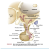
5) thyroid gland
6) adrenal gland
7) pancreas
8) ovary
9) testis

4
Q

All hormones share certain general characteristics, which include:

A

1) have specific rates & rhythms of secretion

  • diurnal: during the day
  • pulsatile and cyclic
  • depending on level of circulating substrates

2) operate within feedback systems - negative or positive - for homeostasis
3) affect only target cells with specific receptors for the hormone and then act on these cells to initiate specific cell functions or activities
4) Excretion/inactivation

  • steroid hormones - excreted via kidneys or metabolized by liver (inactives them and makes them more water soluble for renal excretion)
  • peptide hormones - catabolized by circulating enzymes and eliminated in feces/urine
5
Q

What are the different ways hormones can be classified?

A

structure

gland of origin

effects

chemical composition

6
Q

Why do hormones get released (i.e. what happens in the body that would regulate the release of hormones)?

What mechanisms control this release?

A

Why they get released:

  • 1) released to responsd to an altered cellular environment
  • 2) released to maintain the level of another hormone/substance

Mechanisms regulating hormone release:

  • 1) chemical factors (ex. blood glucose cause insulin release, calcium levels)
  • 2) endocrine factors (ex. hormones from hypothalamus controlling pituitary hormones)
  • 3) neural control (ex. ANS directly stimualting beta cells to release glucose)
7
Q

Negative vs positive feedback

A

Negative feedback: when changes to chemical, neural or endocrine response to a stimulus DECREASE the synthesis/secretion of a hormone - maintains homeostasis/original steady state; most common

Positive feedback: when changes to chemical, neural or endocrine response to a stimulus INCREASES the synthesis/secretion of a hormone - moves system FURTHER away from original status

8
Q

Describe the feedback loop between hypothalamus-pituitary axis (HPA) and the thyroid gland when T4 and T3 levels decrease, and whether this is a positive or negative feedback loop.

A

Negative feedback loop

1) ↓ serum levels of thyroxine (T4) and triiodothyronine (T3) stimulate secreation of thyrotropin-releasing hormone (TRH) from hypothalamus
2) TRH stimulates secreation of thyroid-stimualting hormone (TSH) from anterior pituitary
3) TSH stimulates synthesis and secretion of T3 and T4
4) T3 and T4 levels are now ↑ and generate negative feedback on the pituitary and hypothalamus to inhibit TSH and TRH synthesis

9
Q

Negative feedback loops are possible at three levels, which are:

A

1) target organ - ultra short feedback loop
2) anterior pituitary - short feedback loop
3) hypothalamus - long feedback loop

10
Q

Describe the feedback loop within the female reproductive cycle when estradiol levels increase, and whether this is a positive or negative feedback loop.

A

positive feedback loop

1) ↑ estadiol levels act on hypothalamus to release Gonadotropin Releasing Hormone (GnRH)
2) GnRH stimualtes release of follice-stimulating hormone (FSH) from anterior pituitary
3) FSH (and LH) cause ovaries to produce more estrogen, leading to ovulation

11
Q

Water-soluble hormones vs lipid-soluble hormones

Describe how they are transported in the circulatory system and their half lie.

A

Water-soluble hormones:

  • aka peptide hormones (protein hormones, catecholamines)
  • circulate in free (unbound) forms - only these can signal a target cell (if they are bound to a carrier protein like lipid soluble hormones, they cannot)
  • high MW, cannot diffuse across cell membrane - bind with receptors on or in cell membrane
  • half-life typically seconds to minutes because they are catabolized by circulating enzymes
  • ex. insulin

Lipid-soluble hormones:

  • transported bound to a water-soluble carrier or transport protein
  • can pass freely across plasma and nuclear membranes (via simple diffusion) & bind with cystolic or nuclear receptors (vit. D, retinoice acid, and thyroid hormones can also do this)
  • hormone-receptor complex binds to a specific region in the DNA and stimulates specific gene exprsesion
  • can remain in blood for hours to days
  • ex. cortisol & adrenal androgens
12
Q

What are target cells and what are the two main functions of the hormone receptors of the target cell?

A

Target cells: cells with appropriate receptors for THAT specific hormone

Two main functions:

  • 1) to recognize and bind specifically and with high affinity to their particular hormones
  • 2) to initiate a signal to appropriate intracellular effectors
13
Q

Upregulation vs downregulation

A

Upregulation: low concentrations of hormones increase the number or affinity of receptors per cell

Downregulation: high concentrations of hormone decrease the number or affinity of receptors per cell

14
Q

Cells are able to adjust their sensitivity to the concentration of a signaling hormone. How do they do this and what factors/phsyiochemical conditions may influence this sensitivity?

A
  • sensitivity of the target cell to a specific hormone depends on # of receptors and/or their affinity for the receptors
  • receptors are continuously synthesized and degraded, so their numbers on the cell surface can frequently change as well
  • factors that affect their ability to do this:
    • fluidity and structure of plasma membrane
    • pH
    • temperature
    • Ion concentration
    • Diet
    • Presence of other chemicals (like drugs)
15
Q

Describe how the regulation of hormone receptors on cells for glucose uptake is affected in NIDDM/Type 2 Diabetes.

A

In NIDDM, there is a decrease in insulin receptor sensitivity and hyperglycemia (so they need to be able to quickly increase their numbers of receptors to pick up as much insulin as possible to deal with the high sugar levels)

16
Q

Direct effect vs Permissive Effect

A

ways in which hormones affect target cells

  • Direct effect: obvious changes in cell function that are specifically a result from stimulation by a particular hormone
    • ex. insulin directly affects skeletal muscle via insulin receptors to increase glucose uptake
  • Permissive effect: less obvious hormone-induced changes that facilitate max response/functioning of a cell
    • ex. insulin’s effect on mammary cells - facilitates response of mammary cells to prolactin
17
Q

Where are the two locations that hormone receptors can be found?

A

a) in the plasma membrane
b) in intracellular compartment of the target cell

18
Q

First messenger vs Second messenger

A

First messenger: the hormone that binds to the receptor on the plasma membrane that initiates a cascade of intracellular effects

  • intiates signal transduction: the transmission of molecular signals from a cell’s exterior to its interior

Secondary messenger: intracellular molecules that relay signals received at receptors on the cell surface to the cytoplasm & nucleus of the cell and mediates effects of the hormone on the cell

19
Q

Second messengers include:

A
  • cyclic adenosine monophosphate (cAMP)
  • cyclic guanosine monophosphate (cGMP)
  • calcium
  • inositol triphosphate (IP3)
  • tyrosine kinase system
20
Q

What first messengers activate/increase cAMP levels to cause cell signaling?

A

adrenocorticotropic hormone (ACTH)

thyroid-stimulating hormone (TSH)

both^ cause cAMP levels to increase, which then activate protein kinases leading to phosphorylation of cellular proteins (which then either activates/deactivates intracellular enzymes and causing specific functions)

21
Q

What first messengers result in the production of IP3. Describe the cellular cascade as a result of the first messengers triggering IP3.

A

First messengers: Angiotensin II, ADH

Cellular cascade: first messengers generate IP3 which then triggers a release of intracellular calcium ⇒ forms a calcium-calmodulin complex ⇒ mediates effects of calcium on intracellular activities that are crucial for cell metabolism and growth

22
Q

Insulin, GH, and prolactin are first messengers that bind to surface receptors and activate which second messenger(s)?

A

second messengers of the tyrosine kinase family:

Janus family of tyrosine kinases (JAK)

signal transducers and activators of transcription (STAT)

23
Q

With the exception of thyroid hormones, lipid-soluble hormones are synthesized from ___________________. These lipid-soluble hormones include:

A

cholesterol (i.e. hormones with “steroid” in the name)

androgens, estrogens, progestins, glucocorticoids, mineralcorticoids, vitamind D, retinoid

24
Q

Describe the steroid hormone mechanism causing an effect on its target cell.

A

1) Lipid-soluble steroid hormone molecules detach from carrier protein & pass through plasma membrane
2) Hormone molecules then diffuse into the nucleus where they bind to a receptor to form a hormone-receptor complex
3) Hormone-receptor complex then binds to a specific site on DNA molecule
4) Triggers transcription of genetic information encoded there
5) Resulting mRNA molecule moves to the cytosol where it associates with a ribosome and initiate synthesis of a new protein
6) new protein now produces specific effects on the target cell

25
Q

The ______________ forms the structural and functional basis for central integration of the neurological and endocrine systems, creating the neuroendocrine system.

A

hypothalamic-pituitary axis (HPA)

26
Q

Hypothalamus - Structure and function

A

Structure: at the base of the brain, connected to the pituitary gland by the pituitary stalk

  • connected to the anterior pituitary through hypophysial portal blood vessels
  • connected to the posterior pituitary via nerve tract, hypothalamohypophysial tract

Function:

  • contains special neurosecretory cells that synthesize and secrete hypothalamic-releasing hormones that regulate the release of hormones from anterior pituitary
  • ^ cells also synthesize ADH and oxytocin that are released from posterior pituitary gland
27
Q

Structure of Pituitary gland

A

Structure: located in the sella turcica (saddle-shaped depression of the sphenoid bone at the base of the skull)

  • weights ~0.5g except during pregnancy (weight ↑ ~30%)
  • composed of two distinct lobes (different emrbyonic origins, cell types, function)
    • 1) anterior pituitary: aka adenohypophysis
    • 2) posterior pituitary: aka neurohypophysis
28
Q

Structure of anterior pituitary - how much does it weigh and what are the 3 regions that make up the anterior pituitary

A
  • accounts for 75% of total weight of pituitary gland
  • 3 regions:
    • 1) pars distalis - major component, source of anterior pituitary hormones
    • 2) pars tuberalis - thin layer of cells on anterior and lateral portions of pituitary stalk
    • 3) pars intermedia - lies between those two^ and secretes melanocyte-stimulating hormone in fetus ; disappears in the adult and cells are distributing through pars distalis and pars nervosa of posterior pituitary
29
Q

What are the two main cell types that make up the anterior pituitary?

A

1) Chromophobes: nonsecretory

2) Chromophils: secretory cells, subdivided into 7 cell types - each secretes a specific hormone(s)

30
Q

Anterior pituitary hormones are regulated by what?

A

1) secretion of hypothalamic peptide hormones or releasing factors
2) feedback effects of hormones secreted by target glands
3) direct effects of other mediating NTs

31
Q

What are tropic hormones?

A

hormones that are released to target another endocrine gland; typically secreted by anterior pituitary

32
Q

Tropic hormones released by the anterior pituitary

A

1) Melanocyte-stimulating hormone (MSH)
2) Follicle-stimulating Hormone (FSH) and Luteinizing hormone (LH)
3) Adrenocorticotropic hormone (ACTH)
4) Thyroid-stimulating hormone (TSH)
5) Growth Hormone (GH)
6) Prolactin

33
Q

Melanocyte-stimulating hormone (MSH)

Secreted by which gland

Target Organ

Function

A

Secreted by: anterior pituitary

Target organ: anterior pituitary

Function: promote secretion of melanin and lipotropin by anterior pituitary; makes skin darker

34
Q

Adrenocorticotropic Hormone (ACTH)

Secreted by which gland

Target Organ

Function

A

Secreted by which gland: anterior pituitary (corticotropic cell type)

Target Organ: adrenal cortex

Function: regulates cortisol and androgenic hormones release from the adrenal cortex (increased steroidogenesis); helps maintain adrenal gland

35
Q

Growth Hormone

Secreted by which gland

Target Organ

Function

A

Secreted by which gland: anterior pituitary (somatotropic cell type)

Target Organ: muscle, bone, liver

Function: normal tissue growth and maturation; impacts aging, sleep, nutritional status, stress, and reproductive hormones

36
Q

Prolactin

Secreted by which gland

Target Organ

Function

A

Secreted by which gland: Anterior pituitary (lactotropic cell type)

Target Organ: Breast

Function: Milk production during pregnancy and lactation. Some immune stimulatory effects & modulates immune and inflammatory responses

37
Q

Thyroid-stimulating hormone (TSH)

Secreted by which gland

Target Organ

Function

A

Secreted by which gland: anterior pituitary (thyrotropic)

Target Organ: thyroid gland

Function: increase production and secretion of thyroid hormone; increased iodide uptake, promotes hypertrophy and hyperplasia of thymocytes (immune cells in thymus)

38
Q

Lutenizing Hormone (LH)

Secreted by which gland

Target Organ

Function

A

Secreted by which gland: anterior pituitary (gonadotropic cell type)

Target Organ: in women - granulosa cells; in men - Leydig cells

Function:

  • women: ovulation, progesterone production
  • men: testicular growth, testosterone production
39
Q

Follicle-stimulating hromone (FSH)

Secreted by which gland

Target Organ

Function

A

Secreted by which gland: anterior pituitary (gonadotropic cell type)

Target Organ: in women - granulosa cells; in men - Sertoli cells

Function:

  • women: Follicle maturation, estrogen production
  • men: spermatogenesis
40
Q

IGF regulation of Growth Hormone

A

IGF-1: most biologically active, binds to IGF-1 receptors to mediate anabolic effects of GH & to insulin receptors on skeletal muscle

IGF-2: fetal growth but suppresses GH in the adult

41
Q

Somatotropic hormones secreted by anterior pituitary

A

GH, prolactin

42
Q

Glycoprotein hormones

A

TSH

LH

FSH

β-Lipotropin

β-Endorphins

43
Q

β-Lipotropin

Secreted by which gland

Target organ

Function

A

Secreted by which gland: anterior pituitary (corticotropic cell type)

Target organ: Adipose cells

Function: fat breakdown and release of fatty acids

44
Q

β-Endorphins

Secreted by which gland

Target organ

Function

A

Secreted by which gland: anterior pituitary (corticotropic cell type_

Target organ: adipose cells; brain opioid receptors

Function: analgesia; may regulate body temp, food and water intake

45
Q

Structure of posterior pituitary

A

Structure: derived from hypothalamus, made up of 3 parts:

  • Median eminence: at base of hypothalamus; mostly nerve endings of axons from hypothalamus and contains 10+ hypothalamic-releasing hormones & NTs (dopamine, NE, 5HT, ACh, histamine)
  • Pituitary stalk: contains nerve axons coming from supraoptic and paraventricular nuclei of hypothalamus, connects pituitary gland to the brain
  • Infundibular process (pars nervosa/neural lobe): where the hypothalamus axons terminate & where posterior pituitary hormones are secreted
46
Q

What are the two hormones that are secreted by the posterior pituitary? Where are they synthesized?

A

Hormones:

  • Antidiuretic hormone (ADH) aka arginine vasopressin
  • oxytocin ‘

Synthesis: made in the supraoptic and paraventricular nuclei of the hypothalamus, packaged in secretory vesicle and moved down the axons of the pituitary stalk to the pars nervosa for storage

47
Q

Function and regulation of ADH

A

Function: controls plasma osmolality; increases the permability of distal renal tubules and collecting ducts which leads to increased water reabsorption into the blood, concentrating urine and reducing serum osmolality

Regulation:

Increases:

  • secretion regulated by osmoreceptors in hypothalamus (stimulated when plasma osmolality ↑ which causes increase in ADH secretion, more water reabsorption by kidney, plasma diluted back to baseline)
  • secretion also regulated by intravascular volume changes (via baroreceptors in LA, carotid arteries, aortic arches) - decrease in volume causes increased ADH secretion
  • ADH has no direct effect on electrolytes (but water reabsoprtion will dilute and cause their concentration to decrease)
  • ADH secretion can also be increased via stress, trauma, pain, exercise, nausea, nicotine, heat exposure and drugs like morphine
  • Decreases:*
  • ADH secretion decreases with decreased osmolality, increased intravascular volume, hypertension, alcohol ingestion (alcohol inhibits the secretion of ADH), increase in estrogen, progesterone or angiotensin II levels.
48
Q

Function and Regulation of oxytocin

A

Function: contraction of uterus, milk ejection in lactating women, may affect sperm motility in men. Has an antidiuretic effect similar to ADH.

  • enhances effectiveness of contractions, promotes placental deivery, stimulates postpartum uterine contractions to prevent excessive bleeding

Regulation:

  • In women: secreted in response to suckling and mechanical distension of the female reproductive tract - binds to receptors in mammary tissues to cause contraction of myoepithelial cells, which increases intramammary pressure and milk expression (“let-down” reflex)
49
Q

Describe the structure and function of Pineal Gland, and the regulation of hormone release from this gland.

A

Structure: made of photoreceptive cells, located near center of brain. Innervated by noradrenergic sympathetic nerve terminals controlled by pathways within hypothalamus

Function: secrete melatonin (synthesized from tryptophan, which is first converted to 5HT and then melatonin)

Regulation: stimulated by exposure to dark, inhibited by light exposure.

50
Q

Function of melatonin

A
  • regulates circardian rhythms and reproductive systems (onset of puberty, secretion of gonadotropic-releasing hormones)
  • immune regulation
  • involved in aging process
  • increases nitric oxide release from blood vessels
  • removing oxygen free radicals
  • decreasing insulin secretion
51
Q

Structure and Function of Thyroid Gland

A

Structure: located in neck below larynx and thyroid cartilage. Has two lobes, on either side of the trachea and is connected by the isthmus.

  • Pyramidal lobe is superior to isthmus (just another lobe of the thyroid gland that sticks upwards)
  • Follicular cells: gland consists of follicles that contain follicular cells surrounding colloid (viscous substance) - these cells synthesize and secrete thyroid hormones and can be directly affected by NTs Ach and catecholamines
  • 2 month supply of thyroid hormone stored in the gland
  • Parafollicular/C cells: secrete calcitonin (thyrocalcitonin)

Function: produces hormones that control rates of metabolic processes throughout body

52
Q

Function and regulation of calcitonin

A

Function: lowers serum calcium levels by inhibiting bone-reabsorbing osteoclasts (which is the opposite function of PTH)

  • lowers serum phosphate levels
  • decreases calcium and phosphorous absroption in GI tract

Regulation:

  • stimulated by high calcium levels (major), gastrin, calcium-rich foods, pregnancy
  • release inhibited by lowered serum calcium level
53
Q

Regulation of thyroid hormone (TH)

A
  • regulated via a negative feedback loop
  • initiated by thyrotropin-releasing hormone (TRH) which is synthesized and stored in hypothalamus
    • TRH levels increase with cold exposure or stress, decreased thyroxine (T4)
  • TRH released into hypothalamus-pituitary portal system, circulates to anterior pituitary which is then stimulates release of thyroid-stimulating hormone (TSH)
  • TSH circulates and binds to receptors on thyroid follicular cells → immediate release of stored TH and increase in TH synthesis; also increases growth of gland by stimulating thymocyte hyperplasia & hypertrophy
  • TH levels rise, negative feedback loop on the HPA to inhibit TRH and TSH release leading to decreased TH synthesis and secretion
  • TH synthesis also controlled by iodide levels and deiodinases that inactivate thyroxine
54
Q

How is thyroid hormone synthesized?

A
  1. Uniodinated thyroglobulin is produced by the ER of the thyroid follicular cells.
  2. Tyrosine is incorporated into the thyroglobulin as it is synthesized.
  3. Iodide (inorganic form of iodine) is actively pumped from the blood into the colloid by carrier proteins located in the outer membrane of the follicular cells (active transport system called the iodide trap)
  4. Iodide is oxidized and quickly attaches to tyrosine within the thyroglobulin molecule.
  5. Coupling of iodinated tyrosine forms thyroid hormones. Triiodothyronine (T3) is formed from coupling of monoiodotyrosine (one iodine atom and tyrosine) and diiodotyrosine (two iodine atoms and tyrosine). Tetraiodothyronine (T4/thyroxine) is formed from coupling of two diiodotyrosines.
  6. Thyroid hormones are stored attached to thyroglobulin within the colloid until they are released into the circulation.

Thyroid gland produces 90% T4 , 10% T3

55
Q

Once released in the circulation how are T3 and T4 transported?

A

transported bound to thyroxine-binding globulin, some via thyroxine-binding prealbumin (transthyretin), albumin, or lipoproteins

when bound, acts like a reservoir while unbound form is active

in body tissues, most T4 converted to T3 which acts on target cell

56
Q

Function of thyroid hormone (TH)

A
  • ++effect on growth, maturation and function of cells/tissues
    • needed for normal growth, neuro development
    • affects metabolic, neurologic, CV, and resp functioning over lifespan
  • required for metabolism (so causes increased heat production and oxygen consumption), blood cell function, normal muscle function
  • required for integumentary integrity
  • has permissive effects - i.e. it optimizes the actions of other hormones and NTs
57
Q

Structure and Function of parathyroid glands

A

Structure: two pairs of glands behind the upper and lower poles of the thyroid gland, but can range from 2-6

Function: produce parathyroid hormone (PTH)

58
Q

Regulation and function of parathryoid hormone (PTH)

A

Function: most important in the regulation of serum calcium concentration

  • increases calcium levels and decreases phosphate levels in serum
  • Acts directly on bone to release calcium via stimulating osteoclast activity
  • also acts on kidney to increase calcium reabsorption & decrease phosphate reabsorption

Regulation: via Calcium - stimulated by decrease in serum-ionized calcium levels. Inhibited when serum calcium concentration increases

  • via Phosphate - Increased phosphate levels decrease calcium levels, causing calcium phosphate preciptation in soft tissue and bones ⇒ PTH stimulated
  • via Magnesium - Hypomagnesemia also mildly stimulates PTH
  • Note: intermittent admin of low dose PTH stimulates bone formation (which is opposite of what it normally does) - used for osteoporosis tx (with vit D as a cofactor to promote calcium and phosphate absoprtion to enhance bone mineralization & control inflammation)
59
Q

Structure and Function of pancreas

A

Structure: located behind the stomach, between spleen and duodenum

  • contains islets of Langerhans, which has 4 types of hormone secreting cells:
    • alpha cells: secrete glucagon
    • beta cells: secrete insuiln and amylin
    • delta cells: secrete gastrin and somatostatin
    • F (or PP) cells: secrete pancreatic polypeptide
  • Innervation: both sympathetic and parasympathetic

Function: considered both an endocrine (produces hormones) and exocrine gland (produces digestive enzymes)

60
Q

Endocrine vs exocrine gland

A

Exocrine glands: glands that secrete substances onto an epithelial surface via a duct. Examples: sweat, salivary, mammary, ceruminous, lacrimal, sebaceous, prostate and mucous.

Endocrine Glands: glands that secrete their products directly into the bloodstream.

61
Q

What two organs are endocrine and exocrine glands?

A

The liver and pancreas are both exocrine and endocrine glands; they are exocrine glands because they secrete products—bile and pancreatic juices—into the GI tract through a series of ducts, and endocrine because they secrete other substances directly into the bloodstream

62
Q

Synthesis and regulation of Insulin

A

Synthesis:

  • from beta cells in pancreas, from the precursor proinsulin which is formed from larger precursor preproinsulin
  • Proinsulin: A peptide and B peptide connected by a C peptide and two disulfide bonds
    • C peptide then cleaved by enzymes, and bonded A and B peptides become insulin
  • Insulin circulates freely, no carrier molecule

Regulation: via chemical, hormonal, and neurla control

  • Increases: when beta cells are stimualted by parasympathetic NS usually before a meal, increased BGL, AA (leucine, argining, lysine), GI hormones (glucagon, gastrin, cholecystokinin, secretin)
  • Decreases: in response to hypogycemia, high insulin levels, sympathetic stimulation of beta cells in islets, prostaglandins
63
Q

Function of insulin and what factors alter the sensitivity of insulin

A

Function:

  • once bound to cell surface receptor, it promotes cellular glucose uptake via glucose transports (GLUT) - considered an anabolic hormone that promotes glucose uptake primarily in liver, muscle and adipose tissue (insulin not needed for glucose uptake in brain, RBCs, kidneys, and lens of eye)
  • Increases synthesis of proteins, carbs, lipids, and nucleic acids
  • facilitates intracellular transport of potassium, phosphate, and magnesium
  • Net effect: stimulate protein and fat synthesis, decrease BGL

Factors affecting sensitivity (and thus maintenance of normal cell function):

  • age, weight, abdominal fat, physical activity
  • adipocytes release a number of hormones and cytokines that are altered in obesity which impact insulin sensitivity
  • weight loss and exercise improves sensivity
64
Q

Regulation and function of amylin

A

Regulation: a peptide hormone that is co-secreted with insulin by beta cells in response to nutrient stimuli

Function: regulates blood glucose concentration by delaying gastric emptying and suppressing glucagon secretion after meals. Also has satiety effect (reducing food intake) - considered to have an antihyperglycemic effect

65
Q

Synthesis and regulation of glucagon

A

Synthesis: produced by alpha cells of pancreas and by cells lining GI tract

Regulation: secretion stimulated by AA (alanine, glycine, asparagine), low glucose levels and sympathetic stimulation. Inhibited by high glucose levels

Function: antagonistic to insulin. Acts on liver to increase blood glucose concentration by stimulating glycogenolysis (breaking down glycogen) and gluconeogesis (building glucose) in muscle, lipolysis in adipose tissue.

66
Q

Synthesis and Function of pancreatic somatostatin

A

Synthesis: produced by delta cells of pancreas in response to food intake. Different from hypothalamic somatostatin

Function: essential for carb, fat, and protein metabolism. Involved with regulating alpha and beta cell function within the islets by inhibiting insulin, glucagon, and pancreatic polypeptide

67
Q

Descibe the function of gastrin, ghrelin, and pancreative polypeptide.

A

Pancreatic Gastrin: stimulates secretion of gastric acid. May be necessary for fetal islet cell development.

Ghrelin: stimulates GH secretion, controls appetite, plays a role in obesity and regulation of insulin sensitivity

Pancreatic polypeptide: released by F cells in response to hypoglycemia and protein-rich meals. Inhibits gallbladder contraction and exocrine pancreas secretion. Increased in those with pancreatic tumours or DM

68
Q

Structure of adrenal glands

A
  • paired, pyramid-shaped organs behind the peritoneum and close to upper pole of each kidney
  • each gland surround by a capsule, embedded in fat, and well supplied with blood from aorta, phrenic, renal arteries
  • Venous return:
    • from left gland is to renal vein
    • from right gland to IVC
  • Each gland consists of two separate portions that basically act like separate glands - cortex (outer) and medulla (inner)
69
Q

Structure and zones of the adrenal cortex

A
  • 80% of the weight of the gland, three zones:
    • 1) Zona glomerulosa: outer layer, ~15% of the cortex and primarily produces mineralocorticoid aldosterone
    • 2) Zona fasciculata: middle layer, ~78% of cortex and secretes glucocorticoids cortisol, cortisone, and corticosterone
    • 3) Zona reticularis: inner layer, ~7% of the cortex and secretes mineralocorticoids (aldosterone), adrenal androgens and estrogrens, and glucocorticoids
  • stimualted by adrenocorticotropic hormone (ACTH) from pituitary
  • all hormones synthesized from adrenal cortex are made from cholesterol
70
Q

What are glucocorticoids and what function(s) does it have?

A
  • steroid hormones that have metabolic, neuro, anti-inflammatory, and growth-suppressing effects (which directly affect carb metbaolism)

Functions/effects:

Carbohydrate/lipid metabolism:

  • increase blood glucose by promoting gluconeogenesis in the liver & decreasing glucose uptake into muscle/adipose/lymphatic cells
  • protein catabolism

Immune system: increased glucocorticoids = decreased immunity

  • immunosuppression: decreases T lymphocyte proliferation (T-helper cells)
  • Can also affect innate immunity by decreasing the activity of pattern receptors on the surface of macrophages
  • anti-inflammtory: decreased function of NK cells, supressing inflammatory cytokines and decreasing release of proteolytic enzymes
  • seen in stress conditions

Other: inhibition of bone formation, ADH secretion and stimulation of gastric acid secretion. Thyroid and growth hormone effects on adipose tissue are also potentiated. May also cause mood changes due to fluctuations in steroid levels.

71
Q

The most potent naturally occurring glucocorticoid is

A

cortisol

72
Q

Cortisol - function and regulation

A

Function: main secretory produce of adrenal cortex; needed to maintain life and protect body from stress. Liver is primarily responsible for its deactivation

Regulation: secretion regulated via hypothalamus and anterior pituitary

  • Corticotropin-releasing hormone (CRH) produced by hypothalamus and stored in median eminence. Released to stimulate production of ACTH, β-lipotropin, γ-lipotropin, endorphins, and enkephalins by anterior pituitary
  • ACTH - main regulator of cortisol secretion and adrenocortical growth
    • once it stimulates cortex, cortisol synthesis and secretion is immediate - so secretory patterns of ACTH and cortisol are nearly identical
  • Cortisol may be attached to albumin when circulating, but primarily bound to transcortin protein; some free form
73
Q

Synthesis and regulation of ACTH

A

Synthesis: from precursor proopiomelanocortin (POMC)

Regulation: 3 factors

  • 1) -ve feedback effects of high circulating levels of cortisol and synthetic glucocorticoids (suppresses CRH and ACTH); low cortisol levels stimulate their secretion
  • 2) diurnal rhythms (ACTH peaks 3-5 hrs after sleep begins & declines throughout the day; cortisol levels follow a similar pattern)
  • 3) psychological and physiologic stress (hypoxia, hypoglycemia, hyperthermia, exercise) increases ACTH secretion and thus cortisol
  • rapidly inactivated in circulation followed by removal via liver and kidneys

Function: maintains size and synthesizing functions of adrenal cortex. Also stimulates melanocytes and activates tissue lipase

74
Q

Function of mineralocorticoids

A

affect ion transport by epithelial cells causing sodium retention and potassium and H loss

75
Q

What is the most potent mineralocorticoid?

A

aldosterone

76
Q

Synthesis, function/effects, and regulation of aldosterone

A

Synthesis: initial stages occur in zona fasciculata and zona reticularis. Converted from corticosterone to aldosterone in zona glomerulosa

Function/effects: conserves sodium by increasing activity of sodium pump of epithelial cells. Maintains extracellular volume by acting on distal nephron epithelial cells to increase reabsorption of Na+ and excretion of K+ and H+

  • CV-related effects: enhances cardiac muscle contraction, stimulates ectopic ventricular activity (secondary cardiac pacemakers), stiffens blood vessels with increased vascular resistance, decreased fibrinolysis
  • Pathological elevation of aldosterone: leads to HF, HTN, insulin resistance, inflammation

Regulation: RAAS - activated by sodium and water depletion, increased potassium levels, and diminished effective blood volume

  • primary stimulant: Angiotensin II; others: ACTH
77
Q

How is aldosterone circulated/transported in the blood and how is it excreted?

A
  • When Na+ and K+ levels are WNL, ~50-250mg of aldosterone is secreted daily
    • 50-70% of these bind to plasma proteins
  • A lot are unbound due to high metabolic turnover, low plasma concentration, and short half life (15 min)
  • degraded in the liver, excreted by kidney
  • Aldosterone’s renal effect takes 90 minutes to 6 hours
78
Q

Regulation of the secretion of adrenal estrogens and androgens

A
  • ACTH (major regulator)
  • minimal amounts of estrogen and androgens are secreted by adrenal cortex
  • some weak androgens are converted to stronge ones in peripheral tissues like testosterone
  • These hormones have the same effects as the same hormones produced by the gonads
79
Q

Structure and Function of Adrenal medulla

A

Structure: inner portion of adrenal gland. Innervated by sympathetic division and is emryonically derived from neural crest cells

  • contains chromaffin cells (pheochromocytes) that secrete and store epinephrine and norepinephrine (both synthesized from AA phenylalanine)

Function: only secretes~30% of total epinephrine in body (the other 70% is from nerve terminals)

  • medulla a minor source of norepinephrine
  • acts as a sympathetic ganglion where sympathetic cholingergic preganglionic fibers terminate on chromaffin cells and secrete catecholamines directly into blood stresm
80
Q

Regulation of the secretion of catecholamines from adrenal gland

A
  • release triggered by physiological stress (trauma, hypoxia, hypoglycemia) via ACh (preganglionic sympathetic fibers) which depolarizes chromaffin cells
  • Depolarization causes exocytosis of storage granules from chromaffin cells, and epi and norepi are released
  • Secretion also increases with ACTH and glucocorticoids
  • epi is 10x more potent than norepi in exerting metabolic effects
81
Q

Regulation and excretion of catecholamines once released into the bloodstream

A
  • remain in plasma for seconds to mins & bind to alpha and beta receptors - this activates adenylyl cyclase system
  • rapidly removed from plasma by being absorbed by neurons for storage in granules or inactivated and then excreted in urine
  • Catecholamines also directly inhibit their own secretion by decreasing the formation of tyrosine hydroxylase
82
Q

Effects of catecholamines

A

fight or flight response

promoting hyperglycemia

83
Q

General changes to endocrine system with aging

A
  • changes to HPA
  • altered hormone activity and circulating levels
  • altered secretory response of endocrine glands
  • altered metabolism of hormones
  • loss of circadian control of hormone secretion
84
Q

Changes to pituitary gland due to aging

A
  • Posterior: decreases in size, reduced ADH secretion
  • Anterior: increased fibrosis, moderate increase in size. Decline in GH release
85
Q

Changes to thyroid gland with aging

A
  • atrophy of gland, fibrosis, nodularity, increased inflamamtory infiltrates
  • decreased T4 secretion, T3, TSH secretion
  • reduced response of plasma TSH concentration to TRH
86
Q

Changes to GH and insulin-like growth factors

A
  • decreased GH and IGF amounts (makes sense due to decreased muscle size and function with age, as well as reduced fat and bone mass)
  • changes in reproductive and cognitive function
  • increased visceral fat, decreased lean body mass
87
Q

Pancreatic hormonal changes with aging

A
  • glucose intolerance/diabetes
  • decreased insulin receptor activity
  • decreased betal cell secretion of insulin
88
Q

Adrenal gland changes with aging

A
  • Decreased DHEA (androstenedione) levels lead to decreased synthesis of androgen-derived estrogen and testosterone
  • decreased metabolic clearance of glucocorticoids and cortisol leading to decreased cortisol secretion
  • decreased levels of aldosterone
  • Potential changes to circadian patterns of ACTH and cortisol secretion
89
Q

Gonadal hormone changes with aging

A
  • Women: decreased estrogen and progestorone, increased FSH and androgen levels with postmenopause
  • Men: gradual decrease in testosterone levels - decreased sex activity, muscle strength, and bone mineralization
90
Q

What are potential mechanisms behind hormonal alterations?

A

1) Dysfunction of an endocrine gland - due to failure to produce adequate amounts of hormone or too much (hypo- or hyper-secretion); failure to convert precursors to active hormones

2) Feedback systems - fail to function properly (fail to recognize or may respond to inappropriate signals)

3) Altered metabolism of hormones - may be degraded at an altered rate or be inactivated before reaching targe cell by antibodies that act as hormone inhibitors (i.e. thyroid disease)

4) Dysfunctional delivery system - inadequate blood supply, carrier proteins, ectopic production of hormones (like tumors that lead to abnormal elevations of hormone levels)

91
Q

Alterations in hormone levels/function can also occur with inappropriate response of target cells to hormonal stimulation. What are the two general types of target cell insensitivity to hormones?

A

1) Cell surface receptor-associated disorders: primarily in water soluble hormones like insulin

  • decreased # of receptors (resulting in decreased/defective hormone receptor binding)
  • impairment of receptor function (resulting in insensitivity to the hormone)
  • presence of antibodies against specific receptors (reduces available binding sites or mimic horrmone action)
  • unusual expression of receptor function (i.e. tumor cells)

2) Intracellular disorders:

  • acquired defects in postreceptor signaling cascades
  • inadequate synthesis of a second messenger (ex. cAMP)
  • intracellular enzymes or proteins are altered
  • alterations in nuclear coregulators (proteins that facilitate/inhibit target gene transcription)
  • altered protein synthesis
92
Q

Alterations of the Hypothalamic-pituitary system (HPS) leading to hypothalamic dysfunction is due to:

A

interruption of the pituitary stalk caused by: destrucive lesions, rupture after head injury, surgical transection, or tumor (physical connection interruptions)

93
Q

Decreased hypothalamic function leads to decreased GnRH, TRH, CRH, PIF, and GHRH. Describe what effect each of these would have on their respective target hormones in the anterior pituitary.

A

↓GnRH: cessation of menstruation (women) and hypogonadism & impaired spermatogenesis (men)

↓TRH: hypothalamic hypothyroidism

↓CRH: decreased ACTH response to low serum cortisol levels

↓PIF (prolactin inhibitory factor like dopamine): no inhibitory control of prolactin secretion by dopamine leads to hyperprolactinemia

↓GHRH: GH deficiency and growth failure in children

94
Q

Disease of the posterior pituitary cause abnormal secretion of what hormone that influences osmolarity? Excess or deficient amounts of this hormone results in what?

A
  • ADH (arginine vasopressin)
  • Excess leads to water retention and hypoosmolar state
  • Deficiencies in amount/response to ADH result in serum hyperosmolarity
95
Q

Syndrome of Inappropriate ADH secretion (SIADH)

Definition, Cause, Pathophysiology, Manifestations, Diagnosis, Treament

A

Definition: Abnormally high levels of ADH without normal physiological stimuli to cause its release

Causes: tumors (ectopic production) - small cell carcinomas of duodenum, stomach, & pancreas; bladder, endometrial, prostate cancer; lymphomas, sarcomas.

  • Pulmonary: bronchogenic carcinoma, pneumonia (TB), asthma, CF, resp failure needed mechanical ventilation
  • CNS: encephalitis, meningitis, intracranial hemorrhaging, tumors, trauma
  • Surgery: elevated levels up to 5-7 days post-op (unknown but may be due to fluid and volume changes). May also follow pituitary surgery
  • Medications: hypoglycemic meds, narcotics, general anesthetics, antidepressants, antipsychotics, chemo agents, NSAIDs, synthesis ADH analogs

Pathophysiology: **enhanced renal water retention from ADH acting on kidneys to increase renal collecting duct permeability to water via aquaporin-2 protein = increased water reabsorption)**. Leads to expansion of ECF volume ⇒ dilutional hyponatremia, hypoosmolarity, & highly concentrated urine due to little/no water excreted.

Clinical Manifestations: from hyponatremia - determined by severity and how rapid onset is.

  • thirst, impaired taste, anorexia, dyspnea on exertion, fatigue, dulled ability to sense (with rapid decrease in sodium levels)
  • No peripheral edema
  • GI symptoms: vomiting, cramps
  • weight gain from water retention
  • Slow decrease in sodium levels: confusion, lethargy, muscle twitching, convulsions, potential irreversible neuro damage

Diagnosis: based on the following findings

  • 1) serum hypoosmolality and hyponatremia
  • 2) urine hyperosmolarity
  • 3) urine sodium excretion matches sodium intake
  • 4) normal adrenal and thyroid funciton
  • 5) absence of conditions that can alter volume status (CHF, hypovolemia causes, renal insufficiency)

Treatment: Sx resolve with correction of hyponatremia, fluid restriction. In severe cases, hyponatremia is corrected with hypertonic saline. Resolution usually within 3 days, some weight loss and salt wasting

96
Q

Diabetes Insipidus (DI)

Definition, Types of DI and Causes, Pathophysiology, Manifestations, Diagnosis, Treament

A

Definition: insufficiency of ADH activity leading to frequent urination (polyuria) and frequent drinking (polydipsia). Two forms:

  • Neurogenic/central DI: due to lesion of hypothalamus, pituitary stalk, or posterior pituitary leading to interference with ADH synthesis, transport, or release. Lesions can be from brain tumors, hypophysectomy (pituitary gland removal), aneurysms, thrombosis, infections, immune disorders. TBIs, hereditary disorders that affect ADH genes. Abrupt onset of S/S.
  • Nephrogenic DI: caused by inadequate response of renal tubules to ADH (usually acquired, may be genetic). Often related to drugs that damage the renal tubules or inhibit generation of cAMP in the tubules ie. Pyelonephritis, amyloidosis, polycystic kidney disease. Drugs: lithium carbonate, loop diuretics, general anesthetics. Gradual onset of S/S.
  • Gestational DI: rare form - increased levels of vasopressin-degrading enzyme vasopressinase

Pathophysiology: partial/total inability to concentrate urine. ADH activity is shit therefore lots of dilute urine leaves the body and plasma osmolality increases. Patients may be super thirsty stimulating polydipsia (high fluid intake). No ongoing fluid replacement leads to dehydration and if the patient cannot conserve as much water as is lost, then hypernatremia and hyperosmolality occurs.

Clinical Manifestations: polyuria, nocturia (peeing at night), continuous thirst, polydipsia. Varied urine output but can be higher than daily fluid intake. If chronic, these people can develop large bladder capacities and hydronephrosis (kidney swelling).

Diagnosis: low urine specific gravity, low urine osmolality, hypernatremia, high serum osmolality, continued diuresis (filtering of fluid) despite high sodium levels in blood. Dx done through water deprivation testing.

Treatment: Depends on age, health, lifestyle. ADH replacement but usually fluid replacement (PO/IV). Vasopression. Treat underlying cause (i.e. meds)

97
Q

How is diabetes insipidus different than dipsogenic/primary polydipsia?

A

Primary polydipsia is due to chronically ingesting lots of fluid that washes out the renal medullary concentration gradient leading to partial ADH resistance. This resolves with decreased fluid ingestion; this may be psychogenic in nature.

Can be distinguished from GI because with polydipsia, there will be low ADH levels. In nephrogenic DI, ADH levels are normal or high.

98
Q

Hypopituitarism

A

Definition: absence of one or more anterior pituitary hormones OR complete failure of all anterior pituitary hormone functions

Causes: due to inadequate supply of hormones from hypothalamus (due to damage to pituitary stalk) or inability of pituitary gland to produce hormones. Common causes: infarctions/lesions that take up space (pituitary adenomas which is a benign tumor of a gland, aneurysms).

  • Others: TBIs, removal or destruction of gland, infections (meningitis, syphilis, TB), autoimmune hypophysitis, drugs, gene mutations (PROP-1)

Pathophysiology: pituitary gland is vulnerable to ischemia and infarction due to high vasculature and needs portal blood flow from hypothalamus. Leads to necrosis and edema of gland, further cutting off blood supply. Eventually, fibrosis sets in and hypopituitary S/S occur. Same thing occurs with adenomas and aneurysms compressing pituitary cells and cutting off hormonal output

Clinical Manifestations: variable because it depends on which hormones are affected.

  • Panhypopituitarism: all hormones are deficient and multiple complications occur (lack of ACTH = cortisol deficiency, thyroid deficiency from lack of TSH, loss of 2’ sex traits from lack of FSH and LH). Low GH and IGF-1 affects growth. Lack of prolactin = women cannot lactate for their bb.
  • See more in other flashcards.

Diagnosis: measuring tropic hormone levels (from anterior pituitary or targen glands). Imaging of pituitary gland to determine lesions, tumors, etc.

Treatment: correcting unerlying disorder asap. Hormone replacement. For cases of circulatory collapse, immediate therapy with glucocorticoids and IV fluids.

99
Q

Sheehan syndrome

A

postpartum hypopituitarism caused by necrosis of the pituitary gland

usually due to blood loss and hypovolemic shock

100
Q

Signs and symptoms of ACTH deficiency in someone with hypopituitarism

A
  • associated loss of cortisol which can be life-threatening
  • Within 2 weeks of complete ACTH absence:
    • N/V
    • anorexia
    • fatigue, weakness
    • hypoglycemia from increased insulin sensitivity, decreased glycogen stores, decreased gluconeogenesis
  • limited aldosterone secretion
  • decreased urine outpute due to decreased GFR
101
Q

Signs and symptoms of TSH deficiency in someone with hypopituitarism

A
  • rarely seen in isolation
  • Sx develop 4-8 weeks after hypothryotropinemia (low TSH levels) occur: cold interolerance, skin dryness, mild myxedema (advanced hypothyroidism), lethargy, decreased metabolic rate
  • Sx less severe than those of primary hypothyroidism
102
Q

Signs and symptoms of FSH and LH deficiencies in someone with hypopituitarism.

A
  • in women of reproductive age: amenorrhea & atrophy of the vagina, uterus, breasts
  • in postpuberty men: testicles atrophy, diminished facial hair
  • Both: decreased body hair, diminished libido
103
Q

Signs and symptoms of GH deficiency in someone with hypopituitarism

A
  • In children: growth failure, hypopituitary dwarfism
  • Chronic adult GH deficiency syndrome: increased body fat, decreased strength and lean body mass, osteoporosis, reduced sweating, dry skin, psychological problems (social withdrawal, fatigue, loss of motivation, diminished feeling of well-being)
  • increased mortality with MI and stroke associated with DLD and atherosclerosis
104
Q

Primary Adenomas (Hyperpituitarism)

Definition, Cause, Pathophysiology, Clinical Manifestations, Dx, Treatment

A

Definition: benign, slow growing tumors that arise from anterior pituitary cells. Most are tiny and detected post mortem or incidentally on MRI. Most are hormonally silent and do not pose significant risks to the individual. Large adenomas (macroadenomas) alter hormone secretion & impinge on neighbouring structures.

Cause: unknown, occurs sporadically. Altered gene expression occurs

Pathophysiology: Local expansion of adenoma may impinge on optic chiasma (resulting in visual disturbances). May invade cavernous sinuses which may compromise oculomotor, trochlear, abducens, and trigeminal nerves. May extend to and disrupt hypothalamus (wakefulness, thirst, appetite, temperature). Hypersecretion from adenoma (which secretes hormones of the cell type it was made from) with no regulatory feedback mechanisms, hyposecretion from surrounding pituitary cells (esp. GH, FSH, LH-secreting cells).

Clinical Manifestations: depends on tumor growth and hyper/hyposecretion of hormone

  • Increased tumor size: headache, fatigue, neck pain/stiffness, seizures
  • Visual changes: visual field impairments, temp blindness
  • most often with increased secretion of GH and prolactin
  • Gonadotropin hyposecretion: menstrual irregularity in women, decreased libido, receding 2’ sex characteristics (both men and women)

Diagnosis: MRI, CT

Treatment: goal - protect from effects of tumor growth and to control hormone hypersecretion while limiting damage to appropriately secreting portions of pituitary.

  • Meds to supress tumor growth, tumor resection, radiation therapy
105
Q

Acromegaly

A

Definition: hypersecretion of GH and IGF-1 (during adulthood)

Cause: almost always due to GH-secreting pituitary adenoma (rarely from ectopic production of GHRH)

Prevalence/Demographic: 40-59 age group but often present years before Dx

Pathophysiology: GH secretion pattern and peaks are lost (now totally unpredictable). In children the epiphyseal plates have not yet closed so increased GH levels lead to giantism (excessive skeletal growth). In adults, epiphyseal plates closed so tissue proliferation and increased cytoplasmic matrix + bony proliferation occurs.

  • GH also has ++ effects on glucose, lipid, and protein metabolism. Hyperglycemia results due to GH inhibition of glucose uptake, then compensatory high levels of insulin reuslting in finally insulin resistance (DM may occur).
  • HTN and Left HF
  • acts on renal tubules to increased phosphate reabsorption leading to mild hyperphosphatemia
  • adenoma can also become space-occupying and lead to hypopituitarism

Clinical Manifestations: Connective tissue proliferation - enlarged tongue, insterstitial edema, enlarged & overactive sebaceous and sweat glands (bad BO), coarse skin and body hair. Enlargement of the bones of the face hands and feet. Lower jaw and forehead protrude. Rib bones elongate causing barrel chest, changes in the joints hands. S&S of DM ie polydipsia, polyuria due to insulin sensitivity. Hypertension. CNS symptoms (headache, seizure, visual disturbances, papilledema) due to tumor size. Nerve entrapment leading to weakness, atrophy, footdrop and sensory changes. If hypopituitary compression occurs, leads to decreased gonadotropin secretion so amenorrhea in women and sexual dysfunction in men.

Slow progressive disease that decreases life expectancy if untreated. Death caused by heart disease 2’ to HTN and CAD, stroke, DM, or cancer.

Diagnosis: clinical presentation, MRI, elevated levels of IGF-1 and GH.

Treatment: goal is to normal/reduce GH secretion and relieve/prevent complications related to tumor expansion.

  • *surgical removal of GH-secreting adenoma*
  • Radiation therapy
  • Somatostatin analogs - to lower IGF-1 and GH levels
106
Q

Giantism vs Dwarfism

A

Giantism - hypersecretion of GH (occurs in children who are still growing because their epiphyseal plates are not closed yet. Exaggerated bone growth and an abnormal increase in height occurs.

Dwarfism - hyposecretion of GH

107
Q

Prolactinoma (resulting Hypersecretion of prolactin)

Definition, Cause, Pathophysiology, Clinical Manifestations, Diagnosis, Treatment

A

Definition: pituitary tumors that secrete prolactin - the most common hormonally active pituitary tumors; leads to hypersecretion of prolactin

Cause: tumor*, renal failure, PCOD, 1’ hypothyroidism, breast stimualtion, stress of venipuncture. Meds that block dopamine effects (because it usually inhibits prolactin release) or interfere with delivery of dopamine from hypothalamus pituitary cells (antipsychotics, tricyclic antidepressants, estrogens).

Pathophysiology: *sustained increases in serum prolactin* which leads to amenorrhea (no period), galactorrhea (nipple discharge that’s not milk production), hirsutism (excessive male pattern hair growth), osteopenia (decreased bone density)/osteoporosis due to estrogen deficiency. In men, causes hypogonadism (not enough testosterone) and ED. Adenoma also expands and can compress cells. CNS symptoms from pressure of adenoma within sella turcica (esp in tumors >1cm).

Clinical Manifestations: as above. In men, often present late with symptoms and usually due to increase adenoma size (headache, visual impairment)

Diagnosis: Hx taking, MRI, TSH & prolactin levels

Treatment: dopaminergic agonists (usually helps restore fertility in women). Surgery or radiation therapy if resistant to meds.

108
Q

Primary thyroid disorders

vs.

Central (secondary) thyroid disorders

A

Primary thyroid disoders: result in changes of TH levels with seoncdary feedback effects on TSH from pituitary (i.e. more related to dysfunction of the thyroid gland)

  • i.e. if TH levels are high, TSH will secondarily decrease due to -ve feedback

Secondary thyroid disorders: disorders of the pituitary gland TSH production. TSH elevation leads to TH elevation (and same thing with inadequate TSH production)

109
Q

Subclinical thyroid disease

A

thyroid disease that presents with minimal to no symptoms but has abnormla lab values

110
Q

Thyrotoxicosis/Hyperthyroidism

Definition, Cause, Clinical Manifestations, Dx/Tx

A

Definition: Thyrotoxicosis - condition resulting from any cause of ↑TH levels. Hyperthyroidism - form of thyrotoxicosis where excess amounts of TH are secreted from thyroid gland

Causes: for Primary hyperthyroidism - Graves disease, toxic nodular goiter, adenoma. for Secondary hyperthyroidism - TSH-secreting pituitary adenomas (less common). Others - ectopic thyroid tissue, excessive TH ingestion

Clinical Manifestations: increased metabolic rate with heat intolerance & increased tissue sensitivity to stimulation by the SNS. Enlargement of the thyroid gland (goiter).

  • Thin hair, exophthalmos (bulging eyes(, heart failure, weight loss, diarrhea, warm skin, hyperreflexia, pretibial edema

Diagnostics: elevated T4 and T3 levels and suppressed TSH levels (primary hyperthyroidism). Increased TSH levels despite elevated TH (secondary hyperthyroidism)

Treatment: drug therapy, radioactive iodine therapy (absorbed by thyroid tissue to cause cell death) and surgery. Complication for all forms of tx is excessive ablation leading to _hypothyroidism._Resolving the TH increase underlying issue will allow TSH secretion normally to subside and the thyroid gland returns to its original size. Irreversible changes can occur in some follicular cells so these cells function autonomously and produce excessive amounts of TH. Others may cease to function.

111
Q

Graves Disease

A

Definition: autoimmune disease leading to hyperthyroidism and reuslts from a type II hypsersensitivity. Causes 50-80% of hyperthyroidism cases but exact cause unknown. More common in women.

Pathophysiology: Type II hypersensitivity - stimulation of thyroid by autoantibodies (thyroid-stimulating immunoglobulins is TSIs) directed against the TSH receptor. Overrides normal regulatory mechanisms and causes hyperplasia (goiter) and increased TH synthesis. TSH production by pituitary is inhibited through -ve feedback loop.

Clinical Manifestations:

  • Ophthalmopathy:
    • lag of eyeball on upward gaze and of upper lid on downward gaze (due to hyperactivity of sympathetic division)
    • enlargement of ocular muscles and changes - connective tissue acummulates, inflammation, edema leading to exophthalmos, periorbital edema, and extraocular muscle weakness => all leads to diplopia
    • irritation, pain, lacrimation, photoboia, blurred vision, lots of visual changes.
  • Pretibial myxedema (Graves dermopathy): subQ swelling of anterior portions of legs, hardened and red skin (can also appear on hands with clubbing of fingers - this is due to thyrotropin receptor antigens and recruited T lymphocytes stimulating excessive hyaluronic acid production in dermis & subQ tissue
112
Q

Hyperthyroidism resulting from nodular thyroid disease

Definition, Clinical Manifestations, Diagnosis, Treatment

A

Definition: several hyperfunctioning nodules leading to hyperthyroidism (if it’s just one hyperfunctioning module, it’s just toxic adenoma).

Clinical Manifesations: classic hyperthyroidism S/S but slow onset. No exophthalmos and pretibial myxedema. Nodules may be palpable on physical exam and there is increased uptake of iodine.

Diagnosis: biopsy in case of malignancy

Treatment: radioactive iodine x surgery x antithyroid meds

113
Q

Thyrotoxic Crisis

Definition, Cause/Onset, Clinical Manifestations, Treatment

A

Definition: aka thyroid storm. A rare but dangerous worsening of thyrotoxic state, death can occur within 48 h without treatment.

Cause/Onset: may be spontaneous. Usually in those with undiagnosed or partially treated GRaves disease & are subjected to excessive stress (infection, pulm. or CV disorders, trauma, seizures, surgery, OBS complications, emotional distress, dialysis).

Clinical Manifestations: hyperthermia, tachycardia (esp. atrial tachydysrhythmias), high output HF, agitation or delirium, N/V/D leading to fluid depletion.

Treatment: (1) drugs to block TH synthesis, (2) beta-blockers to control CV symptoms, (3) steroids, (4) iodine, (5) supportive care.

114
Q

Hypothyroidism

Definition and Cause

A

Definition: condition resulting from deficient TH production by thyroid gland. Most common disorder of thyroid function. More common in women. Can be primary or central.

  • Primary hypothyroidism - 99% of cases (related to inadequate production)
  • Secondary hypothyroidism - 1% (related to pituitary/hypothalamic failure)

Cause: Primary hypothyroidism - most commonly due to autoimmune thyroiditis (Hashimoto disease, chronic lymphocytic thyroiditis) where immune system attacks gland leading to gradual inflammatory destruction. Linked to genetic risk factors and associated with other autoimmune conditions.

115
Q

Spontaneous recovery of thyroid function is seen in 3 conditions. What are these 3 conditions?

A

1) Subacute (de Quervain) thyroiditis: rare nonbacterial inflammation of thyroid gland often preceded by a viral infection. Inflammatory process starts with elevated levels of TH via release of store thyroglobulin, then leads to transient hypothyroidism before gland recovers normal activity. S/S: fever, tenderness, thyroid gland enlargement; may last 2-4 months. Tx: NSAIDs, corticosteroids.

2) Painless (silent) thyroiditis: similar course as subacute thyroiditis (initial elevation of thyroid hormone levels and then hypothyroidism) and then normal activity. Pathologically identifical to Hashimoto disease (immune cells attacking thyroid cells).

3) Postpartum thyroiditis: generally occurs up to 6 mos after delivery, similar course as subacute thyroiditis. Initial hyperthyroid phase and then hypothyroid phase.

Spontaneous recovery in 95% of these conditions.

116
Q

Congenital Hypothyroidism

Definition, Pathophysiology, Clinical Manifestations, Dx, Treatment

A

Definition: the absence of thyroid tissue during fetal development or defects in hormone synthesis. TH is essential for embryonic growth so a deficiency will lead to congenital disabilities.

Pathophysiology: Primary hypothyroidism - decreased TH and increased TSH and TRH secretion (most common cause is Hashimoto disease/autoimmune thyroiditis; thyroidal tissue loss 2’ to surgery/radioactive treatment for hyperthyroidism; post head/neck radiation therapy; meds like lithium & amiodarone; iodine deficiency).

Central hypothyroidism -pituitary fails to synthesize enough TSH or lacks TRH (due to compressing tumors, TBIs, aubarachnoid hemorrhages, pituitary infarction). Hypothalamic dysfunction also leads to low levels of TH, TSH, and TRH

Subclinical hypothyroidism - mild thyroid failure. Defined as elevated levels of TSH with normal circulating TH levels.

Clinical manifestations: gradual onset

  • high birth weight, hypothermia, delay in passing meconium, neonatal jaundice.
  • Infants: difficulty eating, hoarse cry, protruding tongue (due to myxedema or oral tissues and vocal cords), hypotonic muscles od abdomen w/ constipation, abdominal protrusion, umbilical hernia, subnormal temperature; lethargy; excessive sleeping; slow pulse rate; and cold, mottled skin. Stunted skeletal growth, nutrient malabsoprtion, lack of bone mineralization. Dwarf, short limbs & delayed teeth growing
  • low heat production and metabolism, low BMR, cold intolerance, lethargy, lower body temp
  • Myxedema: severely advanced hypothyroidism leading to changes in dermis and tissues (produces nonpitting, boddy edema esp around eyes, hands, feet, above clavicles). Also thick tongue and mucous membranes leading to slurred speech and hoarseness.
  • Myxedema coma: decreased LOC with severe hypothyroidism - hypothermia with no shivering, hypoventilation, hypotension, hypoglycemia, lactic acidosis.

Dx: examine cord blood for T4 and TSH levels.

Treatment: hormonal replacement therapy (levothyroxine - Synthroid)

117
Q

Thyroid Carcinoma

Definition, Causes, Clinical Manifestations, Diagnosis, Treatment

A

Definition: thyroid cancer - most common endocrine malignancy.

Cause: ionizing radiation exposure (esp as a child)

Clinical Manifestations: normal T3 and T4 levels. Usually a nodule found in thyroid or a metastatic tumor found in lungs, brain or bone.

  • Voice changes, swallowing and breathing difficulties related to tumor impinging on trachea/esophagus.

Diagnosis: needle aspiration of thyroid nodule.

Treatment: partial/total thyroidectomy, TSH supression therapy (Synthroid), radioactive iodine therapy, post-op radiation therapy, and chemo

118
Q

Hyperparathyroidism

A

greater than normal secretion of PTH and hypercalcemia

classified as primary, secondary, or tertiary

119
Q

Primary hyperparathyroidism

Definition, Causes, Pathophysiology

A

Definition: characterized by inappropriate excess secretion of PTH by one or more of the parathyroid glands.

Causes: It is one of the most common endocrine disorders. Mostly caused by parathyroid adenomas, 10-15% from parathyroid hyperplasia, and ~1% of due to parathyroid carcinoma. Potentially also genetic causes.

Pathophysiology: Increased PTH secretion with no feedback control mechanisms. Calcium level in the blood increases due to increased bone resorption and GI absorption of calcium, but fails to inhibit PTH secretion by the parathyroid gland.

120
Q

Secondary hyperparathyroidism

Definition, Causes, Pathophysiology

A

Definition: compensatory response of the parathyroid glands to chronic hypocalcemia

Causes: Decreased renal activation of vit D (renal failure); dietary deficiency in vitamin D or calcium; decreased intestinal absorption of vitamin D or calcium; and ingestion of drugs (phenytoin, phenobarbital, and laxatives) which either accelerate the metabolism of vitamin D or decrease intestinal absorption of calcium.

Pathophysiology: Elevated PTH secretion but PTH cannot achieve normal calcium levels due to insufficient levels of activated vitamin D.

121
Q

Tertiary hyperparathyroidism

Definition, Causes, Pathophysiology

A

Definition: excessive secretion of PTH and hypercalcemia occurring after long-standing secondary hyperparathyroidism.

Causes: unknown but represents autonomous secretion of PTH from persistent parathyroid stimulation even after withdrawal of calcium and calcitriol therapy. Seen in those with chronic dialysis, renal transplantation, GI malabsorption.

Pathophysiology: Parathyroid chief cells hyperplasia leads to excess secretion of PTH, leader to hypercalcemia.

Treatment: Treatment is surgical removal of one of the parathyroid glands.

122
Q

Clinical manifestations of hyperparathyroidism

A

Primary hyperparathyroidism: *hypercalcemia, hypophosphatemia*

  • fatigue, headache, depression, anorexia, N/V
  • Excessive osteoclastic and osteocytic activity resulting in bone resorption - may lead to pathologic fractures, kyphosis, compression fractures
  • hypercalcemia due to increased renal filtration load of calcium - causes metabolic acidosis and akaline urine
  • high levels of phosphate excretion (hyperphosphaturia)
  • hypercalciuria x alkaline urine x hyperphosphataturia = risk for calcium stones in renal collecting ducts

Secondary hyperparathyroidism: increased bone resorption and S/S of hypocalcemia and hyperphosphatemia

123
Q

Diagnosis and Treatment of Hyperparathyroidism

A

Diagnosis: increased calcium concentration despite elevated PTH concentration (primary hyperparathyroidism). If serum [calcium] is low but PTH levels are elevated (secondary hyperparathyroidism).

Treatment:

  • Mild cases: observation, avoid dehydration and limit dietary calcium intake
  • Primary hyperparathyroidim: surgical removal of adenoma, surgical removel of parathyroid glands (if hyperplasia of gland), meds
  • Secondary hyperparathyroidism: calcium replacement, dietary phosphate restriction, vitamin D replacement, drugs to lower PTH levels
124
Q

Hypoparathyroidism

Definition, Pathophysiology, Clinical Manifestations, Diagnosis, Treatment

A

Definition: abnormally low PTH levels

Cause: most commonly due to damage to parathyroid glands during thyroid surgery (due to how close they are). Other causes: genetic syndromes, hypomagnesemia, idiopathic/autoimmune form of the condition.

Pathophysiology: Lack of circulating PTH results in decreased calcium levels and increased phosphate levels. Calcium resorption from bone and its regulation at renal tubules is impaired. Phosphate resorption at tubules therefore increased leading to decreased phosphate excretion and hyperphosphatemia.

  • Hypomagnesia inhibits PTH secretion (reversible if you restore magnesium levels). May be related to alcoholism, malnutrition, malabsoprtion, increased renal clearance of magnesium due to medications

Clinical Manifestations: S/S of hypocalcemia

  • lowers nerve/muscle excitation - tetany (muscle spasms, hyperreflexia, convulsions, laryngeal spasms, death by asphyxiation if severe)
  • Tests use to evaluate neuromuscular irritability
    • Chvostek sign: tapping the cheek to elicit twitching of upper lip
    • Trosseau sign: sustained inflation of BP cuff above SBP to elicit painful capal spasm
  • dry skin, loss of body/scalp hair, hypoplasia of developing teeth, horizontal ridges on nails, cataracts, basal ganglia calcifications, bone deformities (brachydactyly - short fingers and toes; bowing of long bones)

Diagnosis: Low [calcium], high phosphorus level in the absence of renal failure/intestinal disorders, nutritional deficiencies. Low PTH levels

Treatment: alleviating hypocalcemia (calcium replacement and vitamin D)

125
Q

Diabetes Mellitus (DM) - Definition and Categories

A

Definition: group of metabolic diseases characterized by hyperglycemia due to defects in insulin secretion, action, or both. 4 categories:

  • Type 1: beta cell destruction leading to absolute insulin deficiency
  • Type 2: can be insulin resistance with relative insulin deficiency, or insulin secretory defect with insulin resistance)
  • Other specific types
  • Gestational diabetes
126
Q

Type 1 Diabetes Mellitus (T1DM)

A

Definition: diabetes due to beta cell destruction in pancreas; most common pediatric chronic disease. Two types: idiopathic and autoimmune

Demographics: 10-13% have a first degree relative with type 1 diabetes. Usually diagnosed around 2 years of age.

Pathophysiology:

  • Idiopathic: less common, has strong genetic component and occurs mostly in Asians & Africans.
  • Autoimmune: slowly progressive autoimmune T-cell– mediated disease that destroys beta cells of the pancreas. Gene-environment interactions result in the formation of autoantigens that are expressed on the surface of pancreatic beta cells, which stimulate immunity and beta-cell destruction. Insulin synthesis declines and hyperglycemia develops over time.
  • 80-90% of insulin-secreting beta cells of islets of Langerhands has to be destroyed for insulin production to decline enough to the point of hyperglycemia
  • leads to increased glucagon secretion due to no insulin inhibiting it
  • Glucose accumulates in the blood and appears in the urine once the kidneys are no longer able to filter it out. The osmotic pressure created from this causes the S&S of thirst, frequent urination, etc. Protein and fat breakdown occurs because of the lack of insulin resulting in weight loss. Inreased metabolism of fats and proteins leads to high levels of circulating ketones, leading to DKA.
  • amylin (another beta cell hormone) also decreases which further does not allow suppression og glucagon secretion

Clinical Manifestations: gradual onset

  • Polydipsia - due to intracellular dehydration with elevated BGLs, and stimulation of thirst
  • Polyuria
  • Polyphagia - cell stores of carbs, fats, and protein depleted which increases hunger
  • Weight loss - due to fluid loss in urine
  • Fatigue
  • Recurrent infections - more sugar, more bacteria can feed on & prolonged wound healing
  • Genital pruritis
  • Visual changes - blurred vision, diabetic retinopathy
  • Paresthesia
  • CV symptoms (diabetes contributes to plaque formation)

Diagnosis: children dx when they show S/S of DKA. Polydipsia, polyuria, polyphagia, weight loss, hyperglycemia in fasting/postprandial states

Treatment: insulin therapy x meal planning x exercise

127
Q

DKA

A

Serious complication related to a deficiency of insulin & an increase in the levels of insulin counterregulatory hormones. Characterized by hyperglycemia, acidosis, and ketonuria. Protein and fat breakdown occurs because of the lack of insulin resulting in weight loss. Inreased metabolism of fats and proteins leads to high levels of circulating ketones, leading to DKA.

  • Much more common in type 1 diabetes because insulin is more deficient
  • Acetone (ketones) is exhaled by hyperventilation and gives a sweet/fruity odor.
  • S/S: metabolic acidosis so leads to Kussmaul respirations (hyperventilation), postural dizziness, CNS depression, ketouria, anorexia, nausea, abdo pain, thirst, polyuria
  • Treatment: fluids, insulin, electrolyte replacement
128
Q

Type 2 diabetes mellitus (T2DM)

Definition, Demographics, Cause, Pathophysiology

A

Definition: NIDDM. Ranges from insulin resistance with relative insulin deficiency to insulin secretory defect with insulin resistance

Demographics: prevalence highest in Indigenous. Increased prevalence in children especially obese ones

Cause: Gene x environmental interaction. Risk factors - age, obesity, hypertension, physical inactivity, and family history. Also genetic abnormalities

Pathophysiology: insulin resistance - suboptimal response of insulin-sensitive tissues (especially liver, muscle, and adipose tissue) to insulin, associated with obesity. Pathways affected: abnormalities in insulin molecule, high amounts of insulin antagonists, down regulation of insulin receptor, alteration of glucose transporter (GLUT) proteins

  • Obesity is one of the most important contributors to insulin resistance and diabetes. Mechanisms:
    • Adipokines: leptin and adiponectin produced in adipose tissue. Obesity results in increased leptin and decreased adiponectin levels which contribute to inflammation and decreased insulin sensitivty
    • Elevated levels of free fatty aacids and triglycerides and cholesterol - change insulin signaling and causes decreased tissue responses to insulin
    • Inflammatory cytokines: from adipocytes which induce insulin resistance and toxic to beta cells
    • _Decreased insulin receptor density_
  • _​_Compensatory hyperinsulinemia prevents the clinical appearance of diabetes for many years, but beta cell dysfunction eventually develops and leads to a relative deficiency of insulin activity. Beta cell weight and number go down and remaining cells are exhausted from insulin demand.
  • Increased [glucagon] because pancreatic alpha cells become less responsive to glucose inhibition resulting in an increase in glucagon secretion which stimulates mechanisms to increase BGL
  • Decreased amylin further increases glucagon levels. Amylin increases satiety and suppresses glucagon release from the alpha cells.
  • Ghrelin is a peptide produced in the stomach and pancreatic islets that regulates food intake, energy balance and hormonal secretion. Decreased ghrelin is also associated with insulin resistance.
  • Incretins: class of peptides that are released from GI tract in response to food intake and increase insulin secretion (beta cell’s response to these to make insulin is also reduced in T2DM)
  • Kidneys reabsorb glucose and is an important controller of serum glucose levels.
129
Q

Metabolic syndrome

A

group of disorders (central obesity, dyslipidemia, prehypertension, and an elevated fasting blood glucose level) that increase risk of developing type 2 diabetes and associated CV complications

3 of the following 5 traits:

1) increased waist circumference
2) high plasma triglycerides
3) low plasma HDL levels
4) BP >= 130/85 mmHg
5) fasting BGL >= 5.6mmol/L

130
Q

Type 2 Diabetes Mellitus (T2DM)

Clinical Manifestations

A
  • often overweight, dyslipidemic, hyperinsulinemic, and hypertensive
  • may have classic diabetes symptoms: polyuria, polydipsia, fatigue, pruritus, recurrent infections, visual changes, neuropathy symptoms like weakness and tingling
  • If not tx, then progresses to artery and cerebrovascular diseases
131
Q

Type 2 Diabetes Mellitus (T2DM)

Treatment

A
  • diet and exercise program
  • more complex carbs, foods low in fats, adequate porotein, finer
  • bariatric surgery - improes glycemic control, decreases CV disease risk and promotes weight loss
  • oral hypoglycemic agents (metformin)
132
Q

Maturity-Onest Diabetes of Youth (MODY)

A

Specific type of DM charactierized by 6 autosomal dominant mutations that affect enzymes involved in beta-cell function or insulin action

Similar dx and management as T2DM

133
Q

Gestational Diabetes Mellitus (GDM)

A

any degree of glucose intolerance with onset or first recognition during pregnancy

usually onset after 24 weeks. If found prior to that time, assume mother was undiagnosed type 1 or 2. Treatment is just ongoing monitoring and glucose control around the pregnancy, regular follow ups thereafter

134
Q

Acute complications of DM

A

1) Hypoglycemia: aka insulin shock/insulin reaction. Type 1 more at risk than type 2 as they retain relatively intact glucose counterregulatory mechanisms. May occur in type 2 when they are being treated with insulin.

  • S&S – pallor, tremor, anxiety, tachycardia, palpitations, diaphoresis, headache, dizziness, irritability, fatigue, poor judgment, confusion, visual disturbances, hunger, seizures, and coma
  • Treatment – immediate replacement of glucose orally or IV. Glucagon can be used or dextrose.

2) DKA - serious complication of diabetes that occurs when your body produces high levels of blood acids called ketones

3) Hyperosmolar hyperglycemic nonketotic syndrome - rare but significant complication of NIDDM with high mortality. More often in the elderly who have comorbidities ie infections or cardiovascular or renal disease. HHNKS has very high glucose concentration (higher than DKA) due to volume depletion. S/S: severe dehydration, loss of electrolytes, neurological changes (stupor). Treatment: fluid, insulin, electrolyte replacement

4) Somogyi effect: low BGL during night that may lead to morning rise in BGL

5) Dawn phenomenon: early morning rise in BGL related to release of GH, cortisol, and catecholamines without preceding hypoglycemia

135
Q

Chronic complications of DM

A

1) Microvascular complications: damage to capillaries (esp in the retina, kidneys and distal areas of the body) - leads to hypoxia and ischemia; severity increases with duration of disease

  • Diabetic retinopathy
  • Diabetic nephropathy
  • Diabetic neuropathies

2) Macrovascular complications: damage to larger vessels; increases risk for HTN, atherosclerosis, CV disease, stroke, and peripheral vascular disease

  • coronary artery disease
  • peripheral vascular disease
  • stroke - 2x as common in DM population & shorter survival rate if it occurs

3) Infection

136
Q

Diabetic Retinopathy

A

Definition: an eye condition that can cause vision loss and blindness in people who have diabetes; leading cause of blindness worldwide in those <60 yo

  • develops more rapidly in T2DM due to likelihood of long-standing hyperglycemia before dx

Pathophysiology: reuslts from relative hypoxemia, damage to retinal blood vessels, RBC aggregation, and HTN. 3 stages:

  • Stage 1 (nonproliferative) - increase in retinal capillary permeability, vein dilation, microaneurysm, superficial and deep hemorrhages
  • Stage 2 (pre-__proliferative) - progression of retinal ischemia with areas of poor perfusion that culminate in infarcts
  • Stage 3 (proliferative) - angiogenesis and fibrous tissue formation within retina/optic disc; can cause blurry/loss of vision (macular edema)
137
Q

Diabetic nephropathy

Definition, Pathophysiology, S/S, Treatment

A

Definition: Deterioration of kidney function 2’ to diabetes; most common cause of CKD and end stage kidney disease. ~50% of people with DM develop kidney disease

Pathophysiology: unknown exact mechanism. Contributing factors: hyperglycemia, metabolic pathways, inflammation, advanced glycation end products. Renal glomerular changes occur early in DM (hyperfiltration due to high BGL and eventually decreased blood flow due to sclerosis to glomerular. Leads to fibrosis that causes loss of function.

S/S: microalbuminuria - low albumin secretion in urine

  • hypoproteinemia, reduction in plasma oncotic pressure, fluid overload, anasarca (generalized body edema), and hypertension may occur
  • if renal function continues to decline, eventually hypoglycemia (due to loss of renal insulin metabolism)
  • further decline - nausea, lethargy, acidosis, anemia, uncontrolled HTN

Treatment: early dx and controlling HTN and hyperglycemia decreases nephropathy severity and delays onset of end-stage kidney disease

138
Q

Diabetic neeuropathy

Definition, Pathophysiology, Clinical Manifestations

A

Definition: nerve damage 2’ to diabetes. Most common cause of neuropathy in Western world, most common complication of diabetes

Pathophysiology: metabolic and vascular factors related to chronic hyperglycemia with ischemia and demyelination contributing to neural changes and delayed conduction.

Clinical manifestations:

  • Sensory neuropathies: distal symmetric polyneuropathy (somatic and peripheral nerve cells); focal neuroparhy (wristdrop, footdrop); diabetic amyotrophy (muscle atrophy, weakness, pain in muscles of hip, thigh, butt)
  • Loss of pain, temp, vibration senses
  • Peripheral neuroparhy can lead to Charcot arthropathy - progressive deterioration of weight-bearing joints
  • Autonomic neuropathies include delayed gastric emptying, diabetic diarrhea, altered bladder function, impotence, orthostatic hypotension, HR variability
139
Q

Cardiovascular disease 2’ to Diabetes

A
  • the ultimate cause of death in majority of people with diabetes (most commonly due to CAD)
  • HTN often co-exists with DM, and increases risk for CAD and stroke

Pathophysiology: vessel injury due to insulin resistance and hyperglycemia oxidative stress, accelerated atherosclerosis due to high triglyceride levels, & endothelial cell dysfunction. Also increased CHF incidence in DM patients due to reduced mechanical compliance of the heart during filling with diastolic and systolic failure

140
Q

Peripheral vascular disease 2’ to diabetes

A
  • DM increases incidence of PVD with claudication (pain from reduced blood flow during exercise), ulcers, gangrene, amputation
  • Risk factors: age, duration of diabetes, genetics, smoking, hyperlipidemia, HTN
  • In DM patients, PVD often occurs below the knee - leads to gangrenous changes o fLE and in patchy areas of the feet and toes
  • Lesion begin as ulcers and progress to osteomyelitis or gangrene - amputation needed
141
Q

Why is a patient with DM more prone to infection?

A

1) Senses: impaired vision and touch caused by neuropathy leads to decreased protection with injury and repeated trauma, open wounds, soft tissue, and osseous infection

2) Hypoxia: increased susceptibility to infection due to hypoxia + compromised skin integrity. Glycosated Hb in RBCs also impede release of O2 to tissues

3) Pathogens: more glucose = more growth

4) Blood supply: decreased blood supply results from vascular changes and reduces supply of WBCs to affected area

5) Supressed immune response: Chronic hyperglycemia impaires innate and adaptive immune responses (abnormal chemotaxis, vasoactive responses, phagocytosis)

142
Q

Cushing Syndrome

Definition, Pathophysiology, Clinical Manifestations, Dx, Treatment

A

Definition: syndrome resulting form chronic exposure to excess cortisol regardless of cause (hypercortisolism)

  • Cushing disease: refers to excess endogenous secretion of ACTH (more common in women but worse Sx in men)
  • Cushing-like syndrome: due to side effect of LT admin of glucocorticoids

Pathophysiology: (1) loss of normal diurnal or circadian secretion patterns of ACTH and cortisol; (2) no increase in ACTH and cortisol secretion in response to a stressor

  • in ACTH-dependent hypercortisolism: excess ACTH stimulates excess cortisol production and loss of feedback control of ACTH secretion. Secretion of cortisol and adrenal androgens increased, cortisol-releasing hormone inhibited.
  • ACTH-independent hypercortisolism: due to tumors secreting cortisol

Clinical Manifestations: *weight gain* due to accumulation of adipose tissue in trunk, face, and cervical areas (truncal obesity, moon face, buffalo hump.

  • Glucose intolerance due to cortisol-induced insulin resistance and increased gluconeogenesis and glycogen storage by liver
  • Polyuria
  • Protein wasting leading to muscle wasting and weakness
  • osteoporosis (pathologic & compression fractures, bone and back pain, kyphosis, reduced height) & short stature in children
  • weakened integumentary tissues (vessels susceptible to rupture leading to easy bruising); skin lesions, visible capillaries, purple striate in the trunk area
  • Bronze or brownish hyperpigmentation of the skin, mucous membranes, and hair occurs when there are very high levels of ACTH.
  • Increased hair growth, acne, oligomenorrhea (infrequent periods) in women, vasoconstriction and HTN, immune suppression, irritability and depression, disturbed sleep, schizophrenia like psychosis.

Diagnosis: lab exams show high BGL, glycosuria, hypokalemia, metabolic alkalosis; imaging for tumors

Treatment: specific to the cause of hypercortisol secretion. Surgery, medication, and radiation. If not treatment, 50% will die within 5 years of onset (due to infection, suicide, complications with arteriosclerosis, HTN disease)

143
Q

Congenital Adrenal Hyperplasia

Definition, Pathophysiology, Clinical Manifestations, Treatment

A

Definition: autosomal recessive condition due to enzyme deficiency that is needed for corticol biosynthesis

Patho: cortisol not efficiently produced, leading to [ACTH] to increase and cause adrenal hyperplasia. Overproduction of mineralocorticoids and/or androgens. Most common form is 21-hydroxylase deficiency which is needed for both mineralocorticoid and cortisol synthesis.

Clinical Manifestations: virilization in female children (genital ambiguity, male sex characteristics). Both genders have salt wasting.

Treatment: lifelong treatment of glucocorticoids and mineralocorticoids

144
Q

Hyperadolsteronism

Definition, Pathophysiology, Clinical Manifestations, Diagnosis, Treatment

A

Definition: excesssive adrenal secretion of aldosterone. Two forms:

  • Primary hyperaldosteronism (Conn syndrome): excessive aldosterone secretion from abnormality of adrenal cortex (usually adrenal adenoma but can be bilateral adrenal nodular hyperplasia and carcinomas)
  • Secondary hyperaldosteronism: results from extra-adrenal stimulus of aldosterone secretion most often from angiotensin II via renin-dependent mechanism. Stimuli (decreased blood volume like dehydration, shock; decreased blood delivery to kidneys like renal artery stenosis, HF, hepatic cirrhosis)

Pathophysiology:

  • Primary hyperaldosteronism: fluid electrolyte imbalances. Results in increased renal sodium and water reabsorption which leads to hypervolemia & HTN, and renal excretion of H+ and K+. Manifests as ECF volume overload, HTN, suppression of renin secretion.
  • Secondary hyperaldosteronism: varied effect on increased extracellular volume by renin secretion. If renin secretion is stimulated by other factors than pressure-initiated cellular changes, increased blood volume may not decrease renin secretion via feedback mechanisms (ex. stimulation by increased estrogen levels).

Clinical Manifestations: **HTN, hypokalemia, neuromuscular changes** (for primary hyperaldosteronism). HTN is resistant to tx and can lead to LV dilation and hypertrophy, vascular disease, kidney disease.

Diagnosis: BP (usually HTN). Normal/elevated sodium level, depressed serum potassium level but elevated urinary potassium level. Metabolic alkalosis. Plasma aldosterone:renin ratio increases. Imaging.

Treatment: managing HTN and hypokalemia and correct underlying cause. Surgical removal if adenoma. Meds (aldosterone receptor antagonists)

145
Q

Hypersecretion of Adrenal Androgens and Estrogens

Causes, Clinical Manifestations, Treatment

A

Causes: adrenal tumors (adenomas, carcinomas), Cushing syndrome, defects in steroid synthesis

Clinical Manifestations: depends on hormone secreted, gender, age of hypersecretion onset

  • Feminization: development of female 2’ sex characteristics (due to estrogen hypersecretion)
  • Virilization: develop of male 2’ sex characteristics (due to androgen hypersecretion)
  • gynecomastia (breast development in males), testicular atrophy, decreased libido (in estrogen secreting tumors in males)
  • Androgen-secreting tumor in females: excessive facial and body hair (hirsutism), clitoral enlargement, voice deepends, amenorrhea, acne, breast atrophy
  • In children: leads to early sex development and bone aging

Treatment: surgery to remove tumor

146
Q

Causes of hypocorticolism

A

due to inadequate stimulation of the adrenal glands by ACTH

or

due to primary inability of adrenals to produce and secrete adrenocortical hormones

147
Q

Addison Disease

Definition, Prevalence/Cause, Pathophysiology, Clinical Manifestations, Diagnosis, Treatment

A

Definition: aka Primary Adrenal Insufficiency. Rare autoimmune disorder characterized by low cortisol secretion 2’ to destruction of cortical cells.

Prevalence/Cause: most often 30-60 y.o., more common in women. Most cases due to chronic infections (i.e. TB) in underdeveloped countries.

Pathophysiology: characterizied by inadequate corticosteroid and mineralocorticoid synthesis & elevated ACTH levels (no negative feedback). 90% or more of adrenocortical tissue must be destroyed before S/S of hypocorticolism appears.

  • Idiopathic Addison disease (organ-specific autoimmune adrenalitis) - causes adrenal atrophy and hypofunction. May occur in childhood (type 1) or adulthood (type 2). Has 21-hydroxylase autoantibodies and autoreactive T cells that target the adrenal cortical cells
  • In ^ condition, adrenal glands are smaller than normal and misshapen. Often associated with other autoimmune diseases

Clinical Manifestations: non-specific; primarily due to hypocortisolism and hypoaldosteronism.

  • Mild/mod hypocortisolism: weakness, fatigue, hyperpigmentation, vitiligo (white patches on skin).
  • Progresses to anorexia, N/V./D
  • Severe: adrenal crisis (Addisonian crisis) - hypotension that leads to vascular collapse and shock

Diagnosis: low cortisol levels in serum/urine and elevated ACTH. Low glucose. Elevated eosinophil and lymphocytes. Hyperkalemia which may cause mild alkalosis.

Treatment: Lifetime glucocorticoid and mineralocorticoid replacement therapy, diet modifications (high sodium diet). correct underlying cause.

148
Q

Secondary hypocortisolism

Definition, Pathophysiology, Clinical Manifestations

A

Definition: occurs when the pituitary gland doesn’t make enough of the hormone ACTH. Commonly due to prolonged administration of exogenous glucocorticoids (which suppress ACTH secretion)

Pathophysiology: suppressed ACTH secretion causes adrenal atrophy resulting in inadequatte corticosteroidogenesis once exogenous glucocorticoids are withdrawn. Decreased ACTH secretion can also be due to pituitary infarction, tumors compressing ACTH-secreting cells, or hypophysectomy

Clinical Manifestations: similar to Addison disease but no hyperpigmentation.

149
Q

Hyperfunction of the Adrenal Medulla

A

Cause: pheochromocytomas (chromaffin cell tumors) or sympathetic paragangliomas of adrenal medulla. Rare and ~10% are malignant and metastasize to lungs, liver, bones, paraaortic lymph nodes. Sporadic or inherited.

Pathophysiology: cause excessive production of NE, larger tumors will secrete both E and NE.

Clinical Manifestations: chronic effects of catecholamine secretion ⇒ persistent HTN, headache, pallor, diaphoresis, tachycardia, palpitations. Glucose intolerance due to catecholamines inhibit insulin release from pancreas. HTN results from increased peripheral vascular resistance, and may follow things like exercise, ingestion of tyrosine-containing foods (cheese, wine) or caffeine, external pressure on tumor, induction of anesthesia.

  • extremely vascular tumors that can rupture (can be potential fatal hemorrhaging)

Diagnosis: increased catecholamine production is found in blood/urine. Imaging for site of tumor anbnd metastasis.

Treatment: α- and β-adrenergic blockers. Surgical excision of pheochromocytoma. Chemo for malignant tumors.

150
Q

Peptides (water-soluble protein hormones)

A

GH

insulin

leptin

PTH

Prolactin

151
Q

Polypeptides (water-soluble protein hormones)

A
  • ATH
  • ADH
  • Calcitonin
  • Endorphins
  • Glucagon
  • Hypothalamic hormones
  • Lipotropins
  • MSH
  • Oxytocin
  • Somatostatin
  • Thymosin
  • TRH
152
Q

Amines (water soluble hormones)

A

epinehprine

norepinephrine

153
Q

Lipid soluble hormones

A

thyroxine (T4 and T3)

Steroids: estrogens, glucocorticoids, mineralocorticoids, progestins, testosterone

Leukotrines, prostacyclins, prostaglandins, thromboxane