Path - CNS infectious dz Flashcards
What is the difference between cerebritis and intraparenchymal abscess?
cerebritis is focal inflammation and infection, without capsule
abscess has a capsule
What is the general morphology associated with cerebral abscesses?
capsule is hyperemic (reddish), firm and gray (when mature)
liquefied, tan green, purulent center
What is the general morphology of cerebritis?
involved brain is hyperemic, more poorly defined than with abscess. No capsule
What does the micriscopic view of an abscess and cerebritis have?
- central cavity with necrosis and neutrophils
- collagenous capsule (abscess only)
- exuberant granulation tissue
- surrounding area of gliosis and edema
- macrophages (halo cells)
What makes up the dense fibrotic capsule of a brain abscess? What stain should be used to visualize this?
Reticulin fibers from perivascular fibroblasts
reticulin stain
Where would I find the organisms responsible for an abscess or cerebritis?
within necrotic areas, not capsule or surrouding gliotic brain
What is usually from direct extension from skull bones, middle ear, or air sinuses, but can be from hematogenous spread of bacteria from lung infection?
subdural absecess/empyema
Are epidural abscesses more common in the brain or in the spinal cord?
spinal cord; epidural space does not normal exist in the cranium
Suppurative inflammation from spread of organisms from osteomyelitis or vertebral tuberculosis to the epidural space can lead to…
Epidural space in the spincal column is usually filled with
spinal epidural abscess
fat
inflammatory process of leptomeninges and CSF within the subarachnoid space
meningitis
What will the CBC of someone with acute pyogenic meningitis usually have?
peripheral blood leukocytosis, usually with neutrophilia and left shift
severe inf may cause leukopenia
possible thrombocytopenia
combo leukopenia and thrombocytopenia correlate with a worse prognosis
What labs should you get to work up meningitis?
- CBC
- Coag studies
- chemistry studies
- blood cultures
- LP
What are contraindications for an LP?
no absolute contraindications
use caution if evidence of increased ICP, could cause herniation?
What are CSF findings in acute bacterial meningitis?
- hazy, cloudy, frank pus
- high opening pressure
- increased leukocytes - neutrophils
- increased protein
- decreased glucose
- increased lactate
Why is glucose low in acute bacterial meningitis?
consumed by inflammatory cells, not so much the organisms
What would I find microscopically looking at acute bacterial meningitis? grossly?
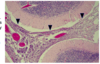
- micro
- acute inflammatory infiltrate (neutrophils) in subarachnoid space
- arrowheads are pointing to inflamed meninges in cerebellum
- gross
- creamy purulent exudate covering teh cerebral hemispheres and settles along the base of teh brain, around the cranial nerves and the openings of the fourth ventricle

What are possible sequelae of meningitis?
cranial nerve deficits
ischemic infarction (invasion of leptomeningeal vessels)
hydrocephalus (after the purulent exudate fibroses and clogs the fourth ventricle)
meningitis with no recognizable organism in culture or gram stain and no pus in CSF is considered … and is usually/…
acute aseptic meningitis
viral
Would you rather have bacterial meningitis or acute aseptic meningitis?
acute aseptic - less fulminant, is self-limiting
Does bacterial or aspetic meningitis have associated mental status changes?
bacterial
What is the MCC of acute aseptic meningitis?
non-polio enteroviruses
What are the morphologic findings of acute aseptic meningitis?
leptomininges/subarachnoid and perivascular spaces by lymphocytes (sparse or uneven distrib)
NO involvement of brain parenchyma
What are typical CSF findings of aseptic meningitis?
- increased WBCs (less than bacterial)
- low number neutrophils and only for 48 hours
- protein moderately increased
- glucose normal
- negative stains and cultures
What is a parenchymal infection of the brain?
viral encephalitis
almost invariably, encephalitis has meningeal inflammation as well. this is called…
if it was the spinal cord involved too, it would be called…
meningoencephalitis
encephalomyelitis
What are CSF findings in viral meningoencephalitis or encephalitis?
- WBC pleocytosis - neutrophil early, mononuclear late
- increased protein (as necrosis increases)
- normal glucose
- fungal and AFB cultures are negative
What are we looking at?

micro appearance of viral encephalitis
- later stages - predominatly lymphocytes and macrophages around blood vessels
- perivascular cuffing (left)
- activation and proliferation of microglia results in formation of ‘microglial nodules’ (right)
- with neuronophagia of infected cells
- necrotizing vasculitis is present in severe cases
- no viral inclusions
MC in teens and young adults, this form of encephalitis presents with alterations in mood, memory, and behavior
What area of the brain does it most often go after?
HSV-1 encephalitis
inferior frontal and temporal lobes (necrotizing and often hemorrhagic)
What type of encephalitis is more likely to affect the brainstem, is seen in adults presenting as meningitis and is more likely the cause of neonatal encephalitis?
HSV-2
Untreated neonatal HSV infection acquired at birth or transplacentally will present as what?
diffuse encephalitis with no prediliction for the frontal and temporal lobes
disseminated systemic disease with necrotizing encephalitis
Seen in immunosuppresion or congenital infections, presenting with microcephaly, often plymicrogyria, and blueberry muffin babies
What cell type will be infected?
CMV encephalitis
any type of cell in CNS, with Cowdry type A (owls eye) intranuclear inclusions
What is pathognomonic for rabies virus encephalitis?
Negri bodies - eosinophilic, well-circumscribed, cytoplasmic inclusion bodies seen in certain nerve cells, esp in pyramidal cells of the hippocampus
HIV microscopically will have…
multinucleated giant cells with no viral inclusions
What is the MCC of chronic bacterial meningitis?
mycobacterium tuberculosis (TB)
If the CNS has a fungal infection, what was the most likely route?
almost always inhaled spores then spread to the brain
What would be two fungal causes of chronic meningitis?
histoplasma capsulatum or cryptococcus neoformans
What fungal CNS infections are really pretty much only found in immunocompromised patients?
candida albicans
rhizopus, mucor
aspergillus fumigatus
What is the greatest risk factor for acquiring aspergillus?
neutropenia
What fungus will appear in the body as spherules?
coccidioides immitis
What fungal CNS infection will be mucicarmine positive?
cryptococcus neoformans
Is histoplasma encapsulated?
no
What am I looking at?

Negri bodies of rabies virus
What am I looking at?

cryptococcus neoformans on india ink stain (left) and mucicarmine stain positive (right)
What am I looking at?

disseminated cryptococcosis
setting of severe immunosuppression showing classic ‘soap bubble’ lesions
large colonies of cryptococci with thick mucoid capsules
What is this? How is it handled in labs?

coccidioides immitus
not opened in labs because of risk of infection
In toxoplasma gondii, what area of the brain is most often affected?
basal ganglia
shows ring enhancing lesions
Primary Amoebic Meningoenceophalitis is most often an infection of what?
How quickly is the onset?
Naegleria fowleri and Balamuthia mandrillaris
death within 2-3 days of symptoms
What am I looking at?

amoebic invasion
Acanthomoeba or balamuthia may cause…
how does this compare to PAM?
GAE - granuloma amoebic encephalitis
acantho - has cysts (unlike naegleria) and is only gets immunocompromised or debilatated host
balamuthia is in person with hx of soil exposure
What is special about prior disease pathology?
path is only in brain and no organism is associated - only misfolded proteins
no inflammation is present and CSF is normal
What is the abnormal protein that is no longer water soluble or degradable by proteinases?
PrP sc
What are we looking at?

Kuru plaques in the molecular layer of the cerebellum
bright pink-red eosinophilic aggregates of PrPsc protein