Neurulation and Development of the PNS Flashcards
(26 cards)
- process by which the neuroectoderm, surface ectoderm, and neural plate are made physically and functionally different
- ectoderm is made into the neuroectoderm and surface ectoderm
- the notochord induces neuroectoderm to form the neural plate (beginning in 3rd week)
neurulation

What is the inducer of neurulation?
notochord
Describe what is happening in these photos:

A: the neural plate is formed superior to the notochordal process
B. cells from the neural plate begin moving upward to create the neural folds and middle neural groove
C. the neural folds begin moving toward each other
D. somites are forming, surrounding the notochord; neural crest are forming on the superior portion of the neural folds; neural groove stays in place as anchor for formation of neural tube
E. the neural folds are nearly closed off to form the neural tube
F. the neural tube is completely closed off now, the neural crest cells get pinched off
TLDR: neural plate > neural folds > neural tube
- gastrulation ends w/ formation of the tail bud
- primary neurulation is complete w/ closure of caudal neuropore (at somite 31)
- the neural tube extends caudal into sacral and coccygeal regions
- the process of _______ _________, is the formation of the neural tube from the tail bud
- closing of caudal neuropore is merged w/ the forming tail bud
- caudal-most notochord, neural crest cells, and somites develop from the tail bud
secondary neurulation
What development are somites important for?
- vertebral column
- axial musculature
- trunk development
What is the role of the tail bud in development?
- the tail bud condenses into a solid mass, the medullary cord
- it then cavitates, forms the lumen, and merges w/ central canal of neural tube
(neural crest cells arise from the roof the neural tube and migrate)
- lateral tail bud cells segment and form caudal somites
(caudal notochord grows into the sacral, coccygeal, and tail regions)

- neuroectodermal cells positioned along dorsal neural folds (bilaterally along entire cranial-caudal neural tube)
- produced by the meduallary chord (tail bud)
- undergo epithelial-mesenchymal-transition (EMT)
- lose contact and migrate ventrally along length of embryo, giving rise to a number of derivatives
- they start migrating before neural tube is completely closed, and finish migrating once it is closed
- vertebrate invention is evolutionarily important
- activate and respond to genetic signaling for differentiation of tissue
neural crest cells

What are the derivatives of neural crest cells?
- in general: cranial (head, neck, jaw structures), cardiac (septation of the heart), trunk, and sacral neural crests
(there is not a single organ/viscera/etc. that does not have some form of derivative of neural crest cells)
- melanocytes, odontoblasts, Schawnn cells (myelinate nerves)
- spinal and autonomic ganglia
- enteric nervous system
- suprarenal (adrenal) medulla
- a class of pathologies, or disorders, in vertebrates, including humans, that result from abnormal expression, migration, differentiation, or death of neural crest cells (NCCs) during embryonic development
- examples: Goldenhar syndrome (hemifacial macrosomia), TCS (mandibulofacial dystosis), Hirschsprung disease, micrognathia, piebaldism
neurocristopathies
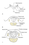
- membranes covering the spinal cord and brain
- develop from the neural crest cells and mesenchyme between 20-35 days
- migrate and surround neural tube to form the primordial meninges, meninx primitiva (primitive meninges)
- vertebrae form in segments and fuse together
meninges

Describe the process of the formation of ectomenix and endomenix:
- primitive meninx differentiates into the ectomeninx and endomeninx from 34-48 days
- between 45-60 days: ectomeninx becomes more compact and forms spaces for future venous sinuses; endomeninx becomes more reticulated and subarachnoid space/cisterns appear
- spinal ectomeninx dissociates from the vertebral bodies and gives rise to the spinal epidural space (essential for epidurals)

Describe the process of neural tube closure:
- does not occur simultaneously the length of the neural tube
- cranial regions are more advanced than caudal regions (still undergoing gastrulation)
- results in the formation of cranial and caudal neuropores
- neural tube closure is initiated at several locations along the AP axis
- various closure sites are involved in neural tube closure
- failure at any of these sites can result in this condition (cranial and/or spinal)
- causes: genetic, nutritional (commonly folate deficiency), and environmental factors
- examples: spina bifida (most common), anencephaly (not viable), meroencephaly (not viable), encephalocele (w/ or w/o brain tissue, prognosis depends on brain tissue involvement), and craniorachischisis (stillborn, premature, fatal)
neural tube defects
(do not need to know numbering or sites of closure)

- most common neural tube defect, happens at closure site 5, most proximal to posterior neuropore
- occulta: most mild form, unfused neural arch, no spinal cord involvement; signs may be a tuft of hair, dermal sinus, or pigmentation over defect
- meningocele: no spinal cord involvement, bulge present w/ subarachnoid space containing CSF
- meningomyelocele: spinal cord involvement, bulge is closed off
- myeloschisis: open spinal cord, prognosis in terms of paralysis is significant
spina bifida
Where is the CNS (brain and spinal cord) and PNS (cranial nerves/ganglia, spinal nerves/ganglia, autonomic nerves/ganglia, and enteric nervous system) derived from?
- CNS: neural tube
- PNS: neural tube, neural crest cells, neuroectoderm, ectodermal placodes
- neural tube is divided into dorsal and ventral regions, separated by ______ ______
- neural tube is comprised of a germinal ___________ which spans the full width of the neural tube from lumen/canal to outer surface
- sulcus limitans
- neuroepithelium (will give rise to a range of cell types in brain and spinal cord)

What are the different zones in neural tube differentation and what do they give rise to?
- ventricular zone: stem cells positioned closest to lumen, will divide and migrate
- mantle/intermediate zone: migrating cells that undergo differentiation into neurons and/or glia; extend axons away from lumen toward outer surface
- marginal zone: poor in neuronal cell bodies; contains axons that will eventually be myelinated

Diagram of cells of the nervous system and their derivatives:

What happens to the mantle layer at the end of week 4?
- mantle layer reorganizes into the alar (dorsal) plate and basal (ventral) plate
- the plates are connected by roof plate and floor plate
- plates extend entire length of the spinal cord
- undergo differentiation due to signaling from roof plate and floor plate

How does sonic hedge hog direct dorsal-ventral specification of the neural tube?
- SHH has a gradient that directs neuron identity
- high levels of SHH = motor neurons induced
- low levels of SHH = multiple interneurons induced, dependent on decreasing SHH
- SHH activates downstream targets NKX2.2 and NKX6.1 for ventral neuron formation

What signaling counteracts SHH?
- BMP/TGF-beta signaling counteracts SHH gradient
- BMP4 activates PAX3 and PAX7
- these genes are responsible for induction of dorsal association neurons
(in general, motor neurons form before sensory neurons)
What cell derivatives form the lateral horns?
- most dorsal cells of the basal plate undergo segregation
- will form the intermediolateral cell columns
- form the lateral horns
- T1-L3: visceral motor neurons of sympathetic division (ANS)
- S2-S4: visceral motor neurons of parasympathetic division (ANS)

What are the derivatives of the peripheral nervous sytstem (sensory and motor neurons of the head, trunk, extremities) during development?
- head: neural crest cells and ectodermal placodes form sensory ganglia of the head
- trunk: neural crest cells form sensory neurons in dorsal root ganglion, Schwann cells, and enteric nervous system
Describe the development of the ventral root, dorsal horn, and caudate equina:
- axons exiting ventral horn pass through cranial sclerotome, forming ventral root
- ventral root then joined by axons from the lateral horn
- neural crest cells form the dorsal root ganglia sensory neurons
- axons extend from the dorsal root ganglia, forming the dorsal horn
- uneven development of the meninges and spinal cord leads to spinal cord “ending” higher than the vertebral end, which is why the caudate equina exists


