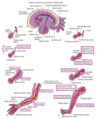
Development of the Limbs Flashcards
(31 cards)
What are the derivatives of the following:
- musculature:
- bones:
- tendons:
- musculature: paraxial mesoderm > myotome
- bones: lateral plate mesoderm > somatic layer
- tendons: lateral plate mesoderm > somatic layer
(tendons in limbs come from lateral plate mesoderm, whereas other tendons come from paraxial mesoderm > somites > syndetome)

When do the limb buds start forming?
What is the importance of the apical ectodermal ridge (AER)?
- formation starts day 24 for upper limbs and days 25-26 for lower limbs
- apical ectodermal ridge (AER) is a thick band of ectoderm, required for proliferation and outgrowth of mesenchyme from the lateral plate mesoderm (causes limb bud to bulge outward)
What are the regions of outgrowth of limb buds and what structures do these regions contribute to?
- stylopod: humerus or femur
- zeugopod: radius and ulna; tibia and fibula
- autopod: carpals, metacarpals, and phalanges; metatarsals and phalanges

What are the axes of the limb bud and their orientation?
- proximal to distal (shoulder to digits)
- cranial to caudal (hallux to phalanx V, aka thumb to pinkie)
- dorsal to ventral (dorsum to palm)

What gene is the outgrowth of AER induced by?
fibroblast growth factor 10 (FGF10)
Describe the FGF10 and FGF8 feedback loop:
- FGF10 induces the outgrowth of AER
- FGF8 is secreted by AER and is a downstream target of FGF10, creating a positive feedback loop
- FGF10 essentially turns on FGF8
- FGF signaling is essential for the initiation of limb development and regulates the outgrowth of the proximodistal axis
TLDR: FGF8 = outgrowth; FGF10 turn on FGF8
What will occur if:
- AER is completely removed or removed early on:
- AER is removed at later stage of development:
- AER is transplanted where it shouldn’t be:
- AER is completely removed or removed early on: arrests limb development
- AER is removed at later stage of development: loss of more distal elements of limb
- AER is transplanted where it shouldn’t be: may result in supernumerary limbs

- limb anomaly
- absence of part of a limb
- intermediate to late loss of FGF signaling
meromelia
- limb anomaly
- absence of an entire limb
- early loss of FGF signaling
amelia
- limb anomaly
- loss of long bones w/ hands and/or feet attached close to the body
- partial loss of FGF signaling or HOX disruption due to thalomide (anti-nausea med they used to give to pregnant women)
phocomelia
- limb anomaly
- absence of digits
- late loss of FGF signaling
adactyly
- limb anomaly
- split-hand or split-foot anomaly
- 1.5:100,000 live births
- partial absence of FGF8 from AER
- usually accompanied by other conditions: ectoderm dysplasia and cleft lip/palate syndrome
ectrodactyly
(EEC: ectrodactyly, ectodermal dysplasia, cleft lip/palate syndrome)
How is digit function differentiated, in what axis does this occur and by what gene?
- zone of polarizing activity (ZPA) exists in the cranial-caudal axis
- sonic hedgedog (SHH) is expressed in the ZPA and is sufficient to provide ZPA functional differentiation
(restriction of cell fates in the limb occurs before limb bud outgrowth)
(SHH also responsible for neural tube patterning and motor neuron differentation)

What would happen if ZPA is grafted to cranial limb bud mesoderm where it should not be?
duplicated digits would emerge (mirrored)
- presence of supernumerary digits
- OE of SHH establishes 2nd ZPA
- inherited as a dominant trait, 2:1000 live births
- extra digit is incompletely formed and lacks normal muscular development
polydactyly
What group of genes regulate limb specificiation?
- HOX genes (HOXA-HOXD)
- regulate the cranial-caudal and proximodistal axes
- distinct clusters are expressed in specific regions of the limb buds at specific times
- organization of physical regions AND the timing this development occurs
What are the nested patterns of expression with HOXD genes?
- D9: scapula
- D9-10: stylopod (humerus)
- D9-11: zeugopod (ulna, radius, and proximal carpals)
- D9-13: autopod (metacarpals and phalanges)

- limb anomaly
- shortening of the fingers and toes due to unusually short bones
- genetic changes in the HOX13 gene or PTHLH gene (parathyroid hormome like hormone)
- autosomal dominant inheritance
brachydactyly
How is the dorsal/ventral axis established within limbs?
- Wnt7a is expressed in the dorsal ectoderm, and is a primary regulator of dorsal fates (overexpression results in dorsalized structures, KO mice have ventralized paws)
- Engrailed (En1) is expressed in the ventral ectoderm, prevents the expression of Wnt7a in the ventral part of the limb and restricts positioning of the AER to establish the dorsal-ventral axis
How, why, and when are digits formed?
- how: removal of interdigital mesenchyme between digital rays by apoptosis;
requires bone morphogenetic protein (BMP) signaling: increased signaling > increased cell death, decreased signaling > decreased cell death (webbed digits)
- why: frees the digits and allows mobility
- when: digital rays form by week 6, foot plates form by week 7, separated digits formed by week 8
- limb anomaly
- fusion of digits because digital rays fail to develop
- 1 in 2,000-3,000 live births
- two different types: cutaneous (disruption in BMP signaling, lack of apoptosis) and osseous (failure of notches to develop between digital rays, autosomal dominant inheritance, HOXD**13 mutation)
syndactyly
How do mygenic precursors go on to develop into limbs as we know them?
- mygenic precursors from the dermomyotome (paraxial mesoderm) migrate into the limb bud
- differentiate into myoblasts, which aggregate and form dorsal/ventral muscles masses in each limb bud

What do dorsal and ventral muscle masses give rise to?
- dorsal: extensors/supinators of upper limb and extensors/abductors of lower limb
- ventral: flexors/pronators of upper limb and flexors/adductors of lower limb
*limb tendons arise from somatic layer of lateral plate mesoderm*

When does limb innervation begin, what is the order of cells to be innervated, and what are the dorsal/ventral structures innervated by?
- begins in week 5
- motor neurons innervate first, then sensory axons, and Schwann cells (from NCC) myelinate the nerves
- dorsal mass muscles are innervated by dorsal branches of ventral rami
- ventral mass muscles are innervated by ventral branches of ventral rami


