Epithelia Flashcards
What are the roles of epithelial tissue?
Characteristics?
What is the think extracellular, felt-like sheet epithelial cells rest on?
Epithelium that is involved in secretion is arranged as:
- roles: physical protection, permeability, secretion, and sensation
- characteristics: cellularity, polarity, attachment (to basement membrane), avascular (get nutrients through diffusion), innervation, and regeneration
- rests on: basement membrane
- secretory glands
Two parts to this card:
Part 1: cells show polarity; organelles and proteins are ________ distributed
Part 2: picture:

- unevenly
- (top to bottom of picture): apical (free) surface, lateral surface, and basal surface
- type of intercellular junction
- impermeable, allows cells to function as a barrier (especially if high number of these are present)
- encircle cells near most apical surface
- proteins: occludins, claudins
- similar to a draw string bag, the higher number of these junctions present, the lower the permeability will be
tight/occluding junctions

- type of intercellular junction
- fluid-filled channels that connect opposed cells
- proteins: connexin aggregates
gap junction (communicating junction)

- type of intercellular junction
- multiple types: adherens, desmosome, hemidesmosomes
- adherens: lateral adhesions involving cadherin:actin interations
- desmosome: lateral adhesions involving cadherins:intermediate filament interactions
- hemidesmosomes: basal adhesions involving intergrins:intermediate filament interactions
anchoring junctions

- clinical relevance of intercellular junctions
- bacteria that cause food poisoning target _____ _______ in the intestine which impairs the junction and causes loss of tissue fuid into intestinal lumen
- H. pylori causes gastric ulcers by binding to _____ _______ in the stomach and increasing permeability, loss of gastric contents (acid) causes the ulcers
tight junctions
- clinical relevance of intercellular junctions
- autoimmune disease pemphigus vulgaris causes abnormal _________ function which reduces cell-cell adhesion and causes blisters in the oral mucosa
desmosomes
- specialized sheet of extracellular material that is located adjacent to basal domain
- selective barrier between tissues permits diffusion of nutrients
- on tissue stains, it can help identify epithelial tissue boundary

basement membrane
- type of apical specialization
- cytoplasmic processes containing an actin core and covered in plasma membrane
- specialized for absorption
- number/shape correlate to the cell’s absorptive capacity
- celiac disease: loss of ______ on absorptive cells in SI
microvilli
- type of apical specialization
- microvilli of unusual length, long and less mobile than cilia
- have an actin core and increase surface area for absorption/secretion
- restricted locations: epididymis (absorption/secretion) and hair cells of inner ear (structural, bend to initiate hearing)
sterocilia
- type of apical specialization
- long, highly motile stuctures containing internal arrays of microtubules (up to 10mm long, 300+ may be present)
- motile: beat in a wave-like fashion to propel substances across the tissue
- primary: immotile, function as chemosensors, osmosensors, and mechanosensors
- nodal: embryonic, have role in L/R axis determination
(critical in airways)
cilia
How do you classify tissue type?
- Determine the amount of layers present: 1 layer = simple, 2+ layers = stratified
- Determine the shape of the cells on the apical surface: squamous cells are flat, cuboidal cells are cube shaped, and columnar cells are column shaped
- tissue type
- location: lining of blood and lymphatic vessels (endothelium), lining of serous membranes (mesothelium), lining alveoli in lungs, loop of Henle in kidneys, various ducts
- function: exchange, barrier, and lubrication
simple squamous

- tissue type
- location: kidney tubules, glands and associated ducts, terminal bronchioles, covering the ovaries
- function: absorption, barrier, secretion
simple cuboidal

- tissue type
- location: auditory tubes, uterus, oviducts, stomach, SI/LI, gallbladder
- function: absorption and secretion
- nucleus can be round/egg shaped, and the cells can be ciliated
simple columnar

- tissue type
- location: lining of nasal cavity, pharynx, trachea, bronchi
- function: absorption/secretion, debris and particulate movement
- restricted to airways, mucus secretion
pseudostratified columnar ciliated

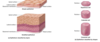
- tissue type
- pseudostratified, transitional epithelium
- location: urinary bladder, ureters, urethra
- function: barrier, distensible property, keep out toxic waste
urothelium
(top of cartoon pic is bladder empty, bottom of cartoon pic is bladder full)

- tissue type
- location: oral cavity, portions of the pharynx, esophagus, anus, vagina, urethra, cornea
- function: barrier, protection
(areas w/ constant/frequent abrasion)
nonkeratinized stratified squamous

- tissue type
- location: epidermis of the skin
- function: barrier, protection
- kertain filaments on outermost surface gives skin a bit of water proofing
keratinized stratified squamous

- tissue type
- location: sweat glands/ducts, ovarian follicles, salivary gland ducts
- function: barrier, passageway
- need to be able to release secretory product w/o losing it all completely
stratified cuboidal/columnar

Epithelial decision tree (study guide):

- type of membrane
- epithelial tissue that secretes mucus
- lines many body cavities and tubular organs, including the gut and respiratory passages
mucous membrane
- type of membrane
- epithelial tissue that lines internal body cavities
- forms a smooth, transparent, two-layered membrane
- lubricated by fluid derived from serum to reduce friction
- includes peritoneum, pericardium, and pleura
- mesothelium: simple squamous epithelium that comprises part of a _____ _______
serous membrane
- Epithelial cells may produce & secrete a product as individual cells or as specialized organs, _____
- Glands are classified as ______ or _______ according to how their products are released
- Classified by arrangement & shape of secretory cells & ductal elements
- Signals released via _______ or _______ signaling
- glands
- exocrine, endocrine
- paracrine, autocrine
- type of gland
- single, secretory cells distributed among non-secretory cells
- globlet cell: mucus-secreting cell lining the intestines and respiratory tract
unicellular glands

- type of gland
- multicellular glands comprised of secretory cells grouped as an acinus (means grape)
- product is secreted into a system of ducts for release
- paranchyma (secretory cells): functional tissue of an organ (not the CT and other supporting tissues)
- stroma: connective tissue support of secretory units; septa partition the gland into separate lobules; may enclose entire gland as a capsule
exocrine glands
(secretions produced in secretory portions (acinus) and released in conducting portions (duct))

- type of exocrine gland
- secretion is delivered in membrane-bound vesicles to apical surface and undergo exocytosis
merocrine

- type of exocrine gland
- secretion accumulates within a cell leading to apoptosis
- secretion and cell debris are released
holocrine

- type of gland
- release of the apical portion of the cell, surrounded by cytoplasm within a plasma membrane
- example: mammilary glands
apocrine

Name these glands:

(from left to right)
- simple tubular
- simple coiled tubular
- simple branched tubular
- simple acinar
- simple branched acinar
Name these glands:

(from left to right)
- compound tubular
- compound acinar (salivary gland)
- compound tubuloacinar (salivary gland)
Tissue type table: (study guide)

