Microanatomy Exam II Material Flashcards

1
Q

Growth Plate: Identify the zones

A
  1. Zone of Reserve Cartilage
  2. Zone of Proliferation
  3. Zone of Hypertrophic Cartilage
  4. Zone of Calcified Cartilage
  5. Zone of Ossification
2
Q

Identify the structure.

A

Growth Plate

3
Q

What type of ossification is occuring here?

A

Intramembranous Ossification

4
Q

Identify the neural tube and the notochord in this H&E section of a chick embryo

A
5
Q

Identify the structures.

A

Mesenchymal cells - stem cells of bone and cartilage

6
Q

What are we looking at here?

A

Uncondensed mesenchymal cells

7
Q

Identify the area where perichondrium is located

A
8
Q

Identify the type of cartilage.

A

Hyaline

9
Q

Identify the type of cartilage.

A

Elastic

10
Q

Identify the type of cartilage.

A

Fibrocartilage

11
Q

What are the arrows pointing to in this hemisected equine tarsus?

A

Articular hyaline cartilage - DOES NOT HAVE PERICHONDRIUM

12
Q

Identify the zone of proliferation in this growth plate of hyaline cartilage.

A
13
Q

Identify the type of cartilage.

A

Fibrocartilage

14
Q

Identify the structures in fibrocartilage

A

Chondrocytes in lacunae

15
Q

Identify the portion that is spongy bone

A
16
Q

Identify the portion that is compact bone

A
17
Q

Identify the osteoblasts in this woven bone

A
18
Q

Identify the circled structure.

A

Osteocyte in lacuna

19
Q

What are we looking at?

A

Neuron - see the cell body (parikaryon) containing nucleus, nucleolus, dendrites, and axon

20
Q

What is this grainy basophilic substance?

A

Nissl substance - dark due to presence of RER and ribosomes

21
Q

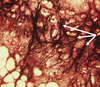
What is the pigment that the top arrow is pointing to?

A

lipofuscin

22
Q

What type of neuron is this?

A

Pseudounipolar

23
Q

What are we looking at here and what does it do?

Where is it located?

A

The golgi organ. It is a proprioceptive sensory receptor/senses stretching

It is located at the insertion of skeletal muscle fibers into the tendons

24
Q

What is the name for this CNS counterpart of the fibroblast?

A

Astrocyte

25
What is the name of this structure (found in the CNS)?
Oligodendrocyte
26
What are we looking at here (in the CNS)?
Microgliocytes
27
Identify the structure.
Choroid plexus
28
Would we find this in the CNS or the PNS?
CNS (note the oligodendroglia)
29
Would we see this in the CNS or the PNS?
PNS (note the Shwann cells)
30
Which part of the brain is this?
Cerebellum
31
What is wrong with the animal that this section cerebellum was taken from?
Cerebellar hypoplasia - granular layer is empty compared to normal
32
What would you call this collection of neurons in the CNS?
a 'nucleus'
33
Identify the circled structures.
motor neurons
34
Which image belongs to an autonomic ganglion and which belongs to a dorsal root ganglion?
Image #1 is an autonomic ganglion. Image #2 is a dorsal root ganglion. If you look at the neuronal cell body, the nucleus is central in a dorsal root ganglion. The nucleus is eccentric in autonomic ganglion.
35
What is this structure found in the small intestine that helps to control peristaltic movement of the GI tract?
Myenteric Plexus
36
Identify the circled structure (this is from an ear)
Ceruminous gland
37
What helps move an egg through the uterine tube?
cilia and muscular contractions
38
where is the acrosome located?
the head of the sperm
39
where does capacitation occur?
uterus, or uterine tubes
40
What is the site of fertilization?
fellopian tubes
41
What maintains the blood testes barrier?
tight junctions between neighboring Sertoli cells
42
In early fetal testes, which cells secrete APDH?
Sertoli cells
43
This duct gives rise to fellopian tube, uterus, and cervix
the mullerian duct
44
This duct gives rise to male anatomy (testes, epididymis, etc..)
the mesonpheric duct
45
Fibrocartilage lacks \_\_\_\_\_\_\_\_\_\_\_\_, and therefore is incapable of regeneration
perichondrium
46
Where would you find fibrous cartilage?
Fibrous cartilage can be found at intervertebral discs, menisci, tendon insertions, and pubic symphysis
47
Where would you find elastic cartilage?
Pinna, spiglottis, external auditory canal, and layngeal cartilage
48
Where would you find hyaline cartilage?
Fetal bones, growth plates, articular cartilage, costochondrial junctions, nasal septum/larynx/trachea/bronchii
49
Appositional growth in cartilage: what is happening?
chondroblasts differentiate into chondrocytes in the chondrogenic layer of the perichondrium. This results in an increase in the diameter or thickness of the cartilage.
50
interstitial growth in cartilage: what is occuring?
chondrocytes divide by mitosis to form isogenous nests, resulting in an increase in cartilage mass
51
From which cell type do both cartilage and bone originate?
mesenchymal cells
52
What are the 3 types of cartilage?
hyaline cartilage, fibrocartilage, and elastic cartilage
53
What is occuring in endochondral ossification?
bone is laid down on a scaffold of pre-formed cartilage, cartilage is removed, and bone remains
54
What is occuring in intramembranous ossification?
mesenchymal cells condense and differentiate into osteoblasts, form bone
55
What are the two basic osteogenic processes?
intramembranous and endochondral
56
What is the zone of the growth plate where chondrocytes multiply and form columns of chondrocytes
Zone of proliferation
57
What is the zone of growth plate comprised of hyaline cartilage and chondrocytes surrounded by a matrix?
Zone of reserved cartilage
58
This is the zone of the growth plate composed of enlarged vacuolated chondrocytes.
zone of hypertrophy
59
What are the two types of bone?
woven (immature) and lamellar (mature)
60
Bone developing from pre-existing cartilage (mesenchymal cells --\> hyaline --\> bone) Process is called:
endochondral ossification
61
What does calcitonin do with regard to serum calcium levels?
Calcitonin decreases serum calcium levels by promoting bone formation
62
What does parathyroid hormone (PTH) do with regard to serum calcium levels?
PTH increases serum calcium levels by increasing osteolysis
63
neurovascular channels in conpact bone are called:
Haversian canals
64
The only bone cells capable of mitosis are:
osteoprogenitor cells
65
These are mature bone cells in lacunae; use gap junctions to transfer nutritional needs to cells; incapable of mitosis:
osteocytes
66
These are immature bone cells only on the surface of bones; secrete collagen and ECM; called osteoid
osteoblasts
67
bone and cartilage are both derived from which germ layer?
mesoderm
68
What are the layers of meninges?
dura mater (outermost layer) arachnoid mater subarachnoid space pia mater
69
These for part of the BBB, form scar tissue, transport nutrients, and are important in antigen presentation:
astrocytes
70
supportive cells that surround neurons are called:
glial cells
71
The nerve layer in the GI tract that regulates peristaltic movement:
Myenteric plexus
72
This is the site of CSF production:
the choroid plexus
73
How often is CSF renewed?
3 times/day
74
These are small nerve cell bodies in the lateral horns:
autonomic neurons
75
These neurons are found in the ventral horns, innervate skeletal muscle, and are only present in grey matter
motor neurons
76
motor neurons are considered what type of neuron?
multipolar
77
What is the principle control center of the pituitary gland?
hypothalamus
78
In which layer of the cerebral cortex will we find purkinje cells?
the ganglionic layer
79
what are the layers of grey matter in the cerebellum?
molecular layer (outermost) ganglionic layer granular layer
80
what is the name for the white matter found in the cerebellum
arbor vitae (tree of life)
81
Which part of the brain is associated with coordination of body movement?
cerebellum
82
Which portion of the brain is associated with memory?
hippocampus
83
These cells surround ganglionic neuron cell bodies and assist in repair
amphicytes (satellite cells)
84
What is the approximate ratio of glial cells to neurons?
approximately 50:1
85
What is the resident macrophage in the CNS?
microgliocytes when active, they are called 'gitter cells'
86
An inhibitory synapse contains which neurotransmitter?
GABA
87
A stimulatory synapse contains which neurotransmitter?
Acetycholine
88
This stimulates muscle contraction when it is releasd into synaptic clefts:
acetylcholine
89
What is the composition of myelin?
~75% lipid and 25% protein
90
\_\_\_\_\_\_\_\_\_\_ myelinate in the CNS.
oligodendrocytes
91
\_\_\_\_\_\_\_\_\_\_\_\_\_ myelinate in the PNS.
Shwann cells
92
What is the location of grey matter in the brain? Spinal cord?
brain = located peripherally spinal cord = located centrally
93
What is the lcation of white matter in the brain? Spinal cord?
brain = located centrally spinal cord = located peripherally
94
This is a granular substance composed of rER and free ribosomes
Nissl substance (or Nissl body)
95
This is the region between cell bodies in the grey matter of the brain and spinal cord
neuropil
96
How can you tell the difference between a sensory ganglia and an autonomic ganglia on an H&E slide?
Sensory ganglia have a central nucleus Autonomic ganglia have an eccentric nucleus
97
What is the name for a group of nerve cell bodies in the CNS?
a nucleus
98
What is the name for a group of nerve cell bodies in the PNS?
a ganglia
99
What are the developmental stages from zygote through gastrulation?
zygote --\> morula --\> blastula --\> early gastrula --\> late gastrula
100
What does the myotome give rise to?
skeletal muscle
101
What induces formation of the neural tube?
the notocord
102
Which cell type layer gives rise to the ectoderm?
Epiblast cells
103
What is responsible for the formation of fetal placental membranes?
The outer cell mass (aka trophoblast)
104
Which germ layer gives rise to the formation of the notochord?
mesoderm
105
Which germ layer gives rise to muscle and CV system?
mesoderm
106
Which germ layer gives rise to the GI tract and respiratory system?
endoderm
107
Which germ layer gives rise to epidermis and nervous tissue?
ectoderm
108
What is a blastomere?
type of cell produced by zygote cell division after fertilization
109
What is the teratogenic influence on development @ the predifferentiation stage?
the embryo dies
110
What are the four divisions of mesoderm?
axial, intermediate, lateral, paraxial
111
At what stage does the zona pellucida shed?
At the blastula stage
112
Which components of the inner ear contain **endolymph**?
The membranous labyrinth: * utricle and saccule * semicircular ducts * cochlear ducts * sensory structures!
113
Which components of the inner ear contain **perilymph**?
Bony labyrinth * vestibule * semicircular canals * cochlea * these are the outer (peripheral) structures, surrounding the internal (endo) sensory structures
114
Which structure in the ear is responsible for detecting angular acceleration?
Crista ampullaris
115
Which structure(s) in the ear is/are responsible for detecting head position and linear movement?
utricle and saccule
116
What are the three bones and two muscles of the ear?
bones: malleus, incus, stapes muscle: tensor tympani m. and stapedius m.
117
What structure in the ear is responsible for pressure equalization?
auditory tube
118
What fluid do you find in the utricle and saccule?
endolymph
119
What fluid do you find in the semicircular ducts?
endolymph
120
What fluid do you find in the semicircular canals?
perilymph
121
What is the structure that brings hair cells back into position after displacement?
kinocilia
122
What structure produces endolymph?
stria vascularis
123
What part of the cochlea responds to high frequency sounds?
the base of the cochlea
124
What do primordial germ cells give rise to?
yolk sac endoderm
125
Identify the ectoderm portion
126
Identify the endoderm portion:
127
Identify the mesoderm portion:
128
Identify the neural fold:
129
What stage of folliculogenesis are we looking at here? (What type of follcle)
PRIMORDIAL FOLLICLES: Primary oocytes surrounded by a layer of squamous follicular cells
130
What stage of folliculogenesis are we looking at? (What type of follicle)
Primary follicle: Zona pellucida acts as an immunologic protective barrier for the developing egg and early embryo
131
Identify the zona pellucida.
132
Identify the granulosa cells
133
Identify the theca interna cells
134
What stage of folliculogenesis are we looking at? (What type of follicle?
Secondary (Secretory) follicle: Primary oocyte with Zona pellucida and Corona radiata cells
135
What stage of folligulogenesis are we looking at? (What type of follicle)
Tertiary (Graafian) = Mature Follicle
136
Identify the circled structure:
The corpus hemorrhagicum
137
Identify the structure that spans the length of the arrows. What does it produce?
Corpus luteum (yellow body). Produces progesterone (and some estrogen)
138
What is the importance of progesterone (produced by the corpus luteum)?
Progesterone helps to maintain the pregnancy, stimulate mammary development, and locally down regulates the immune system
139
What is the name of this connective tissue scar that remains after regression during diestrus?
corpus albicans (white body)
140
What is the name for a follicle that is not selected to complete maturation?
This is called an **atretic follicle**. If a follicle is not selected to complete maturation, it will undergo a programmed detruction, this is the fate of many follicles
141
In this section of testes and epididymis, which white number represents the seminiferous tubules. Hard to see the numbers. Just squint a little.
**1.**
142
In this section of testes and epididymis (10x), which white number represents the efferent ductules?
3.
143
In this section of testes and epididymis (10x), which white number represents the epididymal duct?
4.
144
In this high magnification section of testes and epididymis, what are the arrows pointing to?
interstitial cells of leydig
145
In this high magnification section of testes and epididymis, what are the arrows pointing to?
seminiferous tubules
146
What is the circled structure in this cross-section of a sperm tail?
axoneme
147
What forms bilaminar disc of epiblast and hypoblast; establishes dorsal & ventral axis?
the embryoblast (or inner cell mass)
148
What forms fetal part of placental membranes?
trophoblast (or outer cell mass)
149
gastrulation begins with the formation of the \_\_\_\_\_\_\_\_\_\_\_ in the epiblast.
primitive streak
150
What are the arrows pointing to in this section of elastic cartilage in the epiglottis?
Elastic fibers