Maxillo-facial Trauma 01-22, 24 Flashcards
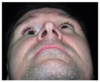
An 18-year-old man presents with right periorbital edema and ecchymosis after an all-terrain vehicle collision. Physical examination shows enophthalmos, diplopia, and pain with eye movements. When asked to look upward from forward gaze, there is upward gaze restriction. A photograph is shown. Which of the following locations is most likely fractured?
A) Greater wing of sphenoid bone
B) Lamina papyracea of ethmoid bone
C) Orbital plate of frontal bone
D) Orbital process of maxillary bone
E) Posterior crest of lacrimal bone

The correct response is Option D.
Limitation with vertical gaze, as described in the vignette, is indicative of extraocular muscle (EOM) entrapment.
The lamina papyracea of the ethmoid bone contributes to the medial orbital wall. Fractures of the medial wall may result in medial rectus entrapment and restriction with lateral gaze. Such injuries can involve the ethmoidal/sphenoidal sinuses.
The greater wing of the sphenoid bone contributes to the lateral orbital wall. Fractures of the lateral wall are less common due to its increased strength, but at the wings of the sphenoid, bone can have serious effects on contents of the superior orbital fissure and may even involve the intraorbital portion of the optic (II) nerve.
The orbital plate of the frontal bone contributes to the superior orbital roof. Fractures of the superior roof may result in superior rectus and/or oblique entrapment. Such injuries can involve the frontal sinus, frontal lobe, supraorbital nerve, and/or supratrochlear nerve (resulting in loss of sensation of the forehead and upper eyelid).
The orbital process of the maxillary bone contributes to the inferior orbital floor. Fractures of the inferior floor may result in inferior rectus/oblique entrapment and restriction with upward gaze. Such injuries can involve the maxillary sinus and/or infraorbital nerve (resulting in malar and superior alveolar numbness).
The posterior crest of the lacrimal bone contributes to the medial orbital wall. Fractures of the medial wall may result in medial rectus entrapment and restriction with lateral gaze. Such injuries can involve the ethmoidal/sphenoidal sinuses.
An otherwise healthy 74-year-old man presents with traumatic brain injury from an open comminuted anterior and posterior table frontal sinus fracture sustained in a motor vehicle collision. Persistent clear fluid rhinorrhea is observed when the patient is upright. The patient is initially medically unstable, and surgical intervention is delayed by 5 days. Which of the following factors represents the greatest increase in risk for a central nervous system infection?
A) Cerebrospinal fluid leak
B) More than 48 hours before operative repair
C) Open fracture injury
D) Patient age
E) Presence of traumatic brain injury
The correct response is Option B.
There are many algorithms for treating frontal sinus fractures. Unfortunately, many of these patients have other injuries that may limit their surgical options. Conservative management has been shown to be successful at managing comminuted, displaced fractures, pre-injury comorbidities, or those with cerebrospinal fluid (CSF) leak. Factors that are not associated with an increased risk for serious infection include: preoperative CSF leak, persistent CSF leak, surgical procedure performed, age, gender, and penetrating or open injury. Factors that do impact the risk for serious infection include: greater than 48 hours from injury to operating room intervention, use of CSF catheter diversion, and soft-tissue infection.
A 20-year-old man has severe hypotension and bradycardia after sustaining multiple facial fractures in a motor vehicle collision. He has no other injuries. Repairing which of the following structures will most likely improve the patient’s symptoms?
A) Mandible
B) Mental nerve
C) Nasal bone
D) Orbital floor
E) Zygomatic arch
The correct response is Option D.
The oculocardiac reflex can precipitate marked bradycardia and hypotension in the setting of trauma with significant orbital and maxillofacial injury. Prompt identification and management with vagolytic agents or definitive surgical intervention may prevent morbidity or mortality. Patients who sustain maxillofacial trauma involving the orbit, most commonly the orbital floor, are at risk for developing the oculocardiac reflex. These patients tend to be young, and common symptoms include nausea and vomiting. The oculocardiac reflex is not static and may evolve during a patient’s clinical course.
Elderly patients are more likely than younger patients to have which of the following facial fractures?
A) Nonoperative Le Fort fracture from a bicycle accident
B) Nonoperative mandible fracture from a motor vehicle collision
C) Nonoperative maxillary fracture from a fall
D) Operative nasal fracture from interpersonal violence
E) Operative orbital fracture from a workplace injury
The correct response is Option C.
Elderly patients are more likely to sustain injury from falls than younger patients and are less likely to be in fights. Nonoperative maxillary fractures and nasal bone fractures are more common in elderly patients, and mandible fractures are more common in younger patients.
A 50-year-old man receives preoperative radiation therapy for a large calvarial osteosarcoma that will require a 9-cm2 craniectomy. Which of the following is the most appropriate material to use for reconstruction?
A) Bone allograft with bone morphogenic protein (BMP)
B) Calcium phosphate
C) Polyether ether ketone (PEEK) implant
D) Titanium mesh
The correct response is Option C.
Any calvarial defect greater than 6 cm2 should be reconstructed. Titanium mesh can be used for craniectomy reconstruction; however, radiologic surveillance of disease recurrence can be challenging with the degree of artifact seen on imaging. Polyether ether ketone (PEEK) implants are radiolucent and therefore do not produce artifact on surveillance imaging and can be customized based on the size of the planned resection. Bone allograft with bone morphogenic protein (BMP) is contraindicated in cases of malignancy since BMP is a growth factor that induces bone formation and is involved in tumorigenesis. Calcium phosphate can be used for smaller calvarial defects, but it is associated with a high risk for infection and would be contraindicated in a patient who received preoperative radiation therapy.
A 50-year-old man sustained multiple visceral injuries, prolonged loss of consciousness, and a fracture of the orbital floor in an accident 3 months ago. He deferred repair of the orbital floor fracture at the time of injury, but is now seeking help for symptoms related to the fracture. The patient is alert and oriented. Orbital floor fracture repair is most likely to achieve correction of which of the following findings in this patient?
A) Blindness
B) Ectropion
C) Enophthalmos
D) Globe volume
E) Vertical restriction
The correct response is Option C.
Orbital floor fractures risk the pathoanatomy of changing the volume of the orbit, which can affect the position of the globe, as well as entrapping the inferior rectus muscle, leading to restriction of globe movement. Therefore, indications for surgery include: vertical globe dystopia (vertical change in globe position from inferior displacement of floor), enophthalmos (retro-positioning of globe from increased orbit volume), and globe entrapment (inability for globe to look up).
For this patient 3 months after unrepaired orbital floor fracture, enophthalmos would be the most likely correctable by delayed orbital floor repair, which reduces the orbital volume closer to its pre-injury state, thereby restoring the globe to its pre-injury position.
Blindness would be from optic nerve injury, which would not be improved by orbital floor fracture repair.
Ectropion would usually be from lower eyelid contracture, which would not be improved by orbital floor fracture repair.
Globe volume refers to volume of the globe itself (not the orbit volume), which would not be improved by orbital floor fracture repair.
Vertical restriction could be due to muscle entrapment, which needs to be repaired urgently otherwise permanent damage to the muscle could result. This patient is presenting 3 months after injury for delayed repair, therefore orbital floor fracture repair would no longer be able to repair a permanently damaged muscle entrapment scenario.
A 30-year-old woman presents with the dental findings shown in the diagram. Which of the following best describes the dental relationship?
A) Angle class I
B) Angle class II
C) Angle class III
D) Negative overjet
E) Overbite

The correct response is Option B.
The images show an Angle class II relationship. The Angle classification system describes the relative positions between the mesial buccal cusp of the maxillary first molar and the buccal groove of the mandibular first molar. Angle class I molar relationship implies that the mesiobuccal cusp is in line with the buccal groove. In an Angle class II molar relationship, the maxillary mesiobuccal cusp is anterior to the mandibular buccal groove. Class II is subdivided into two divisions. In class II, division 1, patients have minimal crowding of the maxillary teeth and proclination of the upper central incisors, and a significantly increased overjet. In a class II, division 2 relationship, the central incisors are retroclined. An Angle class III molar relationship exists when the maxillary mesiobuccal cusp lies posterior to the mandibular buccal groove.
A 23-year-old man sustains a severe right orbital floor fracture in a physical altercation. Reconstruction with a pre-bent orbital floor plate and intraoperative CT scanning is planned. Which of the following is the most likely to be optimized using this imaging modality intraoperatively?
A) Operative time
B) Plate positioning
C) Rate of corneal abrasion
D) Rate of plate extrusion
E) Risk for lid malposition
The correct response is Option B.
Use of intraoperative computed tomography has been gaining traction in maxillofacial trauma. Stated benefits include decreased re-operation rate, improved accuracy of plate positioning, and decreased postoperative enophthalmos. Although the use of intraoperative CT scans may increase time in the operating room, it has no effect on rates of corneal abrasion, lid malposition, or plate extrusion.
Which of the following best represents the likelihood that a patient with a frontal sinus fracture would have a concurrent intracranial injury?
A) 1%
B) 15%
C) 30%
D) 55%
E) 90%
The correct response is Option D.
In an acute trauma setting, the recognition of mild traumatic brain injury (mTBI) is a diagnostic challenge as there are often competing diagnoses that take immediate priority. Furthermore, within this cohort, patients with craniofacial fractures have been shown to be at risk for delayed or missed diagnosis for all degrees of TBI, although with a higher likelihood of missed or delayed diagnosis for mTBI compared with moderate to severe TBI. Previously, it was hypothesized that facial fractures buffered the forces transmitted during blunt head trauma, thereby protecting intracranial structures. This conceptual framework has since been questioned as evidence has mounted that individuals with facial fractures are at increased risk for head injury. The biomechanics resulting in different types of facial fractures and the amount of force required to fracture the different components of the facial bony structure have been well described. The nasal bone has the lowest tolerance for fracture at 25 to 75 lbs, while the frontal bone has the highest tolerance at 800 to 1600 lbs. Recent studies have proposed that craniofacial fractures can serve as clinical markers for brain injury and Mulligan et al. suggest that the prevalence of overall head and cervical spine injuries in the setting of facial fractures is high enough to warrant a change in current protocols.
In this context, the prevalence of mTBI and moderate to severe TBI in patients with isolated facial fractures in the National Trauma Databank (NTDB) was evaluated, and further characterized the association of isolated facial fractures with different degrees of TBI in patients with mild, moderate, and severe TBI. Facial fractures can serve as objective clinical markers for the potential presence of mTBI and moderate to severe TBI in trauma patients. As mTBI patients have been shown to benefit from simple, easy-to-administer educational interventions, trauma patients with facial fractures may benefit from automatically receiving education about mTBI and TBI recovery, given the clinically meaningful prevalence of mTBI and TBI in this population. As one moves up the craniofacial skeleton, the forces are transmitted more reliably to the intracranial space. Therefore, a frontal sinus fracture is at extremely high risk (usually a 45 to 65% chance) of having an associated intracranial injury.
A 27-year-old man sustained multiple facial fractures when he was involved in a motorcycle collision. On arrival to the emergency department, blood pressure is 80/50 mmHg and heart rate is 150 bpm. Significant retropharyngeal bleeding is noted. Trauma workup reveals no other injuries. CT angiography shows active bleeding from the right maxillary artery. Angioembolization is planned and massive transfusion protocol is initiated. Which of the following is the most appropriate intravenous resuscitation in this patient?
A) Fresh frozen plasma (FFP) and packed red blood cells (pRBC) in a 1:1 ratio; discontinuation of crystalloids
B) FFP and pRBC in a 1:1 ratio; crystalloids via rapid transfuser (max rate)
C) FFP and pRBC in a 1:4 ratio; crystalloids at 125 cc/h
D) FFP and pRBC in a 1:4 ratio; discontinuation of crystalloids
E) FFP and pRBC in a 4:1 ratio; crystalloids via rapid transfuser (max rate)
The correct response is Option A.
For initiation of a massive transfusion protocol, transfusing fresh frozen plasma (FFP) and packed red blood cells (pRBC) at a 1:1 ratio and discontinuing intravenous crystalloids is the most appropriate next step in patient management.
Massive Transfusion Protocol guidelines have been set forth by the American College of Surgeons through its Trauma Quality Improvement Program (TQIP). Recommendations for initiating a massive transfusion protocol include:
Beginning universal blood product infusion rather than crystalloid or colloid solutions,
Transfusing universal pRBC and FFP in a ratio between 1:1 and 1:2 (FFP:pRBC),
Transfusing one single donor apheresis or random donor platelet pool for each six units of pRBC.
It is also suggested to deliver PRBC and FFP by a rapid transfuser and through a blood warmer, and that the initial rate of transfusion should restore perfusion while allowing for “permissive hypotension” until the operation or angioembolization to stop the bleeding begins.
A 65-year-old man develops a hemorrhagic stroke requiring decompressive craniotomy. The bone is found to be unusable and a customized polyetheretherketone prosthesis is planned. Which of the following is the most common complication of using this material?
A) Cerebrospinal fluid leak
B) Contour deformity
C) Dehiscence
D) Hematoma
E) Infection
The correct response is Option E.
Reports on using polyetheretherketone (PEEK) as an alloplast for cranial reconstruction vary in terms of outcomes and complications. The larger studies conclude that it is a reliable material compared with other alloplastic alternatives and has the advantage of being custom made for a variety of craniofacial defects. However, infection remains the most common complication, and choosing this material should be weighed against the risk for microorganism seeding through, wound dehiscence, hematogenous spread, or indolent colonization of the wound bed.
A 32-year-old man comes to the emergency department after being hit in the right eye. Examination shows enophthalmos, hyphema, and numbness over the cheek. There is no diplopia. CT scan shows a large orbital floor fracture with herniation of contents into the maxillary sinus. Which of the following findings requires urgent management?
A) Cheek numbness
B) Enophthalmos
C) Hyphema
D) Maxillary sinusitis
E) Orbital floor fracture
The correct response is Option C.
Hyphema is marked by presence of blood in the anterior chamber and is an emergent concern. It can lead to permanent damage to the vision. All the other options are urgent concerns but can be addressed after the hyphema is treated.
A 65-year-old man who wears glasses sustained a massive injury to the left side of the face causing a ruptured globe with total loss of the upper and lower eyelids. Which of the following is the best aesthetic option to recommend?
A) Eye patch
B) Hemifacial prosthesis
C) Ocular prosthesis
D) Orbital prosthesis
The correct response is Option D.
In this case the patient has had severe orbital trauma with loss of lids and globe. Natural-looking and functional total-lid reconstruction is challenging. Lids would be needed to support an ocular prosthesis. An orbital prosthesis would likely provide this patient a comfortable and aesthetically satisfactory prosthesis. Eyeglasses can help mask the seam of the prosthesis. The hemifacial prosthesis is larger than necessary for this patient and has unnatural seams. An eye patch would not improve symmetry or be reconstructive.
In a patient undergoing reconstructive cranioplasty, an increased rate of complications is most likely if which of the following is present?
A) Frontal location
B) Occipital location
C) Parietal location
D) Sphenoidal location
E) Temporal location
The correct response is Option A.
Early decompressive craniectomy is a life-saving maneuver for certain traumatic brain injuries and can be performed far forward in the theater of war. Patients treated with decompressive craniectomy for combat injuries are a unique understudied population. Outcome of treatment of this patient cohort has been previously reported using a standardized cranial defect treatment protocol using custom alloplast implants. Two subgroups of patients (large endocranial dead space and frontal orbital bar injuries) were identified as often having higher rates of complications than other cranial reconstruction cohorts.
A 28-year-old man is brought to the emergency department after sustaining injury during a motor vehicle collision. Cranialization of the frontal sinus is planned. Which of the following best describes the components of cranialization?
A) Removal of the anterior table, reconstruction of the posterior table with a titanium plate, and closure of the dura
B) Removal of the posterior table, sinus mucosa, and closure of the sinonasal tract
C) Repair of both the posterior and anterior tables with bioabsorbable plates, and obliteration of the frontal sinus
D) Repair of the anterior table and obliteration of the frontal sinus
E) Repair of the posterior table with bioabsorbable plates, removal of the sinus mucosa, and closure of the dura
The correct response is Option B.
Cranialization involves removal of the posterior table (not repair), closure of the dura, sinonasal tract, and obliteration of the sinus mucosa. Management of the anterior table is as indicated.
Surgical repair of the anterior table is indicated if there is nasofrontal duct involvement, or, in the absence of nasofrontal duct involvement (such as a minimally displaced anterior table), patient desire for a better aesthetic outcome. If there is nasofrontal duct involvement, the nasofrontal duct and frontal sinus can be obliterated (repair of the anterior table and obliteration of the frontal sinus).
Bioabsorbable or titanium plates can be used to fixate the fractured anterior table. It is not used for the posterior table.
A 30-year-old man sustains significant mid face injuries following a motor vehicle collision, and has a large laceration in the vicinity of the medial canthal region. Canalicular injury is confirmed intra-operatively. Which of the following is the most appropriate method for repairing this patient’s canalicular injury?
A) Delayed dacryocystorhinostomy
B) Direct microsurgical suture repair
C) Healing by secondary intention
D) Immediate dacryocystorhinostomy
E) Placement of silicone canalicular stents
The correct response is Option E.
When canalicular injury is suspected, the lacrimal system should be investigated for patency. Typically, this involves performing a Jones I and II test to determine if fluorescein navigates from the lower lid fornix into the nose. If canalicular interruption is suspected and identified, the proximal and distal stumps of the canaliculus are joined by placing a silicone stent and leaving this in place for 3 to 6 months to allow for healing.
Direct microsurgical suturing is not preferred because of the high likelihood of cicatricial obstruction.
Dacryocystorhinostomy is generally reserved as a “salvage” procedure for patients who have lacrimal obstruction after being treated with a stent. Healing by secondary intention is incorrect since it would likely result in canalicular obstruction.
A 40-year-old man and his 80-year-old father are assaulted. They both have facial fractures. The older victim is more likely to have which of the following?
A) Decreased chance of noncraniofacial injuries
B) Higher mortality
C) Less severe injuries
D) Mandibular body fracture
E) Shorter hospital stay
The correct response is Option B.
In recent years many publications focused on craniofacial injury in the elderly as not only the mode of trauma differs compared with the younger population, but also the associated injuries and morbidities. In general, most related comorbidities in patients older than 60 to 65 (depending on the study) versus those younger are worse, including: longer hospital stays, need for assistance upon discharge, more severe injuries, likely to have noncraniofacial injuries like limb and spine fractures, and, of greatest concern, a much higher death rate. In a recent article though, Mundinger et al, showed that panfacial and mandible fractures were more common in the nongeriatric population, whereas mid face, orbital, and condylar fractures were more common in those older than 60 years of age.
A 20-year-old man desires correction of a depressed, retracted, post-tracheostomy scar. Which of the following is the best recommendation for improving the scar?
A) Perform autologous fat grafting and laser resurfacing
B) Reconstruction tracheal ring and detach adhesions
C) Scar excision and interposition of acellular dermal matrix
D) Scar excision and reapproximation of strap muscles
E) Scar revision
The correct response is Option D.
After decannulation, the tracheostomy site heals by secondary intention. Often the patient is left with a soft, small asymptomatic scar. On occasion, the scar is painful and the skin has adhesions to tissue deep to the strap muscles. This may lead to pulling and retraction with swallowing as well as a scar that is not aesthetically pleasing to the patient. The depressed retracted tracheostomy scar requires reapproximation of platysma and approximation of the sternothyroid and sternohyoid for correction. Fat grafting is unlikely to address retraction or fully correct the depression. Laser resurfacing and fat grafting will have minimal improvement of retraction. Several studies support use of cadaver materials or fascia to support the coverage of the strap muscles when tissue is missing or heavily damaged. The tracheal ring does not need to be reconstructed for routine tracheostomy scar revision. Care must be taken when working around the trachea. Communication with anesthesia about oxygen content and fire risk is important for surgical safety.
A patient underwent open reduction and internal fixation of naso-orbital-ethmoid fractures 12 months ago, and epiphora was noted on follow-up examination. After 6 months of observation and persistent epiphora, which of the following is the most appropriate next step to evaluate the function of the patient’s nasolacrimal system?
A) Conjunctivorhinostomy tube placement
B) Continued observation, as function is likely to return
C) Jones tests
D) Lacrimal system flushing
E) Schirmer tests
The correct response is Option C.
The Jones test is used to evaluate lacrimal drainage. Divided into two parts, the Jones I test investigates lacrimal outflow under normal physiologic conditions. A drop of sterile 2% fluorescein solution or a moistened fluorescein strip is placed into the conjunctival fornix and a cotton-tipped wire applicator is placed into the inferior nasal meatus in the region of the ostium of the nasolacrimal duct at 2 and 5 minutes to check for fluorescein. As this test occasionally yields abnormal results in normal patients, it is not uniformly performed. The Jones II test determines the presence or absence of fluorescein when the residual fluorescein is flushed from the conjunctival sac with clear saline to determine whether there is reflux of fluorescein.
Naso-orbital-ethmoid (NOE) fractures can be challenging fractures, and either through direct instrumentation with transcanthal wiring or from the fractures themselves, the lacrimal drainage system can be affected. Postoperative epiphora can be very common and is present in at least 50% of patients who have undergone open reduction and internal fixation (ORIF) of an NOE fracture. After 3 to 6 months approximately half of this epiphora resolves, with the other half of patients (25%) requiring consideration for other investigations to evaluate lacrimal drainage. Schirmer test is used to evaluate dry eyes and is not appropriate in this patient.
A 22-year-old man is evaluated for multiple facial fractures after he was assaulted. Which of the following fractures is most likely associated with an increased risk of temporomandibular joint dysfunction?
A) Bilateral parasymphyseal mandible
B) Comminuted unilateral condylar mandible
C) Complete Le Fort I maxillary
D) Displaced unilateral subcondylar mandible
E) Unilateral zygomaticomaxillary
The correct response is Option B.
Temporomandibular joint (TMJ) dysfunction symptoms are serious, often overlooked complications of facial fractures and their treatments. They can range from clicking and pain to locking, malocclusion, and trismus. Overt ankylosis can occur in rare circumstances. Fractures that result in significant disruption of the condylar/glenoid apparatus are more likely to result in TMJ dysfunction symptoms than more anatomically remote fractures. Condylar fractures are most susceptible to post-fracture TMJ dysfunction. This is especially true in comminuted condylar head fractures. One recent study demonstrated an increase in TMJ dysfunction symptoms in patients with condylar fractures and concomitant contralateral mandibular body/angle fractures. Le Fort I and zygomaticomaxillary complex (ZMC) fractures are unlikely to be associated with TMJ symptoms.
A 7-year-old boy is brought to the emergency department after being injured in a domestic violence incident. Physical examination shows bruising around the right eye. The patient reports pain and nausea when looking upward. A CT scan shows an entrapped inferior rectus muscle. Three weeks later, the floor of the orbit is repaired with an orbital floor implant. One year later, he continues to have diplopia. Which of the following is the most likely reason for the persistent diplopia?
A) Exophthalmos
B) Location of prosthesis
C) Nerve damage
D) Persistent swelling
E) Timing of surgery
The correct response is Option E.
Timing of pediatric orbital floor fractures is well studied. Unlike adult fractures, significant delays for surgery in children, especially more than 7 days after injury, is associated with varying degrees of diplopia. Many consider this pathology an emergency and should be treated within 24 hours. Assuming a typical, standard of care approach is performed well from a technical standpoint, only delays in time to treat were shown to predict such a poor outcome.
Proper reduction of a zygomaxillary complex (ZMC) fracture requires reduction and realignment of which of the following?
A) Zygomatic arch, infraorbital rim, alveolus
B) Zygomaticofrontal suture, infraorbital rim, alveolus
C) Zygomaticofrontal suture, zygomaticomaxillary buttress, infraorbital rim
D) Zygomaticofrontal suture, zygomaticomaxillary buttress, orbital floor
E) Zygomaticomaxillary buttress, infraorbital rim, alveolus
The correct response is Option C.
A zygoma fracture involves displacement of the zygoma that articulates with the frontal bone, maxilla, and sphenoid. In order to stabilize the fracture after adequate reduction, the zygomaticofrontal, zygomaticomaxillary buttress, and infraorbital rim need to be fixated. If there is a large (>2 cm2) defect in the orbital floor after reduction, reconstruction of the orbital floor is also necessary to prevent enophthalmos.
While the nasomaxillary buttress is one of the vertical buttresses of the face, the zygoma does not articulate with the nasal bones.
A 10-year-old boy is brought to the physician after sustaining a nondisplaced fracture of the mandibular body in a fall. Soft diet is recommended. Two days later, he is brought back to the office and reports pain in the right mandibular lateral incisor when drinking cold liquid. The base of the defect appears yellow and is tender when probed. Examination shows a lingual fracture of the tooth crown. On the basis of these findings, which of the following is the deepest layer of exposed tooth?
A) Cementum
B) Dentin
C) Enamel
D) Pulp cavity
E) Root canal
The correct response is Option B.
This patient has a fracture of the tooth crown that extends through the dental enamel into the deeper parts of the tooth. This is evidenced by the sensitivity to touch and cold, a finding not characteristic of a fracture limited to the enamel. The yellow color to the base of the fracture indicates exposed dentin, which resides just under the hard outer enamel layer of the tooth. If the fracture had extended deeper into the pulp cavity, the area where the vessels and nerves reside, the base of the fracture would appear as a blood-filled cavity. These injuries often challenge the viability of the tooth and often require drilling and packing of the pulp space (root canal). The fracture described is of the crown and there is no indication that it involves the root of the tooth or the surrounding structures. Cementum is a bone-like covering of the tooth root and would not be affected by this injury.
The Ellis classification provides a useful system of categorizing these injuries. There are 9 categories:
Ellis I: enamel fracture. The tooth is non tender and treatment is smoothing of the rough surfaces and, possibly, application of a filling or amalgam.
Ellis II: fracture of the enamel and dentin. Tooth is tender to air, cold, and probing and the base of the defect often appears yellow.
Ellis III: involves the enamel, dentin, and the pulp space. The tooth is sensitive as in Ellis II, but the base of the defect appears red or bloody.
Ellis IV: a nonviable tooth.
Ellis V: luxation of the tooth.
Ellis VI: tooth avulsion.
Ellis VII: displacement without fracture.
Ellis VIII: fracture of entire crown.
Ellis IX: fracture of deciduous teeth.
Which of the following is the most common complication of a fracture to the temporal bone?
A) Cerebrospinal fluid leak
B) Facial nerve injury
C) Hearing loss
D) Meningitis
E) Temporomandibular joint ankylosis
The correct response is Option A.
Cerebrospinal fluid leak is the most common complication of a temporal bone injury. The majority of these will resolve spontaneously within a week. If they persist longer, then there is higher risk for meningitis, but this is not common. Facial nerve injury is the second most common injury and prognosis is dependent on the severity and delay of onset. Incomplete nerve loss or delayed onset is associated with a better prognosis for recovery. Hearing loss is the third most common complication seen with this fracture. Temporomandibular joint ankylosis is an unlikely sequela of this type of injury.