Lower Extremity 01-22, 24 Flashcards
A 46-year-old woman presents with new-onset pain following below knee amputation. Medical history includes a Gustilo IIIB left leg injury and failed limb salvage 3 years ago. The patient reports phantom sensation, pain in her great toe, burning that ascends the limb, and several points along the distal stump that are inappropriately tender.On examination, a well - healed amputation stump without evidence of unstable skin or skin changes consistent with pressure - related trauma is noted.Targeted muscle reinnervation surgery is planned. Which of the following is the most likely evolution of neuropathic pain at 4 weeks and 6 months postoperatively in this patient?

The correct response is Option C.
Targeted muscle reinnervation (TMR), as originally described by Dumanian and Kuiken, is a procedure in which sensory and/or mixed nerves are transferred or coapted to motor nerve branches, in an effort to promote organized nerve growth and also to improve prosthetic control. While initially explored for improvement of prosthetic functionality, researchers observed a concomitant reduction in neuropathic and residual limb pain.
Though more research is needed, targeted muscle reinnervation may be most successful in decreasing or preventing pain when performed at the time of the amputation. The natural history of pain following TMR performed secondarily (not at the time amputation) includes a period of immediate relief (nerves are cut proximal to the neuroma), followed by activation of the nerves and increased pain (3 to 6 weeks), and plateau and reduction of pain (6 weeks to 6 months).
Nearly all patients report a decrease in pain and improvement in quality of life. The degree with which the pain is decreased and life is improved may be related to the timing of the operation. Data suggest that the earlier the operation is performed, the better the results, perhaps because of the centralization of the somatic pain response, though more work is needed to elucidate this mechanism.
A 25-year-old man presents with a comminuted tibia plateau fracture sustained during a self-inflicted gunshot wound. A CT scan is shown. During open reduction and internal fixation of the fracture, a 5-cm gap in the common peroneal nerve is noted. Tendon transfer, nerve repair with grafting, and nerve transfer are planned. Which of the following fascicular nerve transfers is most likely to aid in ankle dorsiflexion?
A) Flexor digitorum longus to soleus
B) Flexor digitorum longus to tibialis posterior
C) Flexor hallucis longus to tibialis anterior
D) Peroneal longus to extensor digitorum longus
E) Soleus to extensor digitorum brevis

The correct response is Option C.
Common peroneal nerve repairs tend to have less favorable results than their upper extremity counterparts. Traction injuries showed only good outcomes in 42% of patients, whereas sharp injuries showed good results in 61% of patients. Gunshot wounds, on the other hand, showed only 49% good outcomes due to the blast injury. This patient had damage of his nerve from penetrating trauma from the fracture fragments as well as damage from the blast injury. Because of the size of the defect, primary nerve repair would not be feasible. Nerve grafts less than 6 cm have been found to have a more favorable result than those greater than 6 cm. When combined with blast injury, a favorable result has only been found in 31% of patients.
In addition to nerve repairs, tendon transfers of the posterior tibial tendon have been useful for more immediate dorsiflexion. Fascicular nerve transfers have found some success in dorsiflexion. Of the options given, only transfer from the flexor hallucis longus (tibial nerve) to the tibialis anterior (deep peroneal nerve) would result in dorsiflexion. Although flexor digitorum longus is a common donor from the tibial nerve, innervating the soleus would result in plantar flexion and the soleus is also innervated by the tibial nerve, so it was not injured in the gunshot. Transfer from the flexor digitorum longus to the tibialis posterior (tibial nerve) would plantarflex the foot and not dorsiflex it. Although transfer to the extensor digitorum longus can lead to dorsiflexion, transfer from the peroneal longus (superficial peroneal nerve) to extensor digitorum longus would not result in a functional nerve repair, since the peroneal longus is innervated by the damaged nerve. Finally, transfer from the soleus to the extensor digitorum brevis (EDB, deep peroneal nerve) would not be a nearby transfer, nor would the EDB dorsiflex the ankle.
A 36-year-old man is evaluated for coverage of a 4 × 3-cm middle-third lower extremity soft-tissue defect with an exposed fracture of the mid tibia. Reconstruction with a proximally based medial hemisoleus muscle flap is planned. Which of the following arteries provides the major contribution to the blood supply of this flap?
A) Anterior tibial
B) Dorsalis pedis
C) Medial sural
D) Peroneal
E) Posterior tibial
The correct response is Option E.
The soleus muscle is a bipennate muscle that is located in the superficial posterior compartment of the lower extremity, deep to the gastrocnemius muscle. The soleus muscle has a medial head that originates from the posterior aspect of the tibia and a lateral head that originates from the posterior surface of the fibula. The soleus functions in plantar flexion of the foot in conjunction with the gastrocnemius as they unite to form the Achilles tendon. Branches from the popliteal artery contribute to the blood supply of the proximal soleus muscle.
The soleus muscle flap is a reliable local flap for coverage of moderate-sized soft tissue defects of the middle-third of the lower extremity. It is customarily elevated as a muscle-only flap and covered with a split-thickness skin graft. Branches from the posterior tibial artery contribute to the blood supply of the medial hemisoleus muscle flap, which is more reliable when based proximally.
Branches from the peroneal artery contribute to the blood supply of the lateral hemisoleus muscle.
The medial sural artery is the dominant blood supply to the medial gastrocnemius muscle flap.
The anterior tibial artery and the dorsalis pedis artery do not contribute to the blood supply of the medial hemisoleus flap
A 51-year-old man presents for evaluation of nerve injury following varicose vein stripping of the left leg. Which of the following findings would be expected with saphenous nerve injury in this patient?
A) Anesthesia around the left medial malleolus
B) Hypersensitivity along the dorsum of the left foot
C) Inability to dorsiflex the left foot
D) Increased insertional activity in the tibialis anterior muscle
E) Numbness near the left lateral heel
The correct response is Option A.
Nerve injury is a relatively rare but significant complication of varicose vein stripping. Knowledge of anatomy can help identify which nerve is involved in most injuries. Injury to the saphenous nerve would cause anesthesia over the medial calf and medial malleolus. Injury to the deep peroneal nerve would cause weakness in dorsiflexion and would result in increased insertional activity on electromyography of the tibialis anterior. Hypersensitivity on the dorsal foot or numbness over the lateral heel would come from an injury to the superficial peroneal nerve and the sural nerve, respectively.
A 20-year-old man presents to the emergency department 6 hours after a bicycle accident with an open tibial fracture. The patient was traveling at 10 miles per hour at the time of the accident. The wound is 5 cm in length, and there is moderate contamination. The fracture is a mid-shaft tibial fracture with moderate comminution, with an associated closed fibula fracture. Which of the following Gustilo classifications is most appropriate for this injury?
A) I
B) II
C) IIIA
D) IIIB
E) IIIC
The correct response is Option B.
Though it was never designed to predict treatment, the Gustilo classification has stood the test of time as a highly utilized grading system for lower extremity trauma. It is often used to predict the need for flap coverage, to estimate the risk for osteomyelitis, and to guide antibiotic use.

A 34-year-old woman presents with a 1-year history of progressive ankle and dorsal foot pain and paresthesias in the first dorsal web space. Electrodiagnostic study is significant for changes in the extensor digitorum brevis muscle. Which of the following nerves is the most likely source of this patient’s symptoms?
A) Deep peroneal
B) Saphenous
C) Superficial peroneal
D) Sural
E) Tibial
The correct response is Option A.
Anterior tarsal tunnel syndrome, also known as deep peroneal nerve (DPN) entrapment, is the result of compression of the DPN at the superior border of the inferior extensor retinaculum at the ankle joint and beneath the extensor hallucis longus tendon. Entrapment can occur as a result of wearing tight-fitting shoes or boots. It is important to rule out exertional anterior compartment syndrome or common peroneal nerve entrapment as the cause of symptoms. The nerve can also experience traction injury caused by chronic ankle instability due to ankle sprains.
The DPN travels in the leg between the extensor digitorum longus (EDL) and tibialis anterior and distally between the EDL and extensor hallucis longus just proximal to the ankle before dividing into the lateral and medial branches, 1.3 cm proximal to the ankle joint. The lateral branch innervates the extensor digitorum brevis (EDB) and the tarsometatarsal (TMT) and metatarsophalangeal joints. The medial branch, a sensory branch, travels to the first dorsal web space and has a dorsomedial cutaneous branch to the second toe and a dorsolateral cutaneous branch to the great toe. Entrapment of the medial branch can occur from the extensor hallucis brevis (EHB) tendon, as it travels over the nerve at the first and second TMT joints. Patients typically present with pain along the dorsum of the foot with intermittent numbness radiating to the first dorsal web space. A Tinel sign may be elicited over the superior and inferior retinaculum along the DPN, resulting in tingling over the first dorsal web space of the foot. The EDB can be weak or atrophied. Patients may also report aching and tightness along the ankle joint or numbness at the first dorsal web space when the ankle is placed in plantar flexion with the toe extended. Electrodiagnostics should be ordered to confirm the diagnosis and location of the entrapment.
A 53-year-old man underwent excision and primary closure with extensive wide subcutaneous undermining for squamous cell carcinoma of the leg 7 days ago. Examination shows margins are positive, and reexcision resulting in a wound with exposed tendon with no peritenon is noted. Which of the following is the most appropriate reconstructive option for this patient?
A) Bipedicled advancement flap
B) Keystone flap
C) Mustardé rotational flap
D) Split-thickness skin graft
E) V-Y advancement flap
The correct response is Option A.
The appropriate reconstructive option from the given choices for this patient would be bipedicled advancement flap. It is important to understand the blood supply to local skin flaps. The bipedicled advancement flap is a random pattern flap with perfusion from the dermal/subdermal plexus. Unlike a standard, single-pedicle random pattern flap, the bipedicled flap maintains skin connections in two directions allowing for a 2:1 length-to-width ratio. The donor defect of the bipedicled flap is typically skin grafted to minimize opposing tension on the flap and allow for greater advancement. The wide subcutaneous undermining 7 days prior would have allowed for the “delay phenomenon,” potentially making the flap more reliable.
The extensive wide subcutaneous undermining during initial primary closure likely injured perforators to the skin. The V-Y advancement flap is typically islandized during closure and would be compromised by adjacent wide undermining. Similarly, a keystone flap would be compromised if the skin was delaminated from the underlying tissues. An adjacent perforator flap is perfused by a cutaneous perforator (septocutaneous or muscular) which likely would have been injured during wide undermining. Split-thickness skin graft is not appropriate because there is no peritenon on the tendon. A Mustardé rotational flap is used for cheek advancement.
A 25-year-old man is brought to the emergency department with a large degloving injury with exposed tibia and ankle joint (Gustilo Type IIIB) sustained during a motorcycle collision. After multiple debridements, the plastic surgeon is consulted 6 days later. Routine laboratory testing shows a platelet count of 1.5 million/mL, increased from normal range on admission. Free flap reconstruction is planned. Which of the following is the most appropriate next step in management in this patient?
A) Consult hematology for bone marrow biopsy
B) Delay reconstruction until platelet count is within normal range
C) Initiate antiplatelet therapy
D) Proceed with free tissue transfer
The correct response is Option C.
Antiplatelet therapy should be initiated to minimize the risk for flap loss in this patient. This patient has a reactive thrombocytosis which can commonly occur following trauma, infection, inflammation, or major surgery (e.g., post-splenectomy). This temporary elevation in platelet count is usually transient and peaks at approximately 2 weeks after injury. The traditional pharmacologic agents for microvascular thrombosis have minimal (e.g., aspirin) or no anti-platelet effects (e.g., heparin, hirudin, and thrombolytic agents). There are no guidelines or standards of care, but Hollenbeck et al. advocate for administration of glycoprotein IIb/IIIa antagonists (e.g., abciximab, tirofiban, and eptifibatide), which block the final common pathway for platelet aggregation. Thienopyridines (e.g., clopidogrel) often reach steady state after 4 to 7 days, which would further delay surgery. Platelet apheresis has also been described to reduce platelet count and secondary flap salvage.
Acute trauma patients with elevated preoperative platelet counts are at higher risk for both intraoperative and postoperative microvascular complications (especially arterial thrombosis). Proceeding with free tissue transfer without any precautions is high risk for failure.
A hematology consult and bone marrow biopsy is not necessary since the hypercoagulable state is usually self-limited but can take several weeks. This is not a chronic medical condition requiring further workup or treatment.
Multiple studies suggest that patients with complex open fractures requiring soft-tissue coverage have improved outcomes with early free flap coverage, so it is not appropriate to wait until after this condition resolves. The platelet elevations can peak at approximately 2 weeks, which would delay reconstruction and increase morbidity. Delaying reconstruction increases the risks for infection, nonunion, and ultimately failure of limb salvage.
A patient presents with a traumatic heel loss. The surgeon decides on an innervated medial plantar artery flap. The medial plantar nerve is a terminal branch of which of the following other nerves?
A) Common peroneal
B) Lateral plantar
C) Saphenous
D) Sural
E) Tibial
The correct response is Option E.
The answer is tibial nerve. The sciatic nerve comes off the sacral plexus, then it branches into sural nerve, common peroneal nerve, and tibial nerve. The tibial nerve terminal branches are the medial sural cutaneous nerve, medial plantar nerve, and lateral plantar nerve. The medial plantar nerve innervates the abductor hallucis, flexor digitorum brevis, flexor hallucis brevis, and first lumbrical muscle. The cutaneous branches innervate the skin of the medial 2/3 of the sole of the foot as well as plantar digital toes. There are also terminal branches to the intertarsal and tarso-metatarsal joints. The flap is supplied by the medial plantar artery off the posterior tibial artery. The artery is found between the abductor hallucis and flexor digitorum brevis. It travels along the medial border of the foot and anastomosis with the first plantar metatarsal artery. Next, the perforators run between the abductor hallucis muscle and the plantar aponeurosis to the skin of the instep.
A 50-year-old man undergoes a total glossectomy for tongue cancer. A microvascular free flap reconstruction is planned with an anterolateral thigh free flap. After a vertical thigh incision is made, no perforators are found between the rectus femoris and vastus lateralis muscles. Exploration of the medial thigh demonstrates a large perforator between the rectus femoris and vastus medialis muscles. Which of the following is the most likely origin of this perforator?
A) Ascending branch of the lateral circumflex femoral artery
B) Common femoral artery
C) Descending branch of the lateral circumflex femoral artery
D) Medial circumflex femoral artery
E) Profunda femoris artery
The correct response is Option C.
An adequate perforator is not found on dissection of the anterolateral thigh (ALT) free flap in up to 5% of cases. When an ALT flap perforator traveling between the rectus femoris and vastus lateralis or through the vastus lateralis to supply the anterolateral thigh skin is not found, there is a higher chance of finding a perforator that supplies the anteromedial thigh skin. This perforator, present in about 50% of thighs, most commonly arises from the descending branch of the lateral circumflex femoral artery, via a branch that supplies the rectus femoris muscle. This perforator can take a course through the rectus femoris or between the rectus femoris and vastus medialis. Alternately, one or more anteromedial thigh perforators may arise directly from the superficial femoral artery.
When an ALT perforator cannot be located, the surgeon may salvage the situation by changing to an anteromedial thigh (AMT) free flap rather than exploring another donor site. Another alternative is to harvest a tensor fascial lata free flap, based on the ascending branch of the lateral circumflex femoral artery. The medial circumflex femoral artery, common femoral artery, and profunda femoris artery do not give rise to previously described cutaneous perforator flaps.
A 63-year-old man presents to the office with a 4 × 4-cm heel defect. A local podiatrist has debrided the calcaneus, and there is healthy granulation tissue present. After extensive discussion with the patient, it is decided to proceed with a distally based sural fasciocutaneous flap for coverage. Which of the following risk factors is associated with the highest rate of flap-related complications?
A) Coronary artery disease
B) Diabetes mellitus
C) Hypertension
D) Obesity
E) Venous insufficiency
The correct response is Option E.
A recent meta-analysis of 61 papers showed that venous insufficiency is associated with a nine-fold increase in risk of developing a complication in a distally based sural flap. Other risk factors, such as peripheral vascular disease, diabetes, obesity, and hypertension, have been associated with increased rates of complications previously in the literature, but venous insufficiency is associated with the highest rate of complications. The pooled data from this series showed a 26% complication rate, a 3.2% flap loss rate, and a 15.3% partial flap loss rate. As the design of the sural flap is distally based, the physiology of the flap requires reversed flow through the venous system.
Advanced age is also associated with an increased complication rate and there is literature to suggest a delay procedure in this patient population. Other papers have found smoking to have a higher risk of complications as well. There is still much debate over which patients should undergo a delay procedure or venous supercharging, but in general, high-risk patients with multiple comorbidities should be considered for this additional surgery. Coronary artery disease has not been found to be associated with increased risk of flap-related complications.
A 56-year-old woman with a traumatic defect of the upper third of the tibia undergoes open reduction and internal fixation with tibial nail. Soft tissue coverage with a gastrocnemius flap is planned. Which of the following arteries provides the dominant blood supply for this flap?
A) Anterior tibial
B) Peroneal
C) Popliteal
D) Posterior tibial
E) Sural
The correct response is Option E.
Each head of the gastrocnemius muscle is supplied by the sural artery: either the medial sural or lateral sural artery for medial and lateral gastrocnemius, respectively. The arteries arise from the popliteal artery about 3-4 cm above the head of the fibula and enter the medial and lateral heads of the gastrocnemius at about the level of the head of the fibula. The flap can be rotated to cover soft-tissue defects of the anterior distal aspect of the knee. The flap ranges from 5 to 9 cm in width and from 13 to 20 cm in length. It provides a vascular bed for a skin graft and improves the delivery of oxygen and systemic antibiotics. The other listed arteries do not supply the gastrocnemius muscles.
A 40-year-old man presents to the emergency department because of severe pain after sustaining a crush injury to the left lower extremity from a forklift. On physical examination, the lower leg is tense and swollen circumferentially. Sensation to the foot is diminished. Distal pulses are palpable. X-ray study does not show any fractures. Which of the following is the most appropriate next step in management?
A) Ace wrap compression
B) CT angiography
C) Emergent fasciotomy
D) MRI
E) Observation and leg elevation
The correct response is Option C.
The patient displays the signs and symptoms of acute compartment syndrome, a surgical emergency requiring emergent fasciotomy. Acute compartment syndrome requires prompt diagnosis and expeditious treatment in order to minimize morbidity. Compartment syndrome can occur following a substantial soft tissue crush injury, even in the absence of a fracture, such as in this clinical scenario. Severe pain is usually the presenting complaint. It may be out of proportion to the injury and unresponsive to analgesics. The presence of paresthesias can signify nerve hypoxia from elevated compartment pressures. Pallor, paralysis, and pulselessness are very late signs. Nerve and muscle do not tolerate long periods of ischemia and may undergo irreversible damage if surgical decompression is delayed.
Compartment syndrome is primarily a clinical diagnosis, but measurement of compartment pressures can provide additional information especially if the diagnosis of compartment syndrome is less obvious. If compartment pressures are greater than 30 mmHg or if the differential pressure (difference between diastolic blood pressure and compartment pressure) is less than 30 mmHg, then fasciotomy is recommended.
Observation and leg elevation would not be appropriate management in the setting of acute compartment syndrome. CT angiography would not be indicated in this case, where there is a low suspicion of vascular injury. MRI has been used in the diagnosis of chronic exertional compartment syndrome but has little value in the setting of acute trauma.
A 35-year-old man presents for evaluation of a laceration to the lateral aspect of the right lower leg 5 cm distal to the knee that he sustained when he fell from a bicycle 2 months ago. Findings on electromyography and nerve conduction studies are consistent with an isolated complete injury of the common peroneal nerve. Which of the following deficits is most likely on physical examination?
A) Dorsiflexion of ankle
B) Plantarflexion of great toe
C) Sensation of lateral foot
D) Sensation of medial foot
E) Sensation of plantar foot
The correct response is Option A.
The common peroneal nerve forms as the sciatic nerve bifurcates at the apex of the popliteal fossa. It then follows the medial border of the biceps femoris muscle and tendon. The nerve then passes over the posterior aspect of the fibular head and winds around the neck of the fibula. The common peroneal then divides into the deep and superficial peroneal nerve branches. The deep branch supplies the anterior muscles of the leg, the dorsum of the foot, and the skin of the first web space. The superficial branch supplies the peroneus longus and brevis muscles and the skin on the distal third of the lower leg and dorsum of the foot. Because of its relatively superficial position, the common peroneal nerve is the most commonly injured nerve of the lower extremity. Transection of the common peroneal nerve results in paralysis of all muscles in the anterior and lateral compartments of the leg (dorsiflexors and ankle evertors). This pattern of injury results in the classic picture of a foot drop. The distribution of sensory loss would include the anterolateral leg and dorsum of the foot.
Sensation of the medial foot is from the saphenous nerve and branches of the medial plantar nerve. Lateral foot sensation is provided by the sural nerve. Sensation of the plantar aspect of the foot is from the terminal branches of the tibial nerve (medial and lateral plantar nerves). All of the muscles of plantar flexion of the ankle and toes (i.e. gastrocnemius, soleus, plantaris, and tibialis posterior, flexor hallucis longus, flexor digitorum longus, and the intrinsic plantar foot muscles) are innervated by the tibial nerve.
When a pedicled sural flap is raised to the heel, which of the following is the origin of the arterial blood supply?
A) Descending genicular artery
B) Lateral sural artery
C) Medial femoral circumflex artery
D) Medial plantar artery
E) Peroneal artery
The correct response is Option E.
The reverse sural flap is a fasciocutaneous flap often used for ankle or heel wounds. The blood supply of the flap can be from a median superficial artery or the arterial plexus that travels with the sural nerve; the origin is a lower peroneal perforator located approximately 5 cm proximal to the lateral malleolus.
The lateral sural artery would be the appropriate blood supply for perfusion of a pedicled lateral gastrocnemius flap. The gracilis flap blood supply derives from the medial circumflex artery. The descending genicular artery provides the blood supply of the medial femoral condyle flap. The medial plantar artery is the blood supply for the medial plantar artery flap.
A 19-year-old man is brought to the emergency department because of an injury to the right heel sustained during a lawn mower accident. After serial debridement is performed, there is a 2 x 2-cm soft tissue defect with exposed calcaneus. Which of the following innervated flaps is most appropriate for coverage of this defect?
A) Anterior lateral thigh flap with anterior femoral cutaneous nerve
B) Medial plantar artery flap with division of posterior tibial nerve
C) Radial forearm flap with superficial branch of the radial nerve
D) Reverse sural artery flap with saphenous nerve
E) Ulnar forearm flap with deep branch of ulnar nerve
The correct response is Option B.
Heel reconstruction is a difficult surgical problem with limited local options, relatively poor vascularity in the region, and weight-bearing requirements. Flap options include a variety of local flaps including transposition or rotation flaps, fasciocutaneous flaps (e.g., medial plantar); local muscle flaps (e.g., abductor hallucis, flexor digitorum brevis, and abductor digiti minimi); reversed fasciocutaneous flaps (e.g., sural); and free flaps. Although innervated (and therefore potentially sensate) free flaps can be performed, these are less predictable than local options. From the above answer choices, the best option is a local flap based on the medial plantar artery, which had sensation from the medial plantar nerve, a branch of a division of the posterior tibial nerve. Another advantage of the medial plantar artery flap is that it covers the heel with glabrous skin, which may better be able to withstand weight-bearing. The other options are not correctly matched with flaps and nerves. Correct pairings of flap and cutaneous innervation are:
Anterior lateral thigh flap - lateral femoral cutaneous nerve
Ulnar forearm flap - medial antebrachial cutaneous nerve
Radial forearm flap - lateral antebrachial cutaneous nerve
Reverse sural artery flap - sural nerve
16-year-old boy with a Gustilo Type IIIB open tibial fracture underwent wound coverage with an anterolateral thigh (ALT) flap including muscle. Which of the following is the most likely muscular complication in this patient?
A) Weakness of knee extension
B) Weakness of knee flexion
C) Weakness of thigh abduction
D) Weakness of thigh adduction
E) Weakness of thigh flexion
The correct response is Option A.
An understanding of the actions of donor muscles is necessary when using muscle flaps for reconstruction. A knowledge of the specific muscles, which are included as part of specific flaps also helps one understand what donor deficits may be produced when using a particular flap.
The anterolateral thigh (ALT) flap perforators often traverse part of the vastus lateralis muscle, and the muscle may need to be dissected and therefore can be injured during flap harvest. It is rare that any long-term sequelae, such as weakness, are noted without muscle harvest. If the muscle is taken, as is often necessary for the filling of a dead space, weakness of knee extension may be noted as the vastus lateralis is a large part of the quadriceps muscle of the thigh, which is primarily responsible for knee extension.
Thigh abduction is accomplished by the tensor fascia lata and sartorius muscles. The pectineus, adductor longus and brevis, gracilis, and adductor magnus are responsible for thigh adduction. Thigh flexion is achieved by the pectineus, adductor brevis, and adductor magnus. Knee flexion is done primarily by the hamstrings-semitendinosus, semimembranosus, and biceps femoris muscles.
A 48-year-old man presents to the emergency department because of spontaneous progressive pain, swelling, cyanosis, and edema of the left lower extremity for the past 24 hours. A photograph is shown. Medical history includes prophylactic inferior vena cava (IVC) filter placement in the setting of prolonged immobilization secondary to traumatic closed head injury sustained 2 years ago. Physical examination shows no dyspnea. Oxygen saturation is 98% on room air. Venous ultrasonography and CT scan show total left deep femoral thrombosis extending into the lower IVC at the indwelling filter. Which of the following is the most appropriate next step in management?
A) Catheter-directed thrombolysis
B) Femoral vein to IVC vascular bypass
C) Isolated extracorporeal membrane oxygenation (ECMO) support to the affected extremity
D) Open thrombectomy
E) Oral anticoagulation

The correct response is Option A.
The patient is presenting with extensive acute thrombotic occlusion resulting in clinically evident symptomatic venous insufficiency of the extremity. If the occlusion is left untreated, progressive cyanosis and secondary ischemia followed by gangrene develop. Locally delivered thrombolytic agents via catheter-directed thrombolysis with or without percutaneous transluminal angioplasty is an effective first line of treatment in this scenario where the patient presents within a few days of symptom onset (ie, prior to clot fibrosis) and is not high-risk for bleeding. In patients who are high-risk for bleeding (eg, acute intracerebral hemorrhage, gastrointestinal bleeding), alternative methods of restoring venous outflow include clot retrieval through other percutaneous or open techniques (eg, transluminal aspiration thrombectomy, open inferior vena cava (IVC) thrombectomy with or without temporizing groin arteriovenous fistula creation). Systemic thrombolysis can be considered when other first line therapies are not available but has been associated with high frequency of major bleeding complications in several randomized trials (14% for streptokinase).
Systemic anticoagulation infusion helps prevent progression but does not restore acute compromised ischemic limb secondary to venous outflow obstruction. Oral anticoagulation is not indicated for acute management of a limb-threatening thrombosis. Femoral vein to IVC vascular bypass is not a described procedure for venous insufficiency. Limb-threatening thrombo-occlusive venous insufficiency resulting in a painful swollen blue leg, such as that pictured (also known as “phlegmasia cerulea dolens,” literally “painful blue edema”) was first described with heparin-induced thrombocytopenia. It has also been associated with cancer or life-threatening critical illness. More recently, a growing population of patients are at risk due to unretrieved IVC filters. While IVC filter placement may protect the pulmonary vascular bed, it does not lessen thrombotic predisposition or incidence in the lower extremities, and IVC thrombosis with or without phlegmasia cerulea dolens has been reported to occur in 3 to 30% of patients following IVC filter placement. Filter retrieval following its initial indicated need can lessen secondary thrombotic complications, but data suggest that only a fraction of retrievable filters are later removed. In a systemic review, overall retrieval was 34% with a high percentage of nonretrieval occurring for a variety of reasons, including loss to follow up (particularly in trauma centers), limited life expectancy, and/or unresolved underlying conditions.
A 62-year-old woman with non-insulin-dependent diabetes mellitus is undergoing lower extremity angiogram to determine her suitability for forefoot reconstruction. Which of the following is the most appropriate therapy for the prevention of contrast-induced nephropathy in this patient?
A) Ascorbic acid
B) Intravenous saline
C) N-acetylcysteine
D) Simvastatin
E) Sodium bicarbonate
The correct response is Option B.
Contrast-induced nephropathy (CIN) is a significant problem in patients undergoing procedures that require contrast administration. The mechanism is believed to be an ischemic injury to the renal medulla. It is the third most common cause of hospital-acquired renal failure. Independent of renal failure, the development of even mild CIN is associated with increased rates of morbidity and mortality. The major risk factor for developing CIN is pre-existing renal dysfunction. This is particularly associated with patients with diabetes and those who have a creatinine clearance less than 60. The best method of prevention is appropriate risk stratification, intravenous hydration with normal saline and withholding of nephrotoxic medications. Intravenous fluid hydration with normal saline is the mainstay of practice in the prevention of CIN. It is low-risk, carries few side effects, and is cost-effective. Randomized trials have found intravenous hydration with normal saline to be consistently effective. The administration of intravenous fluids increases intravascular volume, promotes diuresis, diminishes the overall intravascular contrast load and supports vasodilation. Although intravenous administration of sodium bicarbonate has also gained popularity in the prevention of CIN, recent publications have demonstrated mixed results. The use of N-acetylcysteine, statin drugs and ascorbic acid are not recommended for the prevention of CIN.
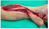
A 40-year-old man is diagnosed with a posterior thigh sarcoma. He undergoes resection of the tumor as well as some of the surrounding muscle. Partial sacrifice of the sciatic nerve is required, leaving a 40% circumferential defect and an 11-cm gap between proximal and distal ends. A photograph is shown. Which of the following is the most appropriate method of nerve reconstruction?
A) Mobilization and primary coaptation
B) Polyglycolic acid nerve tube
C) Processed human allograft conduit
D) Saphenous vein graft
E) Sural nerve cable graft

The correct response is Option E.
Fundamentals of nerve repair include coaptation in a tension-free manner. If there is any tension, nerve grafts or conduits are indicated. In this clinical scenario, there is a large nerve gap that precludes tension-free primary coaptation, even with extensive proximal and distal mobilization. Therefore, a nerve graft is indicated. Common choices include sural, lateral, or medial antebrachial cutaneous. For the size and length of the defect and the fact that multiple cable grafts would be needed, the sural is the most appropriate choice.
Nerve conduits such as PGA tubes and processed human allograft conduits serve as scaffolds to promote nerve regeneration, although these are typically used for gaps less than 3 cm. Given the distance involved, a sural nerve graft using a grouped fascicular or epineurial repair is the most appropriate choice, although a gap this large is almost certain to leave permanent deficits. Appropriate levels of expectation must be set with the patient.

An 18-year-old woman comes to the office because of a large osteosarcoma of the distal shaft of the right femur. A 15-cm bone resection is planned, with a resulting large intercalary segmental defect. The overlying skin and soft-tissue is not involved. The patient is very motivated to proceed with limb preservation. Which of the following is the most appropriate option for reconstruction of this defect?
A) Bone allograft
B) Contralateral vascularized fibula free flap
C) Contralateral vascularized fibula free flap with bone allograft
D) Ilizarov bone transportation
E) Ipsilateral pedicled vascularized fibula flap
The correct response is Option C.
In a young patient who desires limb preservation after sarcoma resection, a contralateral vascularized fibula free flap with bone allograft (Capanna technique) is the most appropriate option for a large intercalary segmental defect. This involves placing the fibula flap within an allograft construct and bridging both osteotomy sites. There are advantages to using the allograft with the fibula flap, as a fibula flap alone may have difficulty with weight-bearing and potential fracture. In select cases a double barrel configuration can be used; however, in this patient the defect is too large. An ipsilateral pedicled flap would have difficulty reaching this large defect and would still have issues with fractures from weight-bearing. Ilizarov bone transportation can be performed for smaller defects (4 to 6 cm), but not in a defect this large. Finally, bone allograft alone is an option; however, this has a high rate of nonunion (34% versus 8 to 10%).
A 45-year-old woman who underwent Achilles tendon repair through a posterior midline incision 3 weeks ago develops a postoperative wound infection and subsequent skin necrosis. Physical examination shows a 3 x 3-cm wound directly overlying the Achilles tendon in the absence of peritenon. A fasciocutaneous propeller flap from the medial leg is designed to cover this defect. The septal perforators to this flap run between which of the following structures?
A) Flexor hallucis longus and gastrocnemius
B) Gastrocnemius and soleus
C) Peroneus longus and peroneus brevis
D) Soleus and flexor digitorum longus
E) Tibialis anterior and extensor digitorum longus
The correct response is Option D.
This defect may be reconstructed with a posterior tibial artery perforator propeller flap. These vessels emerge between the flexor digitorum longus and the soleus muscle. In one anatomic study, there were three clusters of perforators: 4 to 9 cm, 13 to 18 cm, and 21 to 26 cm from the intermalleolar line. The peroneal artery perforators often arise through the posterior peroneal septum, and the anterior tibial artery perforators are often found between the extensor digitorum longus and the peroneus longus or between the tibialis anterior and the extensor digitorum longus.
A 27-year-old woman sustains a Grade IIIB degloving injury of the left lower extremity in a motor vehicle collision. Latissimus dorsi free flap placement is planned. Which of the following is the most likely outcome in this patient in terms of donor site morbidity?
A) Decreased seroma formation but increased hematoma formation
B) Inability to maintain sitting-up position when back is not supported
C) Initial decreased shoulder range of motion that improves by one year
D) Permanent loss of external rotation of the shoulder and inability to reach forward
The correct response is Option C.
Most studies that demonstrate shoulder weakness and loss of motion show that the loss of function is greatest in the early postoperative period and returns to baseline, or close to baseline, at 1 year or more after surgery.
All studies comparing types of latissimus flaps demonstrate less morbidity with perforator or muscle-sparing flaps as compared to traditional or extended latissimus dorsi (LD) flaps. Lower functional morbidity is observed with more native muscle preserved as is other flaps. This assumes that the muscular branches of the motor nerve to the latissimus are spared.
A recent meta-analysis does show higher functional impairment than expected after latissimus flap transfer. The number of patients who required a change in occupation was less than 10%. This was likely because of difficulty with activities such as ladder climbing, painting overhead, and sustained reach overhead.
The function of the latissimus dorsi muscle is shoulder adduction, extension and internal rotation. Other muscles of the rotator cuff perform similar functions and will assist in compensation for the loss of the latissimus. Patients who do develop weakness report it in activities involving shoulder adduction and internal rotation. Paradoxically, limitations in range of motion are mostly in shoulder flexion and abduction possibly related to tight skin closure and internal scarring.
Donor site seroma formation is particularly problematic, with published rates ranging from 3.9 to 79%.
Core muscles such as rectus abdominis, external oblique, gluteus maximus, medius, and minimus, and erector spinae all contribute to rotation, balance, and stabilization during sitting and standing.
A 21-year-old man undergoes reconstruction with a free flap. Photographs are shown. This procedure places the patient at risk for claw toe with loss of active flexion of the great toe. The muscle responsible for this functional loss is located in which of the following compartments in the lower leg?
A) Anterior
B) Deep posterior
C) Lateral
D) Superficial posterior

The correct response is Option B.
Claw toe or loss of active flexion of the great toe interphalangeal joint can result from harvest of the flexor hallucis longus for free fibula flaps. The flexor hallucis longus is present within the deep posterior compartment of the lower leg and should be resuspended to the interosseus membrane and posterior tibial muscles as needed to maintain proper tension. Physical therapy is initiated after adequate wound healing to maintain the mobility of the great toe and ankle. The deep posterior compartment musculature is composed of the tibialis posterior, the flexor digitorum longus, the flexor hallucis longus, and the popliteus.
The superficial posterior compartment musculature is composed of the gastrocnemius, soleus, and plantaris.
The anterior compartment musculature is composed of the tibialis anterior, the extensor digitorum longus, extensor hallucis longus, and the peroneus tertius.
The lateral compartment musculature is composed of the peroneus longus and brevis muscles.