Lecture 2; The Skin Flashcards
(36 cards)
Describe the layers of the skin;
Superficial: Epidermis -basement membrane- Dermis Sub cutaneous fat
Describe the epidermis layers;
Stratum corneum Stratum granulosum Stratum spinosum (hemidesmosomes give appearance) Stratum basilar Basement membrane (anchors stratum spinosum) Melanocytes
Describe the anatomical features found in the skin
Epidermis Dermis Subcutaneous fat Nail Hair Eccrine sweat glands Pilosebacous unit
What are the basic functions of the skin;
Protection Immune Vit D synthesis Temperature regulation Sensory organ Fluid balance Psychology
What is the role of skin colour and disease?
Darker skin protects against folate degradation (the protein that absorbs UV) However, in lower latitude places darker persons can have more skin conditions due to decreased UV absorption.
Vit D Deficiency leads to;
Rickets Osteoporosis
Are there keratinocytes in the skin?
Yes, 10 to 1 melanocytes. They produce keratin which is highly present in the skin.
Describe the basement membrane found between the epidermis and dermis.
Dual layer; Superficial #Hemidesmosomes anchoring cells such as keratinocytes - laminar lucida - laminar densa #Collagens I.e 7 and 3. Deep.
What’s a condition impacting the hemidesmosomes anchoring keratinocytes to the basement membrane?
Bullocks pemphigoid Might be a protein targeting the hemidesmosomes anchoring keratinocytes to the basement membrane Blistering appearance.
What can a gene mutation in collagen type seven cause in the basement membrane
Epidermolysis bullous
What does milignant changes in melanocytes cause? Extra Q; in a melanoma, what is the white in the mole?
Melanoma Extra; white spots can be caused by the body creating antibodies to the cancerous melanocytes.
Describe the embryological origin of melanocytes
Melanocytes are derived from neural crest cells in the formation of the neural tube.
What is the disease where melanocytes are destroyed
Vitiligo Note the clinical symmetrical appearance
Keratin forms;
Hair Nails Skin
Describe the types of keratin and potential keratin related genetic conditions
54 variants of keratin type one (ch. 17) and type two (ch. 12). Diff tissues have dif keratin combinations Pachyonychia congetia
Describe Pachyonychia congetia features
Keratin 6a mutation on ch 12 Clinical notes; Cysts can form Thick nail beds Plantar pain Mucosal involvement
What’s the function of the dermis and what’s found in it?
Structural and nutritional support to the epidermis. Muccopolysaccharides. Elastin Collagen Vessels Cells; - fibroblasts - macrophages - mast cells - dermal dendritic cells
What conditions occurs with excess collagen?
Scleroderma
What is the function of subcutaneous fat?
Protection Insulation Energy sources Biologically active - hormone interaction I.e leptin and generation.
What is the inflammation of subcutaneous fat called?
Panniculitis
Review the diagram of the nail bed from each view.
Do it now.
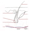
Review hair follicle diagram
Now.
What is folliculitis?
Inflammation of the hair follicle
Describe where deep inflammation of the hair follicle and what it can lead to?
Deep inflammation of the hair follicle can result in damage to the bulge area of the hair follicle containing stem cells. Damage to this area leads to intense scarring; Discoid lupus erythromotsus. More common in pasifika and Maori.