L36. Postural Tone and Locomotion Flashcards
What happens when the brain can no longer influence the spinal cord?
This is an UMN lesion
- Exaggerated segmental reflex
- Brain is no longer in control (because it normally acts to give a net inhibitory influence on the LMN)
What are the two postural signs of damage to the brainstem?
- Decerebrate: where the extensors are overactive in the limbs
- Decorticate: flexors dominating in the upper limbs but the lower limbs are extended

What are the main motor pathways running in the medial brainstem?
What do these tend to control?
- Medial Spinothalamic Tract
- Tectospinal tract (accomodates position to non-conscious vision cues)
- Reticulospinal tract
- Vestibulospinal tracts (position in relation to balance)
Mainly postural control and midline muscles (and tend to be ipsilateral and their effects are on both sides symmetrically)
These tracts synthesise sensory information in relation to our posture

What are the main motor pathways running in the lateral brainstem?
What do these tend to control?
- Rubrospinal tract
- Lateral spinothalamic tract
These tend to control the distal muscles for dextrous and fine movements (not for posture)

What is the rubrospinal tract important for?
What is the major brainstem source of the tract?
To what level does it descend to?
It is one of the pathways for voluntary movement: large muscle movement and fine motor control
It descends from the red magnocellular nucleus of the midbrain (and recieves inhibitory signals from the cortex)
It terminates primarily in the cervical spinal cord, suggesting that it functions in upper limb but not in lower limb control.
It mainly acts as flexor control in upper limbs
What two major extrapyramidal tracts control the extensors of the limbs?
The Reticular formation tracts: they give a net excitation of extensor muscles
- medullary reticular formation tract
- pontine recticular formation tract
(not cortex supplies a tonic inhibition of these)

What major extrapyramidal tract controls the flexors of the limbs?
For the Upper limb: Rubrospinal tract
For the lower limb: corticospinal tract

If a patient is in the decorticate posture what do they look like?
What does this suggest?
They have flexion in the arms and extension in the legs
This suggests that the brainstem centres are intact (but the cortical influence on them is lost)

Explain why the flexors dominate in the upper limb and the extensors in the lower limb for decorticate posture
Because the cortex normally serves to inhibit the rubrospinal tract and thus disinhibition causes flexors to dominate in the arms.
The rubrospinal tract does not descend to the arms and flexion in the lower limbs is from the corticospinal tract which if there is no cortical input is lost.
- Because brainstem tracts are intact the medullary and potine reticular formations take over.
- And also because the cortex is no longer able to provide inhibitory signals to these
If a patient is in the decerebrate posture what do they look like?
What does this suggest?
They have extension of the lower limbs and extension of the upper limbs as well
This suggests that the function of the brainstem rubrospinal tract (the red nucleus) is also now lost.

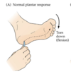
Describe the babinski reflex
A response to the fairly noxious stimuli on the sole of the foot that leads to a flexion of the toes in response (normal plantar response).

What does the Babinski sign (Extensor plantar response) signify?

It suggests a loss of descending cortical control of the spinal cord. This is because there is a loss of the tonic inhibition that is usually intact to prevent this sign from occuring.
How are UMN for cranial nerve lesions identified?
By the pattern of loss of function and innervation rather than looking at LMN/UMN that are commonly looked at for spinal motor output
What is the corticobulbar tract?
A a two-neuron motor pathway connecting the cerebral cortex to the brainstem primarily involved in carrying the motor function of the non-oculomotor cranial nerves.
Describe the path of the corticobulbar tract
- Originates in the primary motor cortex of the frontal lobe,
- The tract descends through the internal capsule
- In the midbrain, the internal capsule becomes the cerebral peduncles. The white matter is located in the ventral portion of the cerebral peduncles, called the crus cerebri. The middle third of the crus cerebri contains the corticobulbar and corticospinal fibers.
- The corticobulbar fibers exit at the appropriate level of the brainstem to synapse on the lower motor neurons of the cranial nerves.

What are the innervations of the corticobulbar tracts? What are the two exceptions?
It innervates all the motor nuclei of the face bilaterally
- Trigeminal V
- Upper Facial VII
- Spinal Accessory XI
- Motor regions of IX, X, XI in the nucleus ambiguus.
The exceptions are the lower half of the facial nerve and the hypoglossal nerve which are innervated contralaterally

Describe the difference between an UMN lesion and a LMN of the facial nerve and what would present
UMN:
Would cause a quadrant phenomena where the lower half of the contralateral side of the face has lost motor control because the top half still has innervation from the ipsilateral side (bilateral)
LMN:
A complete hemiparalysis of the contralateral side because both the UMN and LMNs of the facial nerve are impacted on.

How do we measure muscle movement?
Through electroymyography: measuring electrical discharges of muscles with a high temporal resolution

What are the two basic mechanisms that we control movement?
- Pre-emptive movements: we first stabilise our joints before moving
- Reflex adjustment of movements in response to disturbances in our movement
Imagine a scenario of a person catching a ball from a vertical height and measuring the output.
What would you expect the output of muscle activity to be in the muscles of the arm…
- Just before catching
- Immediately after impact
- Expect muscles to fire before impact: pre-emptive to stablise joints
- Expect reflex muscle activity to account for the new weight/disturbance
What are the first muscles to be activated in a simple voluntary movement of lifting one leg off the floor?
The muscles of the supporting leg will increase activity and allow for weight distribution as they prepare to stabilise the person. This happens before the voluntary movement has happened.
What is the purpose of this pre-emptive stage of voluntary movement?
It is an unconscious program that maintains postural control in anticipation for what the voluntary pathways are going to do
What part of the corticospinal tract is responsible for postural control?
The ventromedial corticospinal tract (responsible for postural control) and also travels ipsilaterally through the spinal cord and gives a bilateral innervation at the spinal cord level (symmetrical innervation)
How are the patterns of activity required for locomotion generated and modulated?
The pattern is present in babies from birth (this is how we know the pattern is spinal generated). It is generated in the lumbar sacral plexus
(note the pattern disappears several weeks later and reemerges when learning to crawl and walk)
Describe the relationship of the brain and spinal cord in relation to locomotor
Humans need the brain to activate the walking process and from there the spinal cord takes over the pattern generation.
What are the two phases of the step cycle?
- Swing Phase (unloaded phase) where the limb is swinging and getting into position
- Stance Phase (loaded phase) where the limb bears all the weight of the body and is pushing off the ground to propel forward

How are the stance and swing phases related for the legs?
The legs are exactly 180 degrees out of sync from one another
What are the major muscles involved in the swing/stance phases?
Swing = Flexion
Stance = Extension

How do decerebrate cats increase their speed when the treadmill they are walking on increases speed?
Through muscle sensors (the intrafusal fibres/spindles and the golgi tendon organs)
Moving the treadmill faster will cause a faster extension of foot during stance phase. The intrafusal spindles sense a lengthening of the muscle that is too much extension and a reflex (segmental) pathway is activated to contract flexors and initiate swing phase sooner.
ie. initation of swing phase is controlled by sensory feedback from extensor muscles
Stimulation of golgi tendon organs in one limb means that it is bearing the whole weight of the animal. What would artificial stimulation of this GTN do to the gait cycle?
It signals that extension phase is not finished yet and thus inhibits the flexor muscles

Describe the role of peripharl inputs in locomotion
The ongoing capacity to walk is dictacted by the feedback given to the system by peripharl inputs (GTN and muscle spindles)
What is meant by there is a need to a flexibilty in the pattern generation of locomotion?
We need to be able to control and modify the pattern in real time in order to respond to environment and obsticales. The motor cortex unit has the ability to stop and alter the pattern
What are some common abnormal gait patterns?
- Ataxic
- Choreaform
- Hemiparetic
- Parkinsonian


