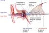
L29. Ear and auditory Tube Flashcards
Describe the main segments of the ear and what is contained within them
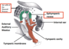
- Outer ear: From the auricle through the external acoustic meatus to the tympanic membrane
- Middle ear: Contains the tympanic cavity, epitympanic recess and the auditory tube
- Inner ear: Contains the bony and membranous labyrinth with the sensory apparatus

What is the auricle? What is the importance of it in terms of function?
The auricle is the skin and cartilage of the outer ear and it collects sound and localises if from different parts of the surrounding space.
What is the canal that connects the outside environment with the inner ear?
The external acoustic meatus
Also termed the ear canal
Label the main seven parts of the outer ear


Describe the external acoustic meatus tube in terms of the components of the walls
The lateral third tube is cartilaginous and the medial two thirds of the tube is boney (temporal bone)

Describe the skin lining the external auditory meatus tube and what function the important secretion has
The tube is lined with hairy skin and cerumen glands that secrete cerumen.
Cerumen is as waxy material that is important for preventing water from macerating/softening the skin
What nerves supply sensory information to the external ear?
- Vagus supplies the posterioinferior walls
- The auricotemporal nerve (a branch of the mandibular division of the trigeminal nerve V3) suppies the anterosuperior of the ear and the external surface of the tympanic membrane
Damage to the tempanic membrane often radiates to other places in the face. Where are these places and why does this happen?
Radiates to the temperomandibule joint, the lower teeth and jaw, and other places.
This is because this is the supply of the manidbular division of the trigeminal nerve (V3) that helps to supply the tempanic membrane
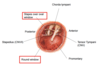
What is the function of the tympanic membrane?
To produce a complete seal around the external acoustic meatus to ensure there is no direct communication between the EAM and the Middle ear.
It also allows pressure waves from sounds to cause it to vibrate and this vibration is the main signal to the inner ear and sensory neurons.
Describe the concave/convexity of the tympanic membrane
Concave outwards (laterall)
Convex inwards (medially)
What is the the “cone of light” seen on the tympanic membrane through an oroscope? Where is this and what would happen in pathology?
The cone of light is found in the anterior-inferior quadrant of the lateral surface of the tympanic membrane.
Shining a light into the ear produces a reflexion in a particular part of the quadrant because of the convexity of the membrane.
In a middle ear problem, pressure changes cause a change in the shape of the membrane causing the position of the light to change

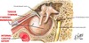
What are the main three compartments of the middle ear?
- Tympanic Cavity proper - directly medial to the tympanic membrane
- Epitympanic recess - to the mastoid air cells
- The Auditory tube (eustacian tube/ pharyngotympanic tube)
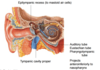
What does the epitympanic recess communicate with?
This recess extends above the tympanic membrane into a divot in the termporal bone above. It has commincations to some spaces and sinuses called mastoid air cells in the mastoid process of the temporal bone.
What is the main function of the eustachian tube?
It is a communication between the inner ear and the outside environmental pressure. Large changes in pressure in the middle ear relative to atmospheric pressure can be potentially dangerous, hence it is important to eqilibrate them.
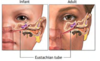
Describe the shape and slope of the eustachian tube and where it connects to.
It projects anterioinferiorly into the nasophayrnx
It has a slope projecting downwards making it difficult for bacteria from the nasopharynx to get to and making it easy for any fluid/bacteria in the inner ear to drain out.
Describe anatomical differences between the eustachian tube of a young child compared to that of an adult.
What is the significance of this difference?
A child has a more horizontal eustachian tube making the middle ear difficult to drain and making it easier for bacterial infections to migrate into the ear. Thus young infants more commonly suffer from otitis media.
Chronic infection can impact on the three bones of the middle ear and lead to impacted hearing and subsequent language development issues
What is a potential pathology and problem faced by the inner ear as a result of the eustachian tube?
Any inflammation or exudate that is able to get into the middle ear through the eustachan tube can potentially travel up through the epitympanic recess into the mastoid process air cells.
These don’t have another easily accessible drainage point (only higher up in the CNS) and therefore exudate becomes very infected and causes a lot of pain
What are the three ossicles? Where in the middle ear do they lie?
They are three very small bones that lie in both the tympanic cavity proper and in the epitympanic recess. They are (from outside to inside)
- Malleus
- Incus
- Stapes

How does the tympanic membrane communicate its vibrating signals to the inner ear?
Through three small bones with synovial joints between them called ossicles
Vibration from sound waves of the tympanic membrane cause vibration of the three bones in turn and these sequentially connect to each other and then to the sensory receptors
Describe the articulations of the bones to the tympanic membrane, with each other and with the inner ear
- The handle of the malleus attaches directly to the medial surface of the tympanic membrane
- The head of the malleus attaches onto the body of the incus
- The lenticular process of the incus attaches to the head of the stapes
- the base of the stapes directly attaches to the oval window of the middle ear

Describe the difference between the vibrations of high/low pitch and high/low volume
PITCH:
- High = High frequency
- Low = Low frequency
VOLUME:
- Loud = High amplitude
- Soft = Low amplitude

What are the two muscles in the middle ear? What is their main function?
- Stapedius muscle
- Tensor Tympani muscle
Contraction of these muscles pulls the ossicles towards the walls and dampens the amplitude of their vibration. This protects the bones and the ear from damage from loud sounds.
Describe the two middle ear muscles in terms of their:
- Attachment to the tube
- Which bone they attach to
- Innervation
Stapedius Muscle
- Attaches to the posterior wall of the canal
- Attaches to the stapes bone
- Innervated by CN VII (facial)
Tensor Tympani Muscle
- Attaches to the anterior wall of the middle ear
- Attaches to the Malleus bone
- Innervated by CN V (Trigeminal)

What kind of pathway controls the muscles of the inner ear in response to loud sounds?
Both muscles are under reflex control
Bell’s palsy is a parlysis of the face as a result of damage to CN VII.
One of the signs of Bell’s palsy is an increased sensitivity to loud sounds. Why is this so?
Because the Stapedius muscle is innervated by CN VII and damage to this nerve means it is unable to reflexively contract.
Loss of contraction means inability to pull on the bone and dampen the amplitude going through the canal. Thus loud sounds travel through unimpeded and are percieved as very loud
What is the promontary of the middle ear?
It is a large swelling on the medial wall of the middle ear in the anterior inferior quadrant. It is formbed by the turn of the cochlear
What are the two windows of the middle ear? Where are they located and what are their functions?
- Oval window is a connection in the superior posterior part of the middle ear and the base of the stapes sits directly on top of it - it recieves vibrational information
- Round window is a connection in the inferior posterior part of the middle ear and acts as an escape for mechanical energy
Are the windows of the middle canal true openings?
No. There is no direct communication between the middle ear and the inner ear.
The windows have membranes over them that allow only mechanical energy (vibration) through
Describe the course of the facial nerve CN VII and especially the chorda tympani through the middle ear
The facial nerve travels over the top of of and behind the middle ear (not in the ear)
The chorda tympani is a branch off the nerve that is given off and has a very tortous course. It travels through the middle ear from the anterior wall and travels through the canal and through the skull towards the tongue. It has no involvement in the ear at all.
Why do people with otitis media often hear a throbbing sound?
Because the internal carotid artery runs in the carotid canal which is close apposition to the middle ear. Fluid in middle ear infections act as a medium for vibrational pulses from the artery to travel and be sensed as sound.

Describe the location of the inner ear
It is embedded in the petrous part of the temporal bone

What are the two labyrinths of the inner ear?
- There is a boney labyrinth which is the cavity/grooves in the temporal bone containing perilymph fluid
- A membranous labyrinth that is a single closed sac, suspended within the boney labyrinth taking its shape, containing endolymph fluid
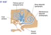
Describe the three main parts of the boney labyrinth

- Vestibule: the main chamber that comminicates with the middle ear through the windows, codes for static equlibrium
- Cochlea: contains the part of the membranous labyrinth that contains the sensory receptors for hearing
- Semicircular canals: three of them orientated in different directions containing membranous labyrinth with sensory neurons for dynamic equilibrium

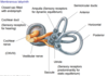
What are the otolith organs? What do they do?
- Utricle
- Saccule
They are small swellings that make up the vestible
They contain sensory receptors predominantly for static equilibrium (and a little bit of dynamic equlibrium)

What are the three orientations of the semicircular ducts?
What is the importance of this?
- Horizontal (lying flat in a plane that is transverse to that of the body)
- Anterior (45 degrees to the horizontal plane)
- Posterior (45 degrees to the horizontal plane)
They are important as they encode for different head movements

What are the ampulla of the inner ear?
The ampulla are swellings at the base of each semicircular ducts that contain sensory receptors for dynamic equilbrium

Describe the nerves coming out of the inner ear
- Cochlear nerve: exits out of the the cochlear duct having taken information from the cochlear about hearing
- The vestibular nerve takes information from the ampulla and otolith organs and out to meet the cholear nerve
The two meet and exit the ear through the internal acoustic meatus as the vestibulocochlear nerve (CN VIII)
There is a topography between the cholea and the brain in processing hearing. What is meant by this?
If the cochlea were to be unwound, the base would be the membranous labyrinth closest to the middle ear and the apex would be the tip.
- High frequency sounds are sensed by the receptors at the base of the choclea
- Low frequency sounds are sensed by receptors at the apex
The fibres of these receptors retain their relative positions as they travel to the cortex
Loudness/amplitude is reflectedin the level of deflextion of the membranous labyrinth and the intensity of the action potential firing.
Describe how the semicircular ducts enable information sense for dynamic equilibrium?
As the head moves in any one direction there is a relative movement of the endolymph fluid past the receptors in the ampulla (the base of the ducts) for that particular plane of movement. The brain encodes this relative change as movement and position
Describe how structures in the vestibule (saccule and utricle) recieve and encode information about dynamic equilibrium
They are heavily influenced by gravity when the head is in a certain position, gravity influences crystals to act on these sensory receptors to give information of head position

