L23. The Retina Flashcards
What are the fundamental limits to Visual Acuity
- Neural Factors (neurons in the retina and their connections
- Optical Factors (how light passes through the eye)
What is Visual acuity?
The ability to see clearly and to resolve fine detail
How do we test for visual acuity? Explain the bases of this test and what the results mean
Visual acuity is tested using the Snellen Chart that gives a fraction result for visual acuity.
VA = Test distance (always 6m)/ Distance where arc subtends 5 minute arc
A result of 6/6 is considered “normal”
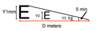
Interperet a visual acuity score of 6/30

The person was only able to read the line labeled 36 and no further down.
This means that what this person is able to see from 6m away is equivalent a letter that a “normal person” would be able to see from 30m away. Thus is this a poor visual acuity
What are the optical factors affecting visual acuity? [3]
- Pupil size (the larger the pupil size, the slighter worse the visual acuity)
- Clarity of the optical media - how light travels through (cornea etc)
- Refractive erros
What is a refractive error?
What are four main refractive errors? Describe them
Vision problems that happen when the shape of the eye keeps you from focusing well.
- Myopia: short sightedness (eyeball is too long)
- Hypermetropia: long sightedness (eyeball too short)
- Astigmatism (imperfection of the curvature of the eye)
- Presbyopia (aging of the lens and ciliary muscles)
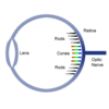
Describe the major differences between photoreceptors: rods and cones
- Detail
- Colour
- Number of them
- Light sensitivity
- Type of vision

RODS:
- See coarse detail
- Only see black and white (only 1 types)
- Majority of photoreceptors 100 million
- Low light sensitivity: Very sensitive
- Peripheral vision and Night vision (scoptic)
CONES:
- See fine detail
- Three types to see three colours
- Only make up 6-7 million receptors
- Less sensitive to low light (photopic)
- Central vision and Detailed Vision
Describe the general distribution of photoreceptors across the retina
- The fovea contains only cones
- Surrounding the fovea there are rods and cones mixed
- Moving out to the peripheral areas of the retina there are more rods than cones
Describe best photoreceptors to use during photopic light levels (well lit conditions).
- Best seen using the cones at the fovea. During photopic light, there is a cone peak at the fovea
- Get the best visual acuity

Describe best photoreceptors to use during scoptic light levels (vision in dim light).
- The best area of the retina to recieve visual input at night is slightly off the centre (fovea) between 5-15 degrees away
- At this point of the retina, there are mostly rods and rod pathways
- However the best visual acuity is 6/60

What are the six neurons that make up the neural retina?
- Cone photoreceptors
- Rod photoreceptors
- Bipolar cells
- Ganglion Cells
- Horizontal cells
- Amacrine cells
Draw the layers/organisation of the retina and their cells

What do the two interneurons connect between?
- Horizontal cells connect the photoreceptors to each other (and bipolar cells)
- Amacrine cells connect the ganglion cells to each other (and bipolar cells)

What are the different layers of the retina? [7]
From the outside in:
- Nerve fibre layer
- Ganglion cell layer
- Inner plexiform layer (synapse layer)
- Inner nuclear layer (bipolar cells)
- Outer plexiform layer
- Photoreceptor layer
- Pigmented epithelium

What are the 2 pathways of the retina?
- Through pathway: direct vertical transmission
- Lateral interactions: interneurons
Describe the through pathway
The standard pathway where light hits the photorecptors which then send signals to the bipolar cell which sends a signal to the ganglion cell.
(1st, 2nd, 3rd order neuron pathway)
Describe how lateral interactions occur in the retina
The standard pathway is modified by the lateral interactions (horizontal and amacrine cells)
What is the importance of the inner nuclear layer of the retina (ie. the bipolar cells)
How many different types of bipolar cells are there?
They are important for spatial vision and colour vision.
There are 10 different types of bipolar cells:
1x Rod bipolar cell
9x Cone bipolar cells
Describe the concept of on and off bipolar cells
There are 2 broad types of bipolar cells (and within that 10 different types)
- OFF bipolar cells hyperpolarise in response to light signals from the photorecptors
- ON bipolar cells depolarise in response to light signals from the photoreceptors

Horizontal cells provide lateral inhibition to photoreceptors only.
- How do they respond to light
- What neurotransmitter do they use?
- Horizontal cells respond to light by hyperpolarising
- They use inhibitory neurotransmitter GABA
Amacrine cells provide lateral inhibition modify biopolar cell and ganglion cell interactions.
- Describe amacrine cell morphology
- What neurotransmitter they use
- There are many different types of amacrine cells; they are axonleses
- They are mostly inhibitory cells that release inhibitory neurotransmitters: glycine and GABA
Ganglion cells are the main output neuron of the retina, their axons form the optic nerve.
What are the [2] types of Ganglion cells? What are the [2] subtypes of Ganglion cells/
- M
- P
and they are subgrouped into either ON or OFF ganglion cells
What neurotransmitter do ganglion cells release?
What makes ganglion cells unique cells in the retina?
Glutamate
The are the only cells in the retina that fire action potentials (all others only release neurotransmitters to communicate).
What is meant by a ganglion receptive field?
(this also applies to the concept of a bipolar receptive field)
Ganglions are ordered in a way that they respond to a small circular patch of photoreceptors delinating a receptor field.
It is an area of the retina that when stimulated with light, causes a change in the cell’s membrane potential.

How do ganglion cells respond to light?
By increasing or decreasing their action potential firing rate
What kind of receptor fields do ganglion cells have? Describe this arrangement and the implications
Ganglions have a concentric-surround receptive field.
The centre of the receptive field leads to one response from the ganglion cell and the periphery of the receptive field wiill have an antagonistic reponse.
This means that the ganglion cells are sensitive to differences in the level of illumination they recieve across the field.

What are the two types of ganglion cell receptor fields?
- ON centre (off surround)
- OFF centre (on surround)

What defines “on” and “off” in terms of action potential/signal firing?
On = depolarises to light
Off = hyperpolarises to light
Describe the action potential firing and level of activity within the an ON-centre ganglion cell when light is shone:
- Outside the receptor field
- Spot in the periphery (surround) of the ganglion field
- Spot in the centre of the ganglion receptor field
- Whole centre illumination
- Whole annular (surround) illumination
- Whole receptor field illumination
- The maximal response of the ganglion occurs when the whole centre is illuminated
- Note that the centre effects are slightly stronger than surround effects when the whole field is illuminated

Describe the action potential firing and level of activity within the an OFF-centre ganglion cell when light is shone:
- Outside the receptor field
- Spot in the periphery (surround) of the ganglion field
- Spot in the centre of the ganglion receptor field
- Whole centre illumination
- Whole annular (surround) illumination
- Whole receptor field illumination

How do photoreceptors respond to light?
Photoreceptors contain photopigments that are activated by light called opsins
The are proteins that bind to vitamin A (retinal) once they have been activated by light and this binding starts the phototransduction cascade

What are the two types of opsins?
- Rhodopsin found in rods
- Coneopsins found in cones
How do photoreceptors respond to light?
What neurotransmitter do they use?
All photoreceptors are hyperpolarised by light
Thye respond to light with graded changes in membrane potential causing a release of the neurotransmitter glutamate
(the amount of glutamate released is determined buy the membrane potential)
Light = hyperpolarisation = less transmitter release

Describe photoreceptor functioning in the dark
In the dark cCMP in the cytoplasm enables a constituative opening of the sodium channels allowing a sodium influx into the cell causing a depolarisation

Describe the phototransduction pathway
Light activates rhodopsin/conopsin molecules and this activates G protein (transducin) that initiates a cascade of events that activates phosphodiesterase and breaks down cGMP back to GMP. This dissociates cGMP from sodium channels (closing them)

What happens to photoreceptors in the light?
cGMP is broken down to GMP by the phototransduction pathway and this leaves no cGMP to bind and keep sodium channels to leave open.
This ceases the flow of Na and the cell is hyperpolarised

The bipolar cell layer is the first stage where a parallel pathway is set up to signal visual information to the brain. How is this set up?
This is set up by the way the levels of glutamate from photoreceptors affect the bipolar cells causes different effects due to the different types (on/off).
Why and how do bipolar cells respond differently to light?
There are two different types of receptors on the different bipolar cells. And depending on receptor the glutamate binds to causes either the ON/OFF response.
Describe the difference between the receptors expressed to respond to photoreceptor released glutamate on the different bipolar cells
OFF bipolar cells express ionotropic receptors (cause ion channel opening and subsequent depolarization)
Eg. AMPA, Kainate, NMDA
ON bipolar cells express metabotopic receptors, which initiate a second messenger cascade to cause hyperpolarization.
Eg. mGluR6
What is the pattern between bipolar cell and ganglion cell communication?
- All on bipolar cells will synapse only with on ganglion cells
- All off bipolar cells will synapse only with off ganglion cells
(Thus ganglion cells mirror the bipolar cells completely)
What creates the surround field (antagonistic response) for the centre-surround response?
Horizontal cells receive input from many photoreceptors and they provide output to other photoreceptors.
The horizontal cells release GABA as their neurotransmitters and inhibit

How does the surround field integrate with central field information?
The surround response results from the action of horizontal cells on the central group of photoreceptors.
The size of the surround is determined by the extend of electrical coupling between horizontal cells
