L22. Contents of the Orbit Flashcards
(37 cards)
What are the two orbital margins? What bones are these margins formed by?
- Supraorbital margin (formed by the frontal bone)
- Infraorbital margin (formed by the zygomatic bone laterally and the maxillary bone medially)

What is the purpose of the supraorbital notch?
It allows the passage of the supraoribital artery and supraorbital nerve to pass through
What kind of shape is the orbit? Orient this shape
The orbit has a base pyramid shape to it with the apex pointing deep into the skull/orbit where the foramen are
What bones make up the:
- Roof
- Floor
- Lateral wall
- Medial wall
… of the orbit
- ROOF: frontal bone and lesser wing of sphenoid
- FLOOR: maxilla, zygomatic bone and palatine bone
- LATERAL WALL: Zygomatic bone, greater wing of sphenoid
- MEDIAL WALL: Maxilla, lacrimal bone, ethmoid bone and body of sphenoid

What part of the orbit is most susceptible to fracture from blunt trauma to the face? Why?
Which two bones in particular are at risk?
The medial wall because these bones are quite thin
- Lacrimal bone is most commonly broken as it is very thin
- Orbital plate of the ethmoid has a region called the papyrus region (paper thin) also commonly broken
The eyeball is roughly covered in three layers or coats. What are these three layers?
- Outer coat (made up of sclera and cornea)
- Middle layer (uvea) made up of the ciliary body, iris and choroid - anterior
- Interior (retina) - posterior
What is the sclera?
A hard layer of collagen for protection that covers 5/6th of the surface of the eyeball.
What is the main functions of the sclera? [3]
- Protection: resistance to internal and external forces
- Maintains the shape of the globe
- Provides attachment for the extraoccular muscles
What gives the sclera its toughness and strength?
It is made of collagen that is laid down in organised whirls that add strength to it.
It is virtually impossible to pierce the globe due to the strength of this sclera.

What is the cornea?
A layer of connective tissue that is continuous with the sclera covering the anterior most 1/6th of the eyeball.
It is CLEAR (transparent) to act as a window for light to allow vision
Describe the cellular features of the cornea
It is avascular
It is arranged in 5 histological layers including: epithelium, connective tissue (like the sclera but more organised) and an endothelium

What makes the cornea transparent?
Because of the way that the collagen fibres are layed down.
They are analogous to packets of spaghetti, extremely ordered, uniform in diameter and evenly spaced apart
They run parallel to each other in bundles lying at angles to each other

What is conjunctivae?
The mucous membrane that covers the front of the eye and lines the inside of the eyelids.

What does the opacity of the sclera depend on? [3 factors]
- Composition of stroma
- Hydration
- Size and distribution of collagen
What would happen if there was a scratch to part of the cornea?
Note: damage/scratch to the epithelial layer of the cornea is the most common problem because the remainder of the slcera is highly resistant to damage.
Damage to the collagen fibres would force them to repair and regrow. But they are at risk of imparied repair and impaired organisation leading to a corneal scar.
What is the anterior chamber and angle?
Chamber: The fluid filled (aqueous humour) space between the cornea and the iris/lens
Angle: The junction between the iris and the cornea

What is the major function of the anterior chamber and the aqueous humour fluid within it?
Where does the aqueous humour fluid drain from?
- Maintaining the right size of the eye
- Maintaining intraoccular pressure
- Maintaining the health of the lens and cornea
Aqueous humour fluid drains out of the eye from the anterior chamber angle
Describe the flow of aqueous humour through the anterior chamber angle
- Fluid is constantly being produced by the ciliary body epithelial cells in the ciliary body
- The fluid is pumped into and through the anterior chamber
- Fluid moves through the trabecular meshwork (a seive like structure) to the anterior chamber angle
- Fluid flows through the canal of Schlemm which eventually joins and drains to episcleral venous systems

Describe the ciliary body
A circular structure just behind the iris composed of the ciliary muscle and ciliary processes which attach to the lens and the ciliary epithelium

What are the major functions of the ciliary body? [3]
- Ciliary epithelium forms the aqueous humour fluid
- Ciliary processes tethers the lens to it
- The ciliary muscle is very important to accomodation
What is meant by “accomodation”? What structures of the eye are involved in this process?
A reflex action of the eye, in response to focusing on a near object, then looking at distant object (and vice versa)
It comprises coordinated changes involving
- The ciliary muscles
- Zonules (also called the suspensory ligaments - attach the ciliary body to the lens)
- The lens

Describe the ciliary muscle and its innervation
It is found within the ciliary body, is a circular muscle surrounding the lens of the eye
Smooth muscle (involuntary)
Innervated by the parasympathetic nervous system through the Edinger-Westphal nucleus (CN III)
It is very important for focusing

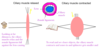
Describe the process of accomodation

All based on the refraction/focusing of objects
- Looking at distances: the ciliary muscles relax and the zonule ligaments pull against the lens causing the lens to flatten to allow focusing on distant objects
- Looking at closer objects: the ciliary muscles contract and the zonule ligaments become relaxed and wavy causing the lens to become thicker and bulge out allowing closer focusing.
What is Presbyopia?
What is it caused by and how is it treated?
The loss of accommodation with age
(the amplitude of accomodation decreases with age meaning the older you are, the further away an object needs to be in order to focus it)
- Caused by reduction in flexibility in the lens capsule and zonules.
- Treated by the wearing of plus lenses.


