Histology: Connective Tissue Flashcards
Define connective tissue.
- Supports and connects other tissues and cells to form organs
- Composed of cells and extracellular matrix (ECM)
Extracellular Matrix
-
Main component of connective tissue
- Protein fibers (collagen, elastic and reticular fibers)
- Ground substance
- Amorphous component (gel-like)
- Binds cells and fibers
Protein Fibers: Collagen

- Family of proteins
- Most abundant protein in the body (Type I, not heavily glycosylated)
- Major component of CT proper
- Resists shearing and tearing
- Various structures
Image:
- Type I collagen stains eosinophilic (light pink)
- Elastic Fibers (dark pink)
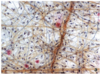
Collagen Fibrils LM

- Stain pink with acidic dyes (eosinophilic)
- Dark pink are nuclei of fibroblasts
Collagen Fibrils EM
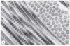
- Banding pattern
- Reflects arrangment of collagel to form fibrils (next card)
Collagen Synthesis
- Made by rough ER of fibroblasts
- Procollagen alpha-chains produced (distinctive repeating sequence of Gly-X-Y; glycine usually in the third position)
- Proline and Lysine residues are hydroxylated (requires Vitamin C)
- Procollagen chains assemble into triple helix
- Stabilized by hydrogen bonds between hydroxyprolines
- Packaged into secretory vesicles in Golgi and exocytosed
- Non-helical ends cleaved to form collagen molecule (tropocollagen) outside of the cell
- Triple helix collagen molecules self-assemble into fibrils with staggered arrangment (67 nm banding pattern)
- Results in fibril
- Fibrils are bundled together to form fibers (seen in LM)
- Requires covalent cross-linking (Cu as cofactor)
Scurvy
-
Vitamin C deficiency
- Results in defective collagen synthesis
- Characterized by:
- Pain/fatigue
- Muscle weakness
- Bleeding gums and tooth loss
Reticular Fibers

- Type III collagen fibrils
- Form branching network
- Requires special stain (PAS, silver stains)
- Because Type III is heavily glycosylated (will not show on H&E)
- Major components in lymphatic and hemopoietic tissues
Elastic Fibers

- Allows tissues to stretch and return to original shape
- Often interwoven with collagen fibers
- Can form fibers or fenestrated sheets (lamellae)
- Requires special staining for LM (Fuchsin)
Image:
- Blackish strands
Elastic Fiber Formation

- Scaffold of fibrillin microfibrils (secreted by fibroblasts or smooth muscle)
- Elastin protein deposited onto scaffold
- Elastin cross-links to form, elastin core
Image:
- Just scaffold on left; dark black is elastin deposit and core
Ground Substance
- Mixture of hydrophilic macromolecules
- Fills spaces between cells and fibers
- Allows diffusion of small molecules
- Barrier to invading substances
3 Main Molecules:
- Glycosaminoglycans (GAGs)
- Proteoglycans
- Multiadhesive glycoproteins
- Fibronectin
Glycosaminoglycans (GAGs)
- Polymers of repeating disaccharide units
- When attached to core protein, form proteoglycans
- Usually sulfated
- Synthesized in Golgi
- Attract water to extracellular matrix (gel-like consistency)
Hyaluronan (Hyaluronic Acid)
- Largest, most ubiquitous GAG
- Synthesized directly in ECM
- Exists as long, free carbohydrate chain
- Link to proteoglycans to form proteoglycan aggregates
- Binds water (responsible for changes in permeability and viscosity within CT)
Proteoglycans
- Core protein bound to sulfated GAGs
- Produced in rough ER of fibroblasts; secreted by exocytosis
- Bind to hyaluronan after secretion
Multiadhesive Glycoproteins
- Stabilize the ECM and link to cell surfaces
- Large molceules with branched oligosaccharide chains
- Examples:
- Laminin in basement membrane
- Fibronectin
Fibroblasts

- Most common cell type in CT
- Elongated cells with oval nuclei
- Difficult to discern cytoplasm
- Produce components of ECM
Fibrocytes

- Inactive fibroblasts (arrows)
- Heterochromatic nucleus
Macrophages

- Phagocytic cells (removal of dead cells, tissue debris)
- Derived from monocytes
- Common in loose connective tissue
- Hard to distinguish: eccentric nucleus
- Easier if recently ingested something (inclusions)
Macrophage EM

- Finger-like projections of cell surface
- Many lysosomes
Monocytes
- Provide defense by ingesting bacteria
- Participate in immune response (Antigen Presenting Cells)
- Inflammatory response
Mast Cells

- Derived from hemopoietic stem cells
- Mediate allergic reactions and anaphylactic shock
- Has receptors for IgE
- Abundant basophilic granules
- Granules contain inflammatory response mediators
Plasma Cells

- Derived from B lymphocytes
- Produce antibodies
- Eccentric nucleus, “clockface nucleus”
- Alternating heterochromatin and euchromatin
Mesenchyme
-
Loose Embryonic CT
- Most adult CT derived from this form
Mucoid CT

- Embryonic CT
- Principle component of fetal umbilical cord
- Gelatin-like ground subtance (Wharton’s Jelly)
- Lots of hyaluronan
Loose (areolar) CT
- Loose arrangment of collagen and elastic fibers, embedded in ground substance
- Many cells present:
- Fibroblasts
- Mast Cells
- Macrophages
- Beneath epithelia
- Fills spaces between fibers of muscles and nerves
- Most abundant type of CT in the body
Dense CT
- Fewer cells than loose CT
- Mostly fibroblasts
-
Abundance of type I collagen in ECM
- Little ground substance
Dense irregular CT

- Collagen fibers randomly arranged
- Resistance to stress in all directions
- Seen in dermis of skin
Dense regular CT

- Collagen fibers and fibroblasts arranged in parallel
- Resistance to stress in one direction
- Tendons, Ligaments
Reticular Connective Tissue

- Loose, located in lymphatic and hematopoietic tissues
- Reticular fibers (type III collagen)
Cells:
- Reticular cells (specialized fibroblasts)
- Immune cells
Adipose Tissue
- Specialized for energy storage
- Most of tissue made of adipocytes (minimal ECM)
- Other Functions:
- Thermal insulation
- Cushioning to organs
- Shape to body surface
- Cushions areas of mechanical stress (palms, heels)
White Adipose Tissue

- Long-term energy storage
- Single lipid droplet (unilocular)
- Ring-like appearance (nucleus/organelles pushed to side)
- Cells surrounded by external lamina
Brown Adipose Tissue

- Specialized to produce heat
- Abundant mitochondria
- Rich vasculature
- Commonly found in newborns/young children
- Multiple lipid droplets (multilocular)
What types of collagen are not principally produced by fibroblasts?
-
Type II Collagen
- Produced by chondrocytes
- Predominant collagen in cartilage
-
Type IV Collagen
- Produced by epithelial cells
- Found in basal laminae of basement membranes
- Most other types of collagen (types I, III, V) are produced by fibroblasts
Where is type I collagen found most predominately?
- Bones
- Endomysium of skeletal muscle
- Tendons
- Dermis of Skin
Most common type of collagen within the body
What type of collagen is hyaline cartilage primarily composed of?
- Type II
Matrix Metalloproteinases
- Class of enzymes are responsible for degradation of ECM proteins, allowing for turnover and renewal (such as collagen fibers)
Eosinophils
- Bi-lobed nucleus with eosinophilic cytoplam (pink) filled with secretory granules
