Histo - Images Flashcards
Where in the respiratory tract would you find this type of epithelium? Type of epithelium?

Nasal cavity
PSCC
What is the function of the cells directly under the PSCC cells?

Basal cells - stem cells for ciliated and goblet cells
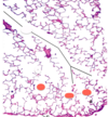
What is the blue-staining area? What does it contain?

Lamina propria
Blood vessels, lymphocytes and other immune cells, seromuocus glands
Where would you find this PSCC epithelium?

Nasal cavity
Name the dark pink, elongated cell and its function

Supporting cell
Provides metabolic and physical support to olfactory cells
Name the yellow elongated cells and their function

Olfactory cells
Basal processes collectively form olfactory nerve for odor transduction
Name the short dark pink cells and their function

Basal cells
Regenerate other cells
Name the outer layer and what it consists of

Adventitia/serosa
Connective tissue w/ or w/o mesothelial layer
Name the layer under the adventitia and what it consists of

Muscularis externa
Smooth muscle in inner circular layer and outer longitudinal layer
Name the layer under the muscularis externa and what it consists of

Submucosa
Connective tissue
Name the layer under the submucosa (duh!) and what it consists of
Mucosa
Epithelium, lamina propria (c.t.), muscularis mucosa
Where are we in the respiratory tract?

Larynx
(notice transition from PSCC on left to simple squamous on right)
What is the yellowish material?

Hyaline cartilage forming C-shaped ring in trachea
What else is surrounding the tracheal lumen besides hyaline cartilage?

Trachealis muscle
What is the narrower lumen at the top of the slide?

Esophageal lumen
Where in the respiratory tract are we?

Trachea
(notice the hyaline cartilage and PSCC)
What is the orange arrow pointing to?

Basement membrane
What is the blue box encompassing? What cells are present?

Lamina propria
Lymphocytes
What is the red box encompassing? Red circle?

Submucosa
Seromucous gland
What is the green box encompassing?

Hyaline cartilage layer
Where in the respiratory tract are we?

Posterior tracheal wall
(notice thick trachealis muscle layer)

Where are we in the respiratory tract?

Trachea
(THICK BM and thick trachealis muscle layer)

Where in the respiratory tract are we?

Intrapulmonary bronchi
(cartilage now in plates)
Where in the respiratory tract are we?

Intrapulmonary bronchi
(Red circle = appearance of MUSCULARIS MUCOSA
Blue box = cartilage plate)

Where in the respiratory tract are we?

Bronchiole
What is no longer present at this level?

Submucosa (and its glands)
We’re looking at a bronchiole
What is present at this level that would not be found in the trachea or upper airways?

Muscularis mucosa
(IS present in intrapulmonary bronchi and down)
What special cell type is present in this structure and what is its purpose?

Clara cell
Secrete a surface-active protein, CC16
Where in the respiratory tract are we?

Respiratory bronchiole
(notice that part of it has alveoli)
What type of epithelium is present?

Ciliated cuboidal
What is the area marked in black box? Orange box?

Bronchiole
Terminal bronchiole
What are the green circles encompassing?

Respiratory bronchioles
What is this? What do “SM” and “A” stand for?

Surface of a respiratory bronchiole
Smooth muscle and alveoli
What is the structure denoted by the black arrow? Red circles?

Alveolar duct (almost no walls; boundaries made up of alveoli)
Alveolar sacs
Which arrow points to the type I pneumocyte? Type II pneumocyte?

Orange
Purple
What is the function of the cell denoted by the blue arrow?

Macrophage - scavenges inhaled material and either travel to the throat where they’re swallowed as mucus or retreat to the alveolar septum where they can remain quiescent for years (think recurrent TB)
Which cell can be known as a heart failure cell?

Macrophage (blue arrow) - scavenge RBCs during heart failure = heart failure cell
What is the function of the area marked by the blue arrow?

Thick region of alveolar septum - contains macrophages, fibroblasts, and fibers that intervene between alveolus and capillary
What makes up the region marked by the purple arrow?

Thin region of alveolar septum - type I pneumocyte, endothelial cell of capillary, fusion of basal lamina from both cell types, thin layer of surfactant
What is the function of the area marked by the purple arrow?

Gas exchange (thin region)
What structure is marked in red? Orange?

Bronchiole
Pulmonary artery
What structure is marker in yellow? Green?

Terminal bronchiole (notice it gives off resp bronchiole)
Respiratory bronchiole (notice the alveoli coming off)
What area is marked in blue? Purple? Grey?

Arteriole
Alveolar duct
Branch of pulmonary vein and capillary bed
At what level do the pulmonary arterioles form the structure marked by the purple arrow?

Level of alveolar duct (arrow = capillary bed)
What structure is marked in black? Yellow?

Alveoli
Connective tissue
What is the function of the small dark holes (ha)?

Alveolar pores of Kohn - allow airflow between alveoli when normal airspace is occluded
What structure is marked by the green arrow? Purple arrows? Yellow arrow?

Type I pneumocyte
Type II pneumocytes
Alveolar pore of Kohn