GU Anatomy/Physiology Flashcards
What is the urinary system composed of?
2 Kidneys
–
2 Ureters
–
Urinary bladder
–
Urethra

What are the functions of the urinary system?
Maintain body’s chemical equilibrium (Nephron)
Excretory – renal cortex
Regulating composition of blood – medullary pyramids
–
Regulates blood pressure
•Volume – Adrenal gland hormone Aldosterone stimulates more Na reabsorption from the kidneys
Vasoconstriction constrict- Hormone Renin
What are the embryologic ducts of the urinary system?
Pronephros (4-5wks) : most primative of the three embryonic excretory organs and is the first to appear.
Mesonephros (4-5wks): Wolffian duct in males, degenerates in females
Metanephros (funct. after 8 wks) – Permanent kidney

what is the most primitive of the embryonic EXCRETORY organs as well as the first to appear?
What duct takes over the exretory function becoming the wolffian duct in males?
What becomes the permanant kidney?
pronephros (4-5 weeks)
mesonephros (4-5 wks)
metanephros (after 8 weeks)

what is the cloaca?
•
In zoological anatomy, opening for the intestinal, reproductive and urinary tracts of certain animal species. The word comes from Latin, and means sewer. All birds, reptiles, and amphibians possess this orifice.

What is the urogenital sinus aka?
what does it do?
persistent cloaca
is a part of the human body only present in the development of the urinary and reproductive organs. It is the ventral part of the cloaca, formed after the cloaca separates from the rectum. It eventually becomes, among other things, the bladder.
what is the function of the alantois?
to collect liquid waste from the embryo, as well as to exchange gases used by the embryo.
what is the urachus?
what is its function?
what space does it lie in?
the obliterated allantois.
it is a solid functionless cord running from the dome of the bladder to the umbilicus.
space of retzius
what is the weeping navel?
It is possible for the urachus to remain open. It can for partially open or it can be fully open along its length. If it is open it can become infected and produce a foul odor from the belly button. This is called a weeping navel.
what do these images show?

urachus
what happens to the kidneys around 6 weeks of devlopment?
they migrate or ascend out of the pelvic cavity into the abdomen. they don’t “migrate” on their own rather ascend due to fetal rowth and the straightening of the fetal curve

explain the anatomy of the renals.

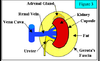
True Capsule – Fibrous, covers renal parenchyma
Perinephric Fat – Surrounds the Kidneys and Adrenal gland
–
Perinephric Fascia (Gerota’s Fascia) – This fascia surrounds the perinephric fat, kidney, adrenal gland and true capsule. Gerota’s fascia cannot be seen with ultrasound.
what is the relationship of the vein, artery and ureter?

vein anterior to artery, artery anterior to ureter.
what are the kidneys and adrenal glands surrounded by?
perinephric fat
(image in long)

pperinephric fat in trans
if a renal hemorrhage is contained in gerota’s fascia what will happen?
renal fascia in this drawing is gerota fascia.
it will cause compression to the kidney.

what is the nehpron?
what does it contain? it’s function?
what does bowmans capsule do?
The functional unit in the kidneys
Renal Corpuscle – Filters out waste via Glomerulus: Capillaries and Bowman’s Capsule
Filters waste products from blood – waste products, excess H2O and other items not needed by the body at that time i.e. excess vitamins…
what does the renal tubule do?
how?
Recycles needed items from waste
via loop of Henle which Recycles necessary minerals, amino acids and other things the body needs at that time.
explain the renal anatomy and it’s collecting system

Parenchyma - made up of nephron (Cortex - Glomerulus and Medulla / Pyramids - loop of Henle)
–
Collecting system: Minor Calyces, Major Calyces, Renal Pelvis
what is antoher name for the renal collecting system?
infundibulum: made up of the major/minor calyces and renal pelvic
location, division, vessels and ureter are variables

what are the 2 major components of the renal collecting system?
the sinus: echogenic. contains: Minor/Major calyxes, Artery / Vein, Fatty fibrous tissue, Nerves, Lymphatics
the hilum - medial portion of the sinus. contains: Arteries enter the kidney, Vein and ureter exit, Renal pelvis

where are the kidneys located?
Retroperitoneal
•
Costal margin
•
Costal Vertebral Angle
•
Flank

what is the kidney size/shape in an avg adult?
9-12 cm, variable
–
Symmetric in size
(within 1.5cm of each other)
–
2 cm thickness (HA)
–
5 cm width (HA)
–
Reniform / Bean
what makes up the posteriomedial border of the renals?
what muscle is posterior to the kidneys?
psoas muscle
quadratus lumborum
what are the sizes and shapes fo the kidneys
neonate?
pediatric?
3.3-5 cm in length
Proportionately > than adult kidneys
Not unusual to see neonate kidneys extending into the pelvis
–
< 1 yo 4.98cm + .155 x age (months)
> 1 yo 6.79cm + .22 x age (years)
what do the ureters do?
how do they run?
how are they supplied?

Ureters transport urine to the bladder through peristaltic action. This explains why a pt. with a kidney stone stuck in the ureter has pain comes in waves i.e. waxes and wanes
–
Ureters run proximately along the psoas muscles.
–The distal ureters pass anteriorly to the psoas muscle, and bifurcation of the common iliac arteries.
–
The ureters are supplied with blood from branches of the renal a, hypogastric a, and inferior vesical arteries.
in the male pelvis, how do the ureters run?
ureters passes posterior to the Ductus Deferens aka Vas Deferens.

in the female pelvis, how do the ureters run?

ureter passes posterior to the uterine arteries
the bladder anatomy

The bladder is a hollow, muscular organ which is posterior to the symphysis pubis.
–
The superior area (dome) is covered with the pelvic peritoneum. The inferior and lateral surfaces are in contact with the pelvic floor muscles.
what is the area of the bladder the urethra is connected to called?
how about the area of the bladder b/t the ureters and urethra?
neck
trigone

what are the 3 tissue layers from inner to outer of the ureter?
Mucosa
•
Circular smooth muscle
•
Fibrous connective tissue (adventitia)
what are teh 4 tissue layers of the bladder from inner to outer?
Mucosa
•
Submucosa
•
Muscularis (3 layers itself)
•
Serosa – Serous membranes line and enclose several body cavities, known as serous cavities, where they secrete a lubricating fluid which reduces friction from muscle movement. Serosa is not to be confused with adventitia, a connective tissue layer which binds together structures rather than reducing friction between them.

where do the renals get their blood supply?

Main Renal artery (MRA) -Originates 1cm inferior to SMA
Segmental artery –MRA divides into approx. 5 segmental art.
Interlobar artery –Division into lobes
Interlobular arteries – Small branches off the arcuate
Arcuate –These vessels run parallel to base of medulla (pyramids)

what does the renal lobe contain?
1 Medullary pyramid
–
Interlobar A/V
–
Cortex

how is the blood drained from the renals?
The RT RV enters the IVC slightly more inferior than the Lt. RV.
The Lt. RV receives the Lt adrenal and Lt gonadal vein. The RT adrenal and gonadal vein enters the IVC.

which renal vein is longer?
the lt renal vein.
where is the vein in relation to the artery?

anterior
What do the renals do?
Filter about 1,200 cc of blood / min
•
Make about 1500 cc urine per day
•
Urine make up: 95% water, 5% nitrogenous waste and inorganic salts
what do increased levels of BUN imply?
of the renal function lab tests BUN (blood urea nitrogen) Increased levels, may mean decreased renal function
it increases when kidney function is low b/c the liver makes urea but the kidney is not able to filter it from the blood.
what do increased levels of serum creatinine imply?
mean deceased in renal function. More specific and more sensitive than BUN
what does the renal’s specific gravity tests tell us?
A measure of the kidney’s ability to concentrate urine. Will generally decrease with certain diseases.
what does renin tell us?
A hormone released by the kidneys which ↑ blood pressure, by vasoconstricting systemic vessels.
what is the urethra?
the conduit which drains the bladder of urine.
when developmental anomalies of the ____ are seen clinically it is common for US to be asked to R/O congenital anomolies of the kidneys.
genitals
where do the kidneys ascend from?
the pelvis.
during migration, the kidney ___ and ___ ?
ascend and rotate
what does the true capsule of the kidney contain?
what is it made up of?
kidney parenchyma
fibrous layer of tissue
the hilum of the kindeys generally point ____.
what is found in th renal hilum?
medial
arteries, lymphatics and veins and teh renal pelvis
if a patient took a deep breath in and held it where would the kindneys move?
inferior
label the kidney

A. = cortex
B. = true capsule
C. = minor calyces
D. = major calyces
E. = renal pelvis
F. = minor calyces
G. = ureter
H. = pyramid or medulla
where are the nephrons of the kidney found?
what does it contain?
the parencyma of the kidneys in the cortex and medulla.
the nephron contains the waste filtering portion of the kidney and the collecting and sorting (reclycle) portion of the kidney
how would you describe the location of the kidney?
retroperitoneal, costal vertebral angle, flank
what action best describes the way urine is transported from the kidney to the bladder?
peristaltic
how mnay major calycies are there typically in each kidney?
2-3
the echogenicity of the renal cortex is _____ compared to the renal medulla
slightly more
a ___ is between each pyramid?
cortex
the cortex found b/t the medulla of the kidney is called ___ ?
column of Bertin
how many pyramids are typically found in each kidney?
What is each pyramid attached to?
8-18
a minor calyx
does the ureter (like the bladder) have an inner layer of mucosa tissue?
yes.
perfusion
the pumping of blood through an organ
the order of arterial vessels that feed the kidney are…
AO, MRA, segmental RA, interlobar arteries, interlobular arteries, arcuate arteries
the MRA typically divides into __ segmental arteries
5
the origin of the MRA is _________
~ 1 cm inferior to the SMA
the ___ areteries help define the renal lobes
interlobar
what is the echogenic center of the kidney?

the renal sinus
label the renal vasculature

A = segmental artery
B = Interlobar arteries
C = Arcuate arteries
D = Interlobular arteries
E = Main renal artery
do you need 2 functioning kidneys to live?
no
what is the phonomena of a single kidney growing in size to increase its filtering power known as?
compensatory hypertrophy
how many cc’s of blood does the kidney filter/minute?
what is the resitance in the kidney to all this volume of blood to filter through?
1200 cc’s
low