Glomerulonephritis Flashcards
pathogenesis
immunologically mediated disorder
- humoral - Ab driven
- cell mediated (T cells)
- involvement of inflammatory cells, mediators and complements
define
immune mediated disorder of the kidneys affecting the glomeruli, with secondary tubulo-interstitial damage
name 3 consequences of inflammation of the glomerulus
- damage to the glomerulus restricts blood flow, leading to compensatory increase in systemic BP
- damage to the filtration mechanism allows protein and blood to enter the urine
- loss of the usual filtration capacity leads to AKI
what does damage to endothelial cells lead to
- vasculitis - most aggressive form of inflammation in the kidney, can lead to AKO
- haematuria

what does damage to mesangial cells lead to
- proliferative lesion, release of angiotensin II which causes vasoconstriction
- chemokines are released which attract inflammatory cells
- haematuria

what does damage to podocytes cause
- non-proliferative lesion - podocytes atrophy, lose filtration barrier
- protein in urine

broad presentation of GN
- specific syndrome eg nephrotic or nephritic
- blood pressure
- urine dipstick: protein/blood
- renal function impairement
compare the presentation of nephritic syndrome to nephrotic
- nephrotic syndrome: Defined as proteinuria >3g/d, hypoalbuminuria (<25g/L) and oedema with severe hyperlipidaemia (and cholesterol). This is indicative of a non-proliferative process affecting podocytes

nephritic syndrome
syndrome comprising signs of nephritis, often occurs in GB
aetiology
- the majority are primary with no underlying drive to disease
- 2y - infections, drugs, autoimmunity, malignancy, systemic disease
define focal and diffuse
- focal <50% glomeruli affected
- diffuse >50%
define global and segmental
all/part of glomerulus affected
focal lesions are often segmental
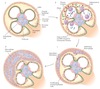
crescenteric
presence of crescents:
- Glomerular disease leads to gaps or holes in the GBM resulting in epithelial cell proliferation with mononuclear infiltration in Bowman’s space. Ultimately, fibrocellular crescent formation
- these are associated with RPGN

how does IgA nephropathy present
- macro or microscopic haematuria ± nephritic syndrome
what is the most common GN in the developed world
IgA nephropathy
pathology of IgA nephropathy
- there is raised abnormal IgA (possibly due to infection of mucosal membrane)
- target by the immune system and forms immune complexes with IgG
- these are deposited in mesangial cells
- accumulate and cause local immune ativation - proinflammatory cytokine release and macrophages come - and injury
- RBS pass through into urine
when does IgA nephropathy typically develop
during infection of mucosal membrane eg resp/GI tract
how does IgA nephropathy progress
- over time due to glomerular inflammation and injury, patients progress to renal failure
- 25% progress to ESRF in 10-30 years
describe a typical patient with IgA nephropathy
- Typical patient: young man with episodic macroscopic haematuria after respiratory/G.I. infection, recovery is rapid between attacks
what is IgA Nephropathy associated with systemically
Henoch Schonlein purpura
biopsy of IgA Nephropathy
mesangial proliferation and expansion
IF and EM of IgA Nephropathy
immune deposits (IgA and C3) are seen in mesangium
H&E stain of IgA Nephropathy
mesangial cell proliferation and expansion
treatment of IgA Nephropathy
the aim is to prevent further damage and avoid ESRF
- control BP: ACEi/ARB, fish oil
- prevent immune complex formation: corticosteroids
common features of RPGN
- aggressive, potential to cause ESRF over days
- severe glomerular injury
- development of crescent shape
crescent formation
- Glomerular disease leads to gaps or holes in the GBM resulting in epithelial cell proliferation with mononuclear infiltration in Bowman’s space.
- Ultimately, crescent is replaced by scar tissue
- lose filtration ability
- quickly leads to AKI


crescent
ANCA positive type of RPGN
- pauci-immune (no anti-GBM or immune complexes)
- cANCA (GPA) and pANCA (microscopic polyangiitis and eGPA)
ANCA negative RPGN
- anti-GBM eg goodpastures disease
- immune complex disease: IgA, HSP, post-strep GN
clinical presentation of RPGN
AKI ± systemic features
what is seen on IF of RPGN
- Anti-GBM – linear as Ab bind to collagen of GBM
- Immune complex deposition: granular
- ANCA – negative on IF (Pauci-immune)
usual age of onset of RPGN
50-60
treatment of RPGN
prompt and supportive, prognosis is poor
- Anticoagulants to reduce fibrin build up in crescent
- Aggressive immunosuppression with high-dose IV steroids and cyclophosphamide ± plasma exchange
ANCA
- circulate throughout bloodstream and adhere to neutrophils in endothelial cells
- PR3 and MPO are induced onto the surface of neutrophils when they are primed
- these promote the inflammatory process and perpetuate the vasculitis process
define nephrotic syndrome
- proteinuria >3g/day
- hypoalbuminuria (<25g/L)
- oedema
severe hyperlipidaemia (total cholesterol>10mmol/l) is often present
why is severe hyperlipidaemia often present in nephrotic syndrome
- may be a by product of the liver trying to produce more albumin
- often resolves with treatment of the syndrome
- the liver also produces more clotting factors, meaning the person is in a pro coagulant state - blood hyperviscosity
renal function and BP in nephrotic syndrome
- renal function usually normal, normal creatinine
- BP nomral-mild increase
pathophysiology of nephrotic syndrome
- non-proliferative process causing injury to podocytes, whichb atrophy
- this lets proteins through the barrier
- proteinuria means there are low protein concentrations in the blood, contributing to reduced oncotic pressure
features of nephrotic syndrome
- periorbital oedema followed by full body oedema
- periorbital oedema drains throughout the day
- a thrombotic compication (eg DVT, PE) may be the first sign

why do you not see peri orbital oedema in patients with oedema 2y to heart failure
they cant lie flat due to fluid in lungs etc
causes of nephrotic syndrome
- primary due to renal disease (listed further on)
- secondary to systemic disorders (hepatitis, SLE, paraneoplastic, drug related, diabetic nephropathy)
treatment aims of nephrotic syndrome
- reduce oedema
- reduce proteinuria
- reduce risk of complications
- treat underlying cause
- find and treat underlying infections
- stop causative drugs
- immunosuppression
the aim is to induce sustained remission (complete remission: <300mg/day, partial: <3g/day)
treatment of nephrotic syndrome: reduce oedema
- high doses of loop diuretics and salt restriction
- gut oedema may prevent oral absorption so IV is useful
- fluid restriction to 1l a day
- IV albumin can be used if volume deplete and diuretics not working
treatment of nephrotic syndrome: reduce proteinuria
start all patients on a ACEi/ARB
treatement of nephrotic syndrome: reduce risk of complications
- anticoagulation
- statin to reduce cholesterol (although often resolves spontaneously when cause treated)
complications of nephrotic syndrome
- infection
- thromboembolism
- volume depletion
- hyperlipidaemia
- vit D def
- subclinical hypothyroidism
complications of nephrotic syndrome: infection
loss of opsonising Ab in urine and IS treatment
complications of nephrotic syndrome: thromboembolism
patient is in a pro coagulant state due to increased clotting factors and platelet abnormalities
- renal vein thrombosis
- PE
- DVT
complications of nephrotic syndrome: volume depletion
- can be due to aggressive use of diuretics, as most of the salt and water is trapped in the tissues and cant move into the vascular space quickly enough to be removed
- patient can be oedematous and volume depleted
complications of nephrotic syndrome: vit D deficiency
- 25OH vitamin D is lost in the urine
complications of nephrotic syndrome: subclinical hypothyroidism
- normal T3/4 and high TSH
- due to increasd urinary excretion of thyroid hormones
- if there is already a low thyroid reserve, the transition into overt hypothyroidism is inevitable
nephrotic syndrome: minimal change disease
cause
- idiopathic
- associated with drugs eg NSAIDs, or paraneoplastic
what is the most common cause of nephrotic syndrome in children
minimal change disease
nephrotic syndrome: minimal change disease
pathophysiology
- cytokine damage to podocyte foot processes
- IL-13 may be implicated
- the flatten out
- negative charge barrier is lost
- albumin passess through
- selective proteinuria (some larger proteins still cant pass through)

nephrotic syndrome: minimal change disease
biopsy
- normal under light microscopy
- IF usually negative as there are no immune complexes
- EM shows fusion of foot processes of podocytes

nephrotic syndrome: minimal change disease
treatment
- 94% experience total remission with oral steroids
- adults respond slower than children
- some are steroid resistant or have multiple relapses
nephrotic syndrome: minimal change disease
how is frequently relapsing or steroid-dependent disease treated
cyclophosphamide or ciclosporin
prognosis of nephrotic syndrome: minimal change disease
1% progress to ESRF

nephrotic syndrome: minimal change disease
what is the most common cause of nephrotic syndrome in adults
FSGS
nephrotic syndrome: FSGS
causes
- primary (idiopathic)
- 2y - HIV, heroin use, sickle cell disease, obesity, reflux)
nephrotic syndrome: FSGS
pathophysiology
- podocytes are damaged and let plasma proteins and lipids through
- some of these are trapped and build up in the glomerulus
- these areas develop focal sclerosis

nephrotic syndrome: FSGS
presentation
usually nephrotic syndrome or proteinuria
around half have impaired rnenal function
nephrotic syndrome: FSGS
biopsy
segmental sclerosis of some glomeruli

nephrotic syndrome: FSGS
EM
effacement of some podocyte foot processes
nephrotic syndrome: FSGS
IF
there may be IgM and C3 (complement) deposits in affected areas
nephrotic syndrome: FSGS
treatment
- there is an inconsistent response to steroids (around 30%)
- cyclophosphamide or ciclopsorin are considered if steroid resistant
nephrotic syndrome: FSGS
prognosis
partially due to the inconsistent repsonse to steroids, and progressive sclerosis of glomeruli, around 50% progress to ESRF in ten years
what is the 2nd most common cause of nephrotic syndrome in adults
nephrotic syndrome: membranous nephropathy
causes
- primary - idiopathic
- 2y to autoantibody generation in response to e.g. malignancy, hepatitis B, drugs (gold, pencillamine), connective tissue diseases (lupus)
nephrotic syndrome: membranous nephropathy
pathophysiology
- basement membrane damaged by subepithelial immune complex deposits
- activate complement system and attract inflammatory cells
- damaged GBM is leaky - thickened in a spike and dome pattern

nephrotic syndrome: membranous nephropathy
biopsy
diffusely thickened GBM

nephrotic syndrome: membranous nephropathy
IF
immune complex deposition in subepithelial area - IgG and C3
granular appearance
nephrotic syndrome: membranous nephropathy
treatment
- underyling cause in secondary
- in primary, general measures eg blood pressure and diuretics
- steroids, alkylating agents, B cell monoclonal Ab
nephrotic syndrome: membranous nephropathy
prognosis
30% progress to ESRF in ten years
which antibodies are found in around 80% of idiopathic membranous nephropathy
phospholipase A2 receptor antibodies
clinical presentation of Goodpasture’s disease
- haemoptysis and haematuria
good pasture’s disease
- anti-GBM disease
- autoimmune diseas primarily affecting the lungs and kidney, where the BM are made up of type IV collagen
- type II hypersensitivity reaction
- auto-antibodioes bind to type IV collagen and activate the complement system
risk factors for Good pasture’s disease
genetic, infection, smoking, oxidative stress