Dermatopathology Flashcards
Four things that spindle cell tumors could be
SCC Melanoma Tumors of nerve derivation Tumors of muscle derivation
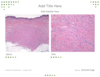
What is this tumor and three pearls.

nevus sebaceous
palliomatosis with acanthosis
Sebacesous glands attaching to epidermis
may be underlying apocrine glands
Name this tumor and 4 pearls

Merkel Cell carcinoma
dense blue dermal infiltrate
usually no epidermal connection
Nuclear molding
CD 20+ paranuclear dots Synaptophysin +
What are the three most common mets to the skin
Breast Prostate Lung
What is this tumor and 4 pearls

sebaceous carcinoma
- malignant epithelial cells with sebaceous differentitation.
- EMA stain +
- Usually around eyelids
- Cytoplasmic vacuoles indenting nuclei
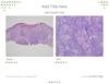
What is this tumor and three pearls.

nevus sebaceous
palliomatosis with acanthosis
Sebacesous glands attaching to epidermis
may be underlying apocrine glands
Name this tumor and 3 pearls

MF 1. CD4+ 2. Pautrier Microabscesses in epidermis 3. Epidermotropism of lymphocytes without spongiosis
Name this tumor and 4 pearls

Merkel Cell carcinoma
dense blue dermal infiltrate
usually no epidermal connection
Nuclear molding
CD 20+ paranuclear dots Synaptophysin +
What is this tumor and 4 pearls

sebaceous carcinoma
- malignant epithelial cells with sebaceous differentitation.
- EMA stain +
- Usually around eyelids
- Cytoplasmic vacuoles indenting nuclei
Name this tumor and 4 pearls

Trichoepithelioma
Dermal proliferation of basaloid cells with no epi connection
stromal stromal clefting
Papillary mesechymal bodies
may have calcified cysts
Name this tumor and 3 pearls

Poroma
Tumor arises from epidermis
Plate like squamous cells
May have many cyst like stuctures
Three things small blue cell tumors could be.
Merkel cell Lymphoma/Leukemia Mets such as lung cancer
What are the five Clark’s levels
- in situ 2. in papillary dermis 3. in papillary reticular dermal interface 4. Reticular Dermis 5. SubQ fat
What is this tumor with two pearls?

Solar lentigo
Even pigmentation without atypical cells
Delicate rete ridge projections that look like puppy feet.
What is this tumor with three pearls

Poroma
Tumor arises from epidermis
Plate like squamous cells
May have many cyst like stuctures
Name this tumor and 3 pearls

MF 1. CD4+ 2. Pautrier Microabscesses in epidermis 3. Epidermotropism of lymphocytes without spongiosis
Bonus lymphocytes intercalulated with basal cells
What is this tumor and 4 pearls

sebaceous carcinoma
- malignant squamous cells with sebaceous differentitation.
- EMA stain +
- Usually around eyelids
- Cytoplasmic vacuoles indenting nuclei
Name this tumor and 4 pearls

Trichoepithelioma
Dermal proliferation of basaloid cells with no epi connection
stromal stromal clefting
Papillary mesechymal bodies
may have calcified cysts
Name this tumor and 2 pearls

Fibroepithelioma of Pinkus
- anastomosing basaloid cells
- Loose Myxoid Stroma
Name this tumor and 4 pearls
Cylindroma
Puzzle like pattern
Hylinized stroma
well circuscribed
Ductal differentiation
Name this tumor and 3 pearls

MF 1. CD4+ 2. Pautrier Microabscesses in epidermis 3. Epidermotropism of lymphocytes without spongiosis
What does this stain show?

CK20 + paranuclear dotting for merkel cell carcinoma
What is this tumor and 4 pearls

sebaceous carcinoma
- malignant epithelial cells with sebaceous differentitation.
- EMA stain +
- Usually around eyelids
- Cytoplasmic vacuoles indenting nuclei
What is this and three other things in differential

Pagets disease of nipple EMA +
R/O melanoma in situ
Bowen’s disease
Sebaceous carcinoma
When breadloafing a specimen what percent of the margin is checked?
<1%
What are the two times you see stratum lucidum.
acral skin rubbed skin - also warts
Name this tumor and 4 pearls

Merkel Cell carcinoma
dense blue dermal infiltrate
usually no epidermal connection
Nuclear molding
CD 20+ paranuclear dots Synaptophysin +
Two finding histologically in MF.
-pautrier Microabscesses -abnormal T lymphocytes tagging along the basal layer in the epidermis without spongiosis, becoming more atypical higher in the epidermis. bonus CD4 +
Name this tumor and 4 pearls
Dermatofibroma
look for dell
cellular blue nodule in dermis
roots of spindle cells going in dermis
Name this tumor and 3 pearls

Dermatofibroma
look for dell
cellular blue nodule in dermis
roots of spindle cells going in dermis
Name this tumor and 4 pearls

Cellular Dermatofibroma
look for dell
cellular blue nodule in dermis
roots of spindle cells going in dermis
Look for Giant Touton cells
Name this tumor and 4 pearls

DFSP
sieve like infiltration of the fat or honeycombing
Storiform pattern
CD 34 +
Name this tumor and 2 pearls

Desmoplastic Melanoma
- spindle cells without pigment
- Cellular pleomorphism
Name this tumor and 2 pearls

Desmoplastic Melanoma
- spindle cells without pigment
- Cellular pleomorphism
Name this tumor and two pearls

Intradermal nevus
1, nests of bland melanocytes
- no deep mitotic figures
What are Kamino bodies
eosinophilic globules found in spitz nevi usually just beyond the tips of the dermal papilla.
They are made from basement mebrane components so are PAS positive.
What is this tumor and four pearls

Spitz Nevus
Nevus cells are spindle shaped and seem to enlongate out stream up in the papillary dermis
They are sharply circumscribed
Maturation of cells in the deeper dermis becoming smaller
Kamino bodies may be present
What is this and 4 pearls

Spitz Nevus
Nevus cells are spindle shaped and seem to enlongate out stream up in the papillary dermis
They are sharply circumscribed
Maturation of cells in the deeper dermis becoming smaller
Kamino bodies may be present
What varient of DFSP is positive for melanin
Bednar varient
+Fontana-Masson stain (Melanin) and + CD34 stain
what are these pallisaded spindle cells with this pattern calls and what tumor are they associated?

Verocay Bodies (Schwannoma) S100+
What is going on in the basal layer here of this tumor?
Basaloid follicular induction over a dermatofibroma
What does induction refer to in regards to dermatopathology
hyperplasia in nearby non-tumoral epithelial structures like basaloid follciular induction with underlying dermatofibroma
What type of glands are these in the eyelid.
Meibomian glands
these are free (not associated with follicles) modified sebaceous glands.
found on the inner aspect of the eyelid
In the eyelid what gland is this?

Glands of Zies
modified sebaceous glands associated with follicles of the eyelashes
What is this gland in the eyelid

modified lacrimal gland
What gland in the eyelid is this?

Moll’s gland
a modified apocrine gland
what is this bluesh substance in this basal cell carcinoma?

Mucin
what is this tumor

chondroid syringoma
ductal structures
Dhondroid looking stroma
This is going on in the lower leg what is happening?

Chronic venous insufficency
look for vertically oriented vessels and scar like changes
What two features might make you think desmoplastic trichoepithelioma over a morpheaform bcc
calcification
horn cysts
What is this in the eyelid

lacrimal duct
what is this tumor

basaloid follicular hamartoma
What is this?

trichostasis spinulosa
retained hair follicles (inside a follicle)
what is this stuff

monsels solution
What tumor is this

Atypical Fibroxanthoma (AFX) if dermal only, Pleimorphic Dermal scarcoma if SubQ
dermal tumor abutting the epidermis
fast growing
very pleiomorphic spindle cells
giant multinuclated cells
What is this tumor

Syringocystadenoma papilliferum
epidermal attachments of endophytic ductal structures lined with cuboidal cells.
What is this tumor

sebaceous carcinoma
What is this tumor

sebaceous carcinoma
Name three lesions that can show the Borst-Jadassohn effect
well defined nests of cells in acantholyic epidermis
SCC Bowen’s disease
Clonal Seb Ker
Hidroacanthoma (an epidermal poroma)
what is this effect known as
Borst-Jadassohn effect
Nests of cells in a clonal seb Ker
can also be in SCC bowens and hidroacanthoma (an epidermal varient of Poroma)
what is this called and what condition is it associated with?

Chornoid lamella
Porokeratosis
There is a column of parakeratois with focal loss of granular layer
what is this tumor

syringoma
What is this tumor

mucinous carcinoma
nests of basal cells floating in mucin separated by thin trabeculae
What is this tumor

mucinous carcinoma
nests of basaloid cells floating in mucin separated by trabeculae
what tumor is this

MAC Microcystic Adenexal Carcinoma
Cysts and calcifications
DEEP (unlike syrigoma)
Tadpole like ducts deep
what tumor is this

Adenoid cystic carcinoma
Looks like a sponge with mucin in the holes (ducts)
normal parotoid gland cells to left (usually arises out of a salivary gland , lacrimal, breast)
What is this tumor and the key features
sebaceous carcinoma
atypical basaloid cells
scattered sebocytes withmitoses necrosis and squamous metaplasia
What is this tumor and what is it associated with?

sclerotic fibroma
Cowdens disease
tricholemmomas
acral keratoses
oral papilomatosis
what is this tumor

mucinous carcinoma
nests of Basloid cells floating in pools of mucin
pools are separated by thin trabeculae
eyelid most common location (scalp neck axillae trunk)
what is this tumor

Microcystic Adenexal carcinoma
paisly tie pattern with cysts
deep
hard to tell from desmoplatic trichoepitheloma without deep bx
What is the average depth of perineural involvement across all body sites ?
2.7 mm
What is the average perineural depth of involvement on the head depth wise.
2.2 mm
What is the depth average for perineural involvement on the back?
4.3
Mesusured from the granular layer to the middle of the most superior nerve involved.
What is this tumor

merkel cell carcinoma
small blue cells groupsed in sheets or trabecular pattern

What is this tumor

trichoadenoma
multiple dermal cysts that have a granular layer (as opposed to seb kers which have more keratinocytes and less cysts)
cystic spaces may look like infundibular portions of hair follicles
Solid epithelial islands may be interspersed among the cyst like structures

when staining for melanoma with MART 1 what are three features used to determine + margins
Melanocytes above the Dermoepidermal Junction
Confluence of nine or more adjacent melanocytes
Nests of 3 or more atypical melanocytes
What is this condition?

Spiny keratoderma (inherited AD no problem)
(aquired over age 50) check for internal malignancy
These are not arsenical keratoses as seen below

Why is breslow thickness not useful in oral melanoma for prognosis.
There is an absense of a granular layer.
What is this tumor

blue nevus
spindled melanocyts with melanophages
look for sclerotic collagen
GNAQ & GNA11 mutations (like uveal melanoma)
What is this tumor

blue nevus
spindled melanocyts with melanophages
look for sclerotic collagen
GNAQ & GNA11 mutations (like uveal melanoma)
in melanoma what are the three most important prognostic factors on histopathologic exam
- Breslow thickness
- Mitotic rate (corolates strongly with breslow thickness)
- Ulceration
What is this tumor and what two features may it share with melanoma

Spitz nevus
Look for large junctional nests raining down
well circumscribed
May share with melanona the below
Pagetoid spread
Mitoses (usually upper dermis not lower)

What is this tumor

Bapoma (BAP1 inactivated Spitzoid nevus)
Dermal neoplasms
Melanocytes have pink glassy cytoplasm with sharp borders.
associated with multiple other cancers
What is this lesion called.
found mostly in young adult females

Reed nevus (benign) Pigmented spindle cell nevus
structureless central area with radical streaming
starburst pattern
What does aluminum choride do to collagen bundles?
turns them a magenta color
When cells are frozen what disappears first?
nuclei
