CPC: Stomach and Colon Flashcards

1
Q
A

gastritis : inflammation of the stomach

  • features of inflammatino: erythema, edema, punctate hemorrhages.
2
Q
A

gastritis

3
Q
A

flat base ulcer

4
Q
A

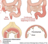
duodenal ulver. can erode into the submucosa

5
Q
A

gastric ulcer: full thickness defect in mucosa. extends into the submucosa.

6
Q

Picture of an ulcer bed: what cells are persent

A

fibrin (scarring)

neutrophils– sign of acute inflammation

7
Q

gross and micropscopic features of a benign peptic ulcer?

A

gross: sharp, punched out border, mucosal margin level with surrounding margin.
microscopic: inflammation (neutrophils), sharp border, no malignant/premature cells.

8
Q

gross and micropscopic features of a malignant peptic ulcer?

A

gross: heaped up margins

microscopic; malignant cells

can be diffuse (signet ring cells), or intestinal (glands present)

9
Q

is this ulcer benign or malignant?

A

benign, has flat edge.

10
Q

is this ulcer benign or malignant?

A

malignant: heaped up edges.

11
Q

is this ulcer benign or malignant?

A

malignant– has heaped up edges.

12
Q

is this ulcer benign or malignant?

A

malignant–has heaped up edges

13
Q

is this ulcer benign or malignant?

A

benign–it has level margins

14
Q

intestinal gastric adenocarcinoma featurs

A

tumor cells form glands, as well as mass/ulceration.

15
Q

diffuse gastric adenocarcinoma features

A
  • signet ring cells because body forms an immune response, preventing stromal flexibility
  • becomes thick nad rigid stomach wall.
16
Q

diffuse or intestinal gastric adenocarcinoma?

A

intestinal. lots of gland formation.

17
Q

diffuse or intestinal gastric adenocarcinoma?

A

diffuse. intense immune/desmoplastic response. lack of glands indicates it’s not intestinal gastric adenocarcinoma.

18
Q

diffuse or intestinal gastric adenocarcinoma?

A

gastric intestinal

19
Q
A
20
Q

diffuse or intestinal gastric adenocarcinoma?

A

diffuse type– firm rubbery gastri wall. prevents inflation in endoscopy– lots of inflammation.

21
Q

diffuse or intestinal gastric adenocarcinoma?

A

diffuse. Dysmoplastic repair causes inflammation. There’s a reduced muscle layer

22
Q

diffuse or intestinal gastric adenocarcinoma?

A

diffuse. expanded layers. dense layers are expanding

23
Q

diffuse or intestinal gastric adenocarcinoma?

A

diffuse. there is desmoplastic strome (very fibrous and dense)– reaction to the cancer cells. There are discohexive malignant cells percolating through the stroma which causes an immune esponse.

24
Q

diffuse or intestinal gastric adenocarcinoma?

A

diffuse type

25
benign or malignant ulcer?
malignant--heaped up edges. probably creates glands that are secreting.
26
benign or malignant ulcer?
malignant
27
what causes chronic gastritis
GERD, helicobacter pylori etc. PUD
28
how is H.Pylori linked to causing duodenal ulcers? (mechanisms of virulence)
1. urease: elevates local gastric PH 2. adhesins: allow adherence to gastric epithelium 3. Toxins: may be involved in ulcer or cancer development 4. flagella: provides motility in viscous mucus.
29
histologic features of H. Pylori Gastritis
1. H.pylori organisms present 2. acute inflammation with intraepithelial neutrophils 3. chronic inflammation (subepithelial plasma cells wtih lymphoid aggregates)
30
IBD --\> \_\_\_\_\_---\> colonic \_\_\_\_\_ (inflammation-dysplasia-carcinoma sequence)
IBD --\> DYSPLASIA---\> colonic ADENOCARCINOMA
31
Two immune mediated diseases that make up IBD
crohns and ulcerative colitis.
32
which one illustrates crohns vs ulcerative colitis
33
which IBD?
crohns-- ileum is affected. crohns can start anywhere in the GI whereas UC starts at the rectum and progresses upwards.
34
picture of terminal ileum. which IBD?
Crohns.
35
crohns disease with ileal ulcers.
36
what lesion pattern is this called
skip lesions seen in crohns disease.
37
which IBD? what is the characteristic feature?
cobblestone appearance of mucosa in Crohns.
38
what is the instrument poking out of?
a fistula-seen in crohns disease. fistula is an abnormal connection between two epithelialized surfaces (colon-bladder), (colon-colon), (colon-small intestine) (colon-uterus)
39
transmural inflammation causing a fistula (between colon and bladder) seen in crohns disease.
40
fistula. seen in crohns disease.
41
which IBD? what is the characteristic feature?
ulcerative colitis. there is continuous involvement without skip areas.
42
Which IBD? Is there transmural inflammation occuring in this speciman?
UC. no transmural inflammation-- continous mucosal erosion.
43
the intact portions of the colonic mucosa creates a pseudopolyp appearance.
44
fill out this table for crohns an UC
45
wall appearance for UC vs Crohns
crohns can cause transmural inflammation and breakdown so crohns causes thickened walls, where as UC still has thin walls because it only affects the mucosa.
46
Why Does UC not cause fat malabsorption
because it usually affects hte lower colon which isn't involved in nutrient absorption. Crohns can cause marked malabsorption because it can involve the small bowel.
47
meaning for dysplasia
disordered growth. can be pre-malignant, althouhgh doens't always progress to acncer.
48
If UC is occuring in someone with \_\_\_\_, it increases the risk of maliganncy. It requires endoscopic surveillance with mucosal biopsies.
if co-existing with primary sclerosing cholangitis (PSC), colitis associated neoplasia risks are increased.
49
what is happening to the colon?
low-grade dysplasia. Ducts are still apparent but there is extensive nuclear crowing, stratification and marked nuclear hyperchromasia. this pic below is normal
50
what is happening to the colon?
high -grade dysplasia. more complex architecture is happening and the normal organization of the ducts is disappearing. htere is a loss of nuclear polarity, and increased nuclear pleomorphism
51
metaplasia vs dysplasia.
metaplasia is a change in structure but still normal (ex/ squamous to columnar), but dysplasia is irregular growth.
52
polyp-cancer sequence is aka
adenoma-carcinoma seuqence. - differs from inflammation sequence (ex/ IBD--\> dysplasia--\>adenocarcinoma, or hepatitis --\> HCC) because inciting factor is genetic rather than inflammation.
53
two types of neoplastic polyps
1. conventional adenomas (tubular and villous) 2. serrated adenoma
54
T/F hyperplastic polyps can become dysplastic and then malignant
false. hyperplastic polyps to not have dysplasia ever.
55
villous or tubular adenomatous polyp?
tubular
56
villous or tubular adenomatous polyp?
villous
57
what type of adenomatous polyps is associated with more advanced lesions
villous polyps
58
59
type of adenoma/polyp
tubular
60
tubular adenoma
61
where are you most likely to see this type of polyp?
this is a sessile serrated adenoma (SSA) - more common on the right colon - they lack histological features of dysplasia, but still have malignant potential. they do not follow traditional adenoma-carcinoma sequence previously described.
62
what type of adenoma/polyp and where is it usually found?
hyperplastic polyp-- usually sigmoid/rectum (left colon) - no malignent potential. serrated morphology but only on the surface. (SSA extends to the base)
63
T/F p53 tumor suppressor gene is mutated in adenomas
false. it is mutated in 75% of colon cancer, but isn't mutated in adenomas (therefore a late step in the sequence) Loss of function of p53 causes a lack of tumor suppression, and the polyp advances into a carcinoma.
64
colon cancer
65
66
67
histological features of colon cancer: • Composed of varying complexity of glands (differentiation) – Lots of glands – ____ differentiated – Few glands – ____ differentiated • Glands often filled with debris (“\_\_\_ \_\_\_\_”) • Invasive component causes ____ response (scarring and reaction to the malignant cells) • Many different morphologies make many different subtypes, not necessary to know at this point
• Composed of varying complexity of glands (differentiation) – Lots of glands – **well** differentiated – Few glands – **poorly** differentiated • Glands often filled with debris (“**dirty necrosis**”) • Invasive component causes **desmoplastic** response (scarring and reaction to the malignant cells) • Many different morphologies make many different subtypes, not necessary to know at this point
68
invasice adenocarcinoma
69