Chapter 7 Flashcards
HSV 1 –>
HSV 2 –>
HSV 3 –>
HSV 4 –>
HSV 5 –>
HSV 6 –>
HSV 7 –>
HSV 8 –>
Oral HSV
STD HSV
VZV
EBV
CMV
?
?
Kaposi’s Sarcoma
HSV 1
Saliva or active perioral lesions
Age affects clinical presentation of symptomatic primary infections
Most commong site of latency –> Trigeminal Ganglion

HSV 1
Recurrent (secondary, reactivated) herpes
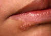
HERPES LABIALIS
Prodrome for HSV 1
Stage of intiail presentation of HSV expression
Pain, Itching, Tingling
6-24 hrs before lesion develops

Primary Herpes Gingivostomatitis (HSV 1)
Primary HSV 1 infection before age 5
Moveable and attached oral mucosa –> YELLOW LESION WITH A RED HALO
Self inoculation – Leading infectious cause of BLINDNESS
Primary Herpes Pharyngotonsillitis
Primary HSV 1 or HSV 2 infection
Sore throat
Fever -indicative of viral infection
Headache
18+ years old
Reactivation of Herpes Simplex
Most common site –> Vermillion border, adjacent to skin of lip
HERPES LABIALIS (HSV 1)
Appearance –> Small erythematous papules
* Fluid filled vesicles
* Vesicles rupture and crust within 2 days
* Heals without scarring in 7 - 10 days
Symptoms are most sever in the first 8 hours
Herpes Labialis
Reactivation of HSV 1
“Cold Sore”

Intraoral reccurent HSV
Keratinized bound mucosa (palate, attached gingiva)
Vessicles rapidly collapse
Form a cluster of erythematous macules that coalece
Damaged epithelium is lost
Central yellowish area of ulceration
Common reasons for HSV reactivion
STRESS
pregnancy
allergies
trauma
illness,
UV LIGHT
immunocompromised

Herpetic Whitlow
HSV 1 infection of thumb and fingers
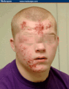
Herpes Gladiatorum
Scrumpox
Herpetic infection found in wrestlers HSV1
Contaminated abrasions

Herpes Barbae
HSV1 infection spread to bearded regions during shaving

HSV Histology
Multinucleation
Ballooning Degeneration
Tzanck Cells
HSV Histology –> Ballooning Degeneration
Acantholysis (separation of keratinocytes)
Nuclear clearing
Nuclear enlargement


Tzanck Cells
Free floating (clump of cells) epithelial cells
Cells detached
Caused by acantholysis
Pemphigous vulgaris
HSV
Two infections that involve Tzanck Cells
HSV
Pemphigous vulgaris (detached desmosomes)
How to diagnosis HSV
Clinical presentation
Cytologic smear (tzanck smear)
Tissue biopsy
Serologic testing (4-8 days after intial exposure)
Latent in TRIGEMINAL ganglion
HSV 1
Latent in DORSAL SPINAL GANGLION
VZV - “chicken pox”
HSV treatment
Acyclovir (systemic or topical cream)
Early introduction of antiviral – accelerated clinical resolution
Leading cause of infectious blindness
HSV1
Primary herpes gingivostomatitis
Varicella Zoster Virus
HSV 3
Primary infection –> Chicken Pox
Recurrent infection –> Herpes zoster (shingles)
Spread through AIR DROPLETS
Most individuals infect by 15 if not vacinnated
Latency –> DORSAL ROOT GANGLION

Chicken Pox
Primary infection of Varicella Zoster

Herpes Zoster – SHINGLES
Reccurant infection of Varicella Zoster
Limited to dermatome of infected dorsal root ganglion
Single occurance - reactivation
ORAL LESIONS – Movable or ound tissue, unilateral
White opaque vesicles that rupture and form shallow ulcerations
May cause permanent blindness
VZV on tip of nose

Sign of ocular involvment
REFER TO OPTHALMOLOGIST

Ramsay Hunt Syndrome
VZV infection
Cutaneous lesions of external auditory canal
Involvment of ipsilateral face and auditory nerves
Facial Paralysis (CN VII - facial nerve)
Hearing defects (CN VIII)
Vertigo
Infectious Mononucleosis
EBV – Epstein Barr Virus
HSV 4
Diagnosis Test –> Paul-Bunnel Heterophil Antibodies
Treatment:
Most cases resolve in 4 -6 weeks
No steriods of ABX
NSAIDS can be given
No antivirals (not clinically beneficial)
Prodrome of EBV
Fatigue
Malaise
Anorexia
EBV Oral Lesions
Oropharyngeal tonsillar enlargement
Petechiae on hard palate
Necrotizing ulcerative gingivitis

Other manifestations caused by EBV infection (3)
Oral Hairy Leukoplakia (HIV patients)
Lymphomas/lymphoproliferative disorders
Nasopharyngeal carcinoma
Cytomegalovirus
HSV 5
Resides in:
- Salivary Gland cells
- Endothelium
- Macrophages
- Lymphocytes
90% asymptomatic –> fever, joint and muscle pain
Common in AIDS patients
CHRONIC mucosal ulcerations

CMV –> HSV 5
“owl eye”
Enteroviruses (3)
Echovirus
Coxsackievirus
Poliovirus
Infection of one strain confers immunity to rest
Fecal oral transmission
Coxsackievirus manifestations/infection
Herpangina
Hand, foot, and mouth
Acute lymphonodular pharyngitis

Herpangina – COXSACKIE VIRUS (enterovirus)
Oral lesions in POSTERIOR MOUTH (Soft palate, tonsillar pillars)
Red macules –> fragile vessicles rupture –> ulcerations
Resovle in 10 days
Dysphagia, sore throat, Fever (viral infection)
Hand Foot and Mouth

Coxsackievirus (enterovirus)
Skin rash on hands and feet (ventrual surfaces, palms)
Rash in mouth
Oral lesions – ARISE FIRST with no prodrome
Resemble herpangina but larger and ore numerous
Buccal mucosa, labial mucosa, tongue (most common)
Acute lymphonodular pharyngitis

Sore throat
Fever
Mild headache
1-5 yellow to dark pink nodules on soft palate or tonsillar pillars
Represents hyperplastic lymphoid aggregates
Resolves in 10 days
Coxsackie virus (enterovirus)
Rubeola
“Measles”
Paramyxovirus
Spread via respiratory droplets
Lymphoid hyperplasia
Nine Day measles –> 3 days in each 3 stages
Rubeola First stage
3 C’s –> Cough, Coryza, Conjuctivitis
KOPLICK’S SPOTS - necrosis of epithelial cells

Koplick’s Spots
Necrosis of epithelial cells
Small blue-white macules (grains of salt on red background)
Pathognomonic – specefic characteristic of Measles
Second Stage of Reubeola
Erythematous rash begins
** Downward progression of rash **
Blanches on pressure
Third stage of Rubeola
Rash and koplick spots resolve
Everything RESOLVES
Rubella
German Measles
TOGAVIRUS
THREE DAY MEASLES – mild symptoms
Arthritis
Forchheimers’ sign (oral manifestation)

Forchheimers Sign
Oral manifestation of Rubella
Small discrete dark red papules on the palate
Congential Rubell
Triad of effects:
Deafness
Heart Defect
Cataracts
Mumps

Epidemic Parotitis
Diseas of exocrine glands – SALIVARY glands is the best known site
Glandular edema and lymphatic infiltration
Salivary gland changes: Discomfort and swelling, saliva stimulation increase in pain
Epidiymorchitis – swollen testicles in males
Epidiymorchitis
Swollen testicles in male due to MUMPS
HSV 6 & 7
Little known
Latent form in CD4 T lymphocyte
HIV/AIDS
Target cell –> CD4 T helper cell
Oral manifestations:
Candidiasis
Hairy leukoplakia (EBV - HSV 4)
Vascular malignancy
Non-Hodgkin’s lymphoma
Periodontal disease

Hairy Leukoplakia (EBV ~ HSV 4)
Most commonin HIV immunosuppressed patients
White mucosal plaque that DOES NOT RUB OFF
**Lateral border of the tongue**
No treatment is necessary

Kaposi’s Sarcoma – HSV 8
Multifocal neoplasm of vascular endothelial cell origin
Oral Lesions: Large red, blue, purple overgrowth lesions
Most commonly found hard palate, gingiva, tongue
** BIOPSY IS REQUIRED**
HIV patient’s
Eczema Herpeticum
Patients with chronic skin conditions may develop diffuse, life-threatening infection
HSV 1
Congenital Rubella
Deafness
Cataracts
Heart Disease
Rubella –>
Rubeolla –>
Togavirus
Paramyoxyvirus
HIV Periodontal Disease Presentation
Linear Erythema Gingivitis
Necrotizing Ulcerative Gingivitis
Necrotizing Ulcerative Periodontitis
Linear Erytham gingivitis
Doesn’t respond to plaque control and is more erythematous than normal
Treatment –> systemic antifungals