Acute Inflammation Flashcards
What is the purpose of inflammation?
A protective response to injury that is essential to survival. Aims to rid the body of the initial cause of the injury and the consequences of such injury
What is acute inflammation?
The body’s initial tissue reaction to injury
What is the characteristic cell involved in acute inflammation?
The neutrophil polymorph (i.e. neutrophils)
What are the 5 main physical characteristics of acute inflammation?
- Redness (rubor) - Heat (calor) - Swelling (tumor) - Pain (dolor) - Loss of function (functio laesa)
Causes of acute inflammation?
- Physical agents; thermal injury, burns, frostbite - Infections - Hypersensitivity reactions - Chemicals - Tissue necrosis; MI
What causes redness?
Dilatation of small blood vessels in the overlying skin
What causes heat?
Increased blood flow through the area within these distended vessels Can also be result of systemic fever due to chemical mediators
What causes swelling?
Oedema (accumulation of fluid in extracellular space)
What causes pain?
Distortion of tissues (swelling), pus etc
What is cholecystitis? What is it normally caused by?
Inflammation of the gallbladder. Typically caused by gallstones
What is empyema?
Pus within pleural cavity
What would be seen down microscope when looking at pus?
- Neutrophils - Necrotic material
What are the 3 major components of acute inflammation (regarding changes in vessel)?
- Changes in vessel calibre 2. Increased vascular permeability and fluid exudate formation 3. Cellular exudate formation
What is an exudate?
Extravascular fluid with high protein concentration, containing cellular debris. Implies inflammation.
What is a fluid exudate formation?
Fluid leaking into extracellular space due to increased permeability
What is a cellular exudate formation?
Cells leaking into tissues
What is the purpose of the vessels undergoing these changes?
To maximise the movement of plasma proteins and cells into the site of infection/injury so noxious agents can be destroyed
What is transudate?
Extravascular fluid with low protein, little or no cellular component
What is oedema?
Excess fluid in interstitial tissue/ serous cavities – exudate or transudate
What is pus?
Inflammatory exudate rich in neutrophils, dead cell debris and microbes
Fluid can be classified as a transudate or an exudate. Transudate vs exudate causes?
Transudate: occurs due to increased hydrostatic pressure or low plasma oncotic pressure Exudate: occurs due to inflammation and increased capillary permeability
Examples of a transudate fluid? Examples of an exudate fluid?
Transudate: congestive heart failure, cirrhosis, nephrotic syndrome, PE Exudate: pneumonia, cancer, TB, viral infection, PE, autoimmune
Contents of a transudate fluid? Of an exudate fluid?
Transudate: low protein and LDH Exudate: high protein and LDH
What are the 2 types of fluid?
Transudate or exudate
Describe the changes in the vessel calibre during acute inflammation
Initial transient vasoconstriction then vasodilation
Explain changes in colour of skin when you drag nail across
Initial white - vasoconstriction Red - vasodilation
How does vasodilation affect blood flow?
Increases it up to 10x
What is effect of increased blood flow?
Heat and redness
What are changes in vessel calibre mediated by?
Histamine and NO which act on vascular smooth muscle to relax it
Explain:
1) redness
2) swollenness
3) heat
seen in these crusted skin lesions?

1) Red = vascular dilatation
2) Swollen = inflammatory exudate into surrounding tissues
3) Hot = vascular dilatation
What causes the formation of fluid exudate after changes in vessel calibre?
Increased permeability of microvasculature results in the escape of protein rich fluid into the tissue. Causes:
- Chemical mediators e.g. histamine, NO, leukotriene
- Direct vascular injury e.g. trauma
- Endothelial injury e.g. bacteria and toxins
How does hydrostatic pressure inside vessel change during acute inflammation?

Normal vessel:
- High hydrostatic pressure inside vessel due to plasma proteins forces fluid out but returns at venous end where hydrostatic pressure is low
Acute inflammation:
- Hydrostatic pressure is increased and plasma proteins escape into extravascular space
- This increases colloid osmotic pressure –> more fluid leaves vessels (exudation) into extracellular tissue
Effect of fluid exudate on toxins?
Dilution of toxins
Effect of fluid exudate on antibodies?
Allows entry of antibodies
Effect of fluid exudate on drugs?
Allows transport of drugs so drugs can get to affected area
Effect of fluid exudate on fibrin?
Fibrin formation as fibrinogen escapes into extracellular tissues
Effect of fluid exudate on nutrients and oxygen?
Increased delivery of nutrients and oxygen to damaged tissue
Effect of fluid exudate on immune response?
Stimulation of immune response
How is cellular exudate formed? (i.e. what causes cellular components to leak out of blood vessels?)
- Loss of fluid into tissues and increased calibre of vessels –> slower blood flow and increased viscosity of blood –> stasis
- This causes neutrophils to line up along vascular endothelium, stick to endothelium and migrate through wall into tissues

What is margination?
Margination refers to the prolonged transit of neutrophils
Explain process of cellular exuvate formation
- Margination of neutrophils
- Pavementing of neutrophils
- Neutrophils pass between endothelial cells
- Pass through basal lamina and migrate into adventita

Is the movement of neutrophils into extracellular space an active or passive movement?
How does this differ from the process by which RBCs leave the vessels?
Active emigration
RBCs leave via diapedesis - this is a passive process which depends on the hydrostatic pressure forcing the RBCs out
How do chemical mediators promote the leukocyte endothelial adhesion?
Selectin - ligand interaction –> ‘rolling’ of neutrophils
- Endothelial expression of selectin
- Neutrophil expression of selectin ligand
ICAM - integrin interaction –> neutrophils stop ‘rolling’ and stick to vascular wall
- Endothelial expression of ICAM
- Neutrophil expression of integrin

What is endothelium ‘selectin’ and neutrophil ‘integrin’ expressed in response to?
IL-1, TNF, LPS and C5a produced at site of inflammation
Under normal conditions, what do neutrophils and endothelial cells express?
Neutrophils - Selectin Ligand
Endothelial cells - ICAM
These are incompatible so the neutrophils circulate freely
When the tissue is infected and inflamed, what do activated macrophages release?
Cytokines - IL-1, TNF
What do IL-1 and TNF stimulate expression of?
Selectin on the endothelial cell membrane which binds to Selectin Ligand on neutrophils
What is result of selectin binding to selectin ligand?
Neutrophils slow down (but Selectin-Selectin Ligand interaction is not enough to stop neutrophils completely, but they instead ‘roll’)
What else is secreted in inflamed and infected tissue?
C5a from increased complement activation
LPS from bacteria
LPS and C5a accumulate
What does this accumulation of LPS and C5a induce?
Integrin expression on neutrophil surface
Integrin then binds to ICAM on endothelial cells lining blood vessel
What does combination of Selectin-Selectin Ligand and Integrin-ICAM interaction cause?
Neutrophils stop ‘rolling’ and stick to vascular wall close to site of infection
How is C5a helpful?
Anaphylatoxins - increase vascular permeability which helps neutrophils reach inflamed tissue
What is chemotaxis?
Neutrophils attracted towards certain chemical substances
Image of neutrophils adhering to walls of blood vessel

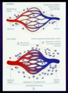
Comparison of normal and inflamed ECM
Inflamed:
- Arteriole dilation, venule dilation and expansion of capillary bed –> increased blood flow
- Deposition of fibrin and other plasma proteins
- Neutrophil emigration –> oedema expands ECM
Where are neutrophils produced?
Bone marrow
What is the most common white cell in the blood?
Neutrophils
Describe the lifespan of neutrophils
Short (hours in tissue)
How do neutrophils destroy microorganisms?
- Microorganisms are opsonised first by immunoglobulins or complement components
- Neutrophils involved in phagocytosis and intracellular killing
- Neutrophils release lysosomal products which attract other leukocytes, increase vascular permeability, some act as pyrogens
What are pyrogens?
substances that can produce a fever by acting on hypothalamus
Cell derived vs plasma derived mediators of acute inflammation?
Plasma derived:
- Complement system
- Kinin system
- Coagulation system
- Fibrinolytic system
Cell derived:
- Histamine
- Prostaglandins
- Lysosomal components
- Leukotrines
- Cytokines
What is histamine released by?
Mast cells
Effect of histamine?
increases vascular dilation and permeability
What are prostaglandins?
long chain fatty acids that come from arachadonic acid
Effect of prostaglandins?
When tissue is damaged or infected, they create the reactions that cause pain, fever and inflammation. Prostaglandins also stimulate the formation of a blood clot and the contraction of the blood vessel wall when your body is bleeding.
What are cytokines mainly produced by?
predominantly by macrophages and lymphocytes
Examples of cytokines?
Interleukins 1–10, tumor necrosis factor α (TNF-α), and interferon γ (INF-γ)
Which part of the arachidoniac acid pathway do;
a) steroids
b) apsirin inhibit?
Certain drugs can inhibit different parts of the pathway:
- Steroids inhibit phospholipases
- Prevents phospholipids turning into arachidonic acid
- Aspirin etc inhibit cyclooxygenase
- Prevents arachidonic being converted to prostaglandins

What are the most commonly measured inflammatory markers?
- C-reactive protein (CRP)
- Erythrocyte sedimentation rate (ESR)
- Procalcitonin (PCT)
What is ESR?
- ESR is an indirect measurement of plasma protein concentrations and is influenced by a number of disease states.
- Because the ESR depends on several proteins with varying half-lives, the rate rises and falls more slowly than CRP
- Normal ESR values are specific to age and sex
- The rate increases steadily with age and is higher in women than in men.
What is CRP?
A substance produced by the liver in response to inflammation
Benefits of testing CRP in acute inflammation?
Concentrations change rapidly within the first 6-8 hours after injury, peak after 48 hours, and return to normal levels once the issue has resolved.
When can inflamation start to become harmful to the body?
- Digestion of normal tissues e.g. abscess cavities
- Swelling e.g. acute epiglottitis
- Inappropriate inflammatory response e.g. hayfever
What is fibrinous inflammation? What can it result in?
- Increased vascular permeability can result in large molecules such as fibrinogen entering tissues and forming fibrin
- Characteristic of inflammation of linings of body cavities e.g. pericardium, pleura.
- Can be removed by firbrinolyis and removal of debris by macrophages.
- If not can lead to scarring (organisation) obliteration of pericardium
Fibrin impedes movement of microorganisms, trapping the and facilitating phagocytosis, acts as a scaffold for granulation tissue formation
What is systemic inflammatory response syndrome?
Systemic inflammation and widespread tissue injury (can be due to sepsis)
What is acute (adult) respiratory distress sydrome?
A type of respiratory failure characterised by rapid onset of widespread inflammation in the lungs.
Causes: Direct causes include pneumonia (including bacterial and viral), aspiration, inhalational lung injury, lung contusion, chest trauma, and near-drowning. Indirect causes include sepsis, shock, pancreatitis, trauma
What is chronic granulomatous disease of childhood?
- An inherited condition of the immune system. It’s known as a “primary immunodeficiency.”
- The body’s immune system fights infections and anything it sees as foreign invaders to the body.
- In CGD, neutrophils aren’t able to make the hydrogen peroxide our bodies need to fight certain invaders like bacteria or fungus.
- Since the neutrophils can’t control the bacteria or fungus, they can spread causing severe infections. The body realises the immune system isn’t clearing the infection, so the granulomas keep growing.
What is hereditary angiodema?
Recurrent episodes of severe swelling (minor trauma or stress may trigger an attack, but swelling often occurs without a known trigger)
What is amyloidosis?
A group of rare, serious conditions caused by a build-up of an abnormal protein called amyloid in organs and tissues throughout the body.
What is ideal result of acute inflammation?
Resolution
What is resolution dependent on?
- Only occurs if there is minimal cell death
- Dependent on regenerative capacity of the organ/tissue
Some patients get excessive exudate in acute inflammation. What can this lead to?
Suppuration - formation of pus which can discharge (naturally) or may have to be done surgically
Tissue then goes on to repair and organise (repaired by granulation tissue and then fibrosis)
Some patients experience excessive necrosis during acute inflammation. What can this lead to? What is this due to?
- Due to a persistant causal agent causing chronic inflammation
- Healing and repair via fibrosis –> scarring
What are the 3 major steps that acute inflammation can be broken down into?
- Vasodilation leading to increased blood flow
- Increased vascular permeability
- Formation of exudate
What are the cells most prominent in AI? In CI?
AI - neutrophils
CI - macrophages
What is vasodilation? What is it induced by?
- The opening up of blood vessels
- Induced by several mediators: notably histamine and nitric oside on vascular smooth muscle
What is the effect of vasodilation?
- Increased blood flow –> rubor (redness), calor (heat)
- Increased permeability of microvasculature
What is the result of increased vascular permeability?
There is a loss of intravascular fluid due to increased intravascular hydrostatic pressure and reduced intravasular osmotic pressure
- Proteins and fluid leak into extravascular space
- Oedema forms

How is exudate formed?
Protein-rich fluid leaks into the extravascular tissue –> forms exudate
As fluid is lost into the tissue (exudate), how does this affect viscosity of blood?
Increases viscosity of blood and stasis occurs
As blood becomes more viscous due to exudate forming, what begins to accumulate along the endothelium?
Leukocytes, especially neutrophils
What then happens to these leukocytes that have accumulated along the endothelium?
They are able to travel from the lumen of the blood vessel into the extravascular tissue by extravasation

Exudate vs transudate:
- Protein content?
- Cellular constituent?
- Causes?
Protein content:
- Exudate - high
- Transudate - low
Cellular constituent:
- Exudate - present
- Transudate - none/minimal
Causes:
- Exudate - Infection, malignancy, pancreatitis
- Transudate - Heart failure, cirrhosis, nephrotic syndrome
What type of fluid is pus?
Exudative that contains leukocytes (mostly neutrophils), cellular debris and micro-organisms
What type of cells are neutrophils?
- Polymorphonuclear granulocyte
- Most common leukocyte (40-75%)

What is the primary function of neutrophils?
Acute inflammatory response - highly mobile and phagocytic
How are neutrophils attracted to site of inflammation?
Chemotaxis - then undergo amoeboid movement
How do neutrophils adhere to micro-organisms?
Able to bind onto microbes either by immunoglobulins or complement proteins
Describe process of phagocytosis?
- Neutrophils send pseudopodia, which fuse and form a phagosome
- The phagosome fuses with lysosomes that contain materials needed to kill the microbe

What are the 3 outcomes of inflammation?
- Complete resolution
- Fibrosis
- Progression into chronic inflammation
If inflammation is not regulated, what can it result in?
- Can result in delayed wound healing
- Reduced clearance of infections
- Exaggerated response: Sepsis/SIRS
- Improper activation of inflammatory response –> allergies
What 3 major blood flow changes occur during acute inflammation? What chemical is responsible for this?
- A transient vasoconstriction** of arterioles followed by **vasodilation of arterioles and capillaries.
- This results in an increased blood flow to the tissue – causing the signs of rubor and calor.
-
Increased permeability of blood vessels, which results in an exudate (protein rich fluid) forming within tissues
- Results in tumor
- The circulation is slowed
- Results in tumor
This is mainly mediated by release of histamine from mast cells, basophils and platelets.
What is the effect of the fluid exudate forming in acute inflammation on RBCs?
This results in an increased concentration of red blood cells within circulation, due to exudation of fluid, causing stasis of circulation in the area.
Where is histamine released from?
mast cells, basophils and platelets
Exudation of fluid occurs due to which law?
Exudation of fluid occurs due to Starling’s Law.
How does fluid exudation occur due to Starling’s Law?
- Vasodilation of arterioles leads to an increase in hydrostatic pressure which leads to increased fluid movement out of vessels.
- The increased permeability of vessels results in protein moving to the interstitium, leading to an increase in colloid osmotic pressure, acting to further increase fluid movement out of vessels.
Function of the formation of fluid exudate?
- Increased lymphatic drainage, which can help to remove damaging substances.
- This fluid allows plasma proteins to be delivered directly to the site of injury; antibodies, fibrin formation as fibrinogen escapes into extracellular tissues
- Dilution of toxins
- Allows transport of drugs so drugs can get to affected area
- Increased delivery of nutrients and oxygen to damaged tissue
- Stimulation of immune response
Describe and explain the movement of neutrophils during acute inflammation
- Margination - neutrophils line up along the endothelium near the site of injury due to stasis of circulation
- Rolling - neutrophils roll along the endothelium, sticking intermittently.
- Adhesion - neutrophils they stick more avidly to the endothelium
- Emigration - Finally, the neutrophils emigrate through the blood vessel walls due to relaxation of inter-endothelial cell junctions and digestion of the vascular basement membrane.
What change in blood flow enables margination of neutrophils?
Stasis of circulation due to exudation of fluid
What is chemotaxis?
Movement of a motile cell or organism in a direction corresponding to a gradient of increasing or decreasing concentration of a particular substance.
What process attracts neutrophils to areas of damage? Which chemicals are involved?
Chemotaxis - often following a concentration gradient of chemotaxins, which include C5a, LTB4 and bacterial peptides.
Function of neutrophils in acute inflammation?
Neutrophils are necessary as they are able to phagocytose pathogens and cellular debris to remove them, which is facilitated by opsonins.
4 major functions of acute infmlammation?
- Exudation of fluid helps deliver plasma proteins to sites of injury, dilutes toxins and increases lymphatic drainage
- Infiltration of neutrophils leads to removal of pathogens and cellular debris
- Vasodilation much like exudation helps to increase delivery of necessary proteins and cells, as well as increasing tissue temperature
- Pain and loss of function help to enforce rest and lower the risk of further tissue damage
Hydrostatic pressure:
- The pressure of the blood against the blood vessel wall - is the opposing force to osmotic pressure
- Increased hydrostatic pressure forces fluid out of the capillary
Osmotic pressure:
- Osmotic pressure draws fluid back into the vessel
- Osmotic pressure is determined by osmotic concentration gradients; the difference in the solute-to-water concentrations in the blood and tissue fluid.
