42: Orthopedics Flashcards
What is the treatment for fibrous dysplasia of bone?
Benign bone tumor treated with curettage +/- bone graft
[UpToDate: Fibrous dysplasia is a lesion in which portions of the bone are replaced by fibrous connective tissue and poorly formed trabecular bone. The process originates in the medullary cavity. It is caused by a postzygotic mutation in the guanine nucleotide stimulatory protein (GNAS1) gene. It is more of a skeletal dysplasia than a true neoplasm.
Fibrous dysplasia most commonly presents in the teens or 20s. It may occur in any bone but is most common in the proximal femur, tibia, ribs, and skull . Fibrous dysplasia affects slightly more males than females.
The treatment of fibrous dysplasia depends upon the presence of symptoms. Asymptomatic patients may be observed every six months with serial radiographs. Children with large lesions or lesions in the proximal femur or other weight-bearing bones are observed more frequently.
Curettage, bone grafting, and stabilization may be warranted for fibrous dysplasia that is associated with symptoms (pain, deformity) or fracture; however, there is a high rate of recurrence. Autograft should not be used because it will be resorbed. Bisphosphonate therapy is another alternative for symptomatic patients.
The deformity of fibrous dysplasia may progress with skeletal growth. Fibrous dysplasia usually is static after growth ceases but may be reactivated with pregnancy. Fibrous dysplasia often recurs after curettage and bone grafting.]

What is the treatment for Legg-Calve-Perthes disease?
Maintain range of motion with limited exercise
[Femoral head will remodel without sequelae. Surgery if femoral head is not covered by the acetabulum]
[UpToDate: Children diagnosed with LCP should be made nonweight bearing and referred to an experienced pediatric orthopedist for management. Therapy for LCP is poorly defined, because no large controlled trials are available, and long-term consequences become evident only after decades of follow-up. Treatment focuses on containing the femoral head within the acetabulum through the use of splints or occasionally surgery.
Almost all children do well in the short term. However, long-term outcome depends upon age at time of disease onset and degree of involvement of the femoral head. Children who are younger than six to eight years have a better prognosis, perhaps because more time is permitted for femoral remodeling and because before eight years of age the acetabulum is plastic and can mold to the deformed femoral head, maintaining congruity.]

What is the typical treatment for an ankle fracture?
Cast and immobilization
[UpToDate: Emergent conditions, such as an open fracture or neurovascular impairment, require immediate surgical consultation and treatment. Fracture dislocations must be reduced immediately to prevent severe complications, such as avascular necrosis.
Once emergent conditions are excluded, clinicians should evaluate the fracture more closely, focusing on any malalignment or instability, to determine proper management and follow-up. The ankle should be splinted at 90 degrees (ie, neutral position) to provide support and control pain. Usually, a short-leg posterior splint is sufficient. A sugar-tong (ie, coaptation) splint can be added for additional mediolateral support. If significant swelling or deformity is present, adequate padding should be placed prior to application of the splint to allow for further swelling, while maintaining stability.
Clinicians should instruct the patient to call immediately for:
- Pain that is severe or increasing
- Numbness that is new or worsening
- Skin discoloration (eg, dusky toes) distal to the splint
These complaints may represent vascular compromise or some other serious complication and should be investigated immediately. Any patient complaint of skin irritation, a splint which has become excessively tight or loose, or a splint which has gotten wet should also be assessed. An examination and repeat radiographs to check for acceptable alignment are generally performed during the first follow-up visit at 10 to 14 days.
For stable, nondisplaced, isolated malleolar fractures, the patient should rest, elevate the involved ankle above the level of the heart, and apply ice, while keeping the splint dry. If the injured leg is placed in a prefabricated splint able to withstand ambulation, the patient may bear weight as tolerated. The importance of elevating the leg should be emphasized to patients, as complications with splint treatment often stem from allowing the foot to remain in a dependent position for too long.
Patients awaiting orthopedic consultation or surgery should remain nonweightbearing in a splint (as described above), apply ice while keeping the splint dry, and use pain medication as needed. If surgery is planned in the acute setting, excessive use of narcotic analgesics should be avoided, if possible, until the orthopedic surgeon is able to explain the procedure and obtain informed consent. Management of specific fracture types is discussed immediately below.]

Which nerve roots contribute to the ulnar nerve?
C8-T1

Anterior shoulder dislocation is associated with a risk of injury to what?
Axillary nerve
[UpToDate: Clinicians perform a neurovascular examination paying particular attention to distal pulses and the function of the axillary nerve, which is most commonly injured in anterior shoulder dislocations. Axillary nerve dysfunction manifests as loss of sensation in a “shoulder badge” distribution, although this finding is not reliably present. Deltoid muscle weakness may also be present, but is impractical to assess during the acute injury. Some degree of axillary nerve dysfunction is present in 42% of patients with an anterior dislocation, but most patients recover completely without intervention. In many cases, dysfunction resolves with reduction.
Associated fractures identified on plain radiographs include Hill-Sachs deformities, Bankart lesions, and greater tuberosity fractures. A Hill-Sachs deformity is a cortical depression in the humeral head created by the glenoid rim during dislocation. They occur in 35% to 40% of anterior dislocations and are seen on the AP radiograph with the arm in internal rotation. Bankart lesions occur when the glenoid labrum is disrupted during dislocation and a bone fragment is avulsed. Bony Bankart lesions are present in 5% of patients, while soft tissue Bankart lesions (no bone is avulsed) occur in approximately 90% of patients less than 30 years old with an anterior shoulder dislocation. Greater tuberosity fractures are present in 10% of patients. Indications for orthopedic referral, including selected Bankart and Hill-Sachs lesions, are discussed separately.]

What is the biggest risk factor for non-union following a fracture?
Smoking
[UpToDate: Common reasons for nonunion and malunion include a tenuous blood supply to the fractured bone (eg, scaphoid, proximal fifth metatarsal, talar neck), behaviors that interfere with bone healing (eg, smoking), poor bone fixation (ie, excessive movement at the fracture site), poor apposition of bone fragments (ie, fragment ends too far from one another), and infection.]
Fractures in which 3 areas of the body are associated with avascular necrosis?
- Scaphoid
- Femoral neck
- Talus
[UpToDate: A variety of traumatic and atraumatic factors contribute to the etiology of osteonecrosis. A definitive etiologic role has been established for some of these factors, based upon longitudinal cohort studies or meta-analyses, but not for the majority, which are considered associated risk factors. Use of glucocorticoids and excessive alcohol intake are associated with more than 80% of atraumatic cases.
The pathogenesis of osteonecrosis is an area of controversy. Most experts believe that it is the result of the combined effects of genetic predisposition, metabolic factors, and local factors affecting blood supply, such as vascular damage, increased intraosseous pressure, and mechanical stresses. The early stages of the natural history are unclear, as these stages are largely asymptomatic and the patient does not present until later. It is generally agreed that there is an interruption of the blood circulation within the bone; subsequently, the adjacent area becomes hyperemic, resulting in demineralization, in trabecular thinning, and, later, in collapse.
The histopathologic finding of bone marrow infarction has been noted in marrow samples from patients with some of the same disorders that cause clinically apparent osteonecrosis, but neoplastic disorders, particularly hematologic and lymphoid malignancies and metastatic cancer with associated coagulopathy, are other potential etiologies. The causes of bone marrow infarction (bone marrow necrosis) are discussed elsewhere.]

What is the treatment for a displaced calcaneus fracture?
Open reduction and internal fixation (ORIF)
[UpToDate: Emergent (ie, immediate) surgical referral is required for open fractures, fractures associated with neurovascular injury, fractures associated with dislocation (which must be reduced immediately), and suspicion or diagnosis of acute compartment syndrome. Virtually all intraarticular calcaneus fractures should be assessed and managed by a surgeon, and urgent referral is indicated. In addition, calcaneal fractures that are comminuted or involve noticeable displacement warrant urgent referral. As a general rule, it is best to contact the surgeon at the time of diagnosis. Initial management includes:
- Elevating the affected foot above heart level and applying ice.
- Providing adequate analgesia.
- Assessing the skin and swelling.
- Evaluating for other associated injuries of the feet, ankles, legs, and thoracolumbar spine.
- Possible admission for observation and pain control depending upon the severity of the fracture, and consequent risk of major complications (eg, compartment syndrome), and the presence of concomitant injury.
When classifying calcaneus fractures, the most important step is to distinguish extraarticular from intraarticular injuries. Extraarticular fractures generally have a good prognosis, and if nondisplaced may not require referral. The major types of extraarticular fractures are reviewed in the text.
Intraarticular fractures virtually always require referral. Their management is controversial and surgery may be performed.]

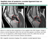
What is the operative treatment for Dupuytren’s contracture?
Transverse carpal ligament release
[UpToDate: Surgery has been the treatment of choice for advanced stages of disease, if function is impaired or if a contracture is progressing. At present, typical interventions are a transection of cords (fasciotomy) or an excision of diseased fascial bands (fasciectomy) with or without excision of the overlying skin. Surgery (limited palmar fasciectomy) should be considered only with functional impairment or in the presence of a contracture that is progressive. The initial results are generally good, but the recurrence rate is high. Flexion deformities >30 to 40 degrees at the metacarpophalangeal (MCP) or >20 degrees at the proximal interphalangeal (PIP) joint have been suggested as indications for surgery. PIP joint contractures are less likely to achieve full correction of the contracture and are less likely to respond to surgery in advanced stages (eg, contractures >60 degrees). The aims of surgery are to reverse digital contractures and to restore hand function. Surgery in younger patients has a much higher recurrence rate than in older patients, but this is likely due to the increased severity of the disease in patients who present with contractures at a younger age.
The likelihood of recurrence after surgery may be related to the degree of cellularity of the lesion. A study of 10 patients found that signal characteristics on magnetic resonance imaging (MRI) predicted cellularity and, thus, may provide prognostic information regarding the likelihood of recurrence after surgery, although further study is warranted before this is used in clinical practice. A second operation can be performed in patients who have a recurrence after the first procedure.
The specific surgical technique used depends upon the individual characteristics of the patient and upon the preferences of the surgeon. More aggressive techniques such as radical fasciectomy or dermofasciectomy do not appear to offer an advantage over limited fasciectomies.]

Which nerve roots contribute to the median nerve?
C6-T1

What is the last clinical sign of compartment syndrome to manifest itself?
Loss of distal pulses
[UpToDate: Several common misconceptions exist pertaining to the clinical diagnosis of ACS. Clinicians should be aware that ACS can occur in the presence of open fractures as these may not necessarily decompress elevated compartment pressures. Moreover, ACS can occur without a fracture (or a crush injury). The diagnosis is often delayed because clinicians fail to consider ACS in patients without a fracture. Arterial pulses and normal capillary refill can persist despite the presence of a prolonged, severe ACS. Pulse oximetry is an insensitive instrument for diagnosis and should not be relied upon.]
What are the non-operative treatments for nerve root compression?
- NSAIDs
- Heat
- Rest
[UpToDate: For patients with acute lumbosacral radiculopathy and painful radicular symptoms caused by disc herniation or foraminal stenosis, the following interventions may be useful:
We suggest short-term treatment with either a nonsteroidal anti-inflammatory drug (NSAID) or acetaminophen (Grade 2C).
We suggest temporary activity modification, including avoidance of provocative activities (Grade 2C).
Physical or manual therapies are often tried for patients with persistent symptoms that are mild to moderate in nature, but there is no convincing evidence that they are effective for this indication. Physical therapy in the first one to two weeks is not recommended because patients with mild symptoms are likely to improve on their own while patients with very severe symptoms cannot participate in exercise therapy.]
What would be the physical manifestation of a nerve root compression of L3 nerve (L2-L3 disc)?
Weak hip flexion
[UpToDate: There is marked overlap of the L2, L3, and L4 innervation of the anterior thigh muscles, making it difficult to differentiate these spinal nerve root levels based on symptoms, neurologic examination, or electrodiagnostic testing. Thus, these radiculopathies are generally considered as a group. These nerve roots are most commonly involved in older patients with symptoms of spinal stenosis.
Acute back pain is the most common presenting complaint, often radiating around the anterior aspect of the leg down into the knee and occasionally down the medial aspect of the lower leg as far as the arch of the foot. On examination, there may be weakness of hip flexion, knee extension, and hip adduction. Higher lesions may result in greater weakness of the hip flexors. Sensation may be reduced over the anterior thigh down to the medial aspect of the lower leg. A reduced knee reflex is common in the presence of moderate weakness.
Electromyography and nerve conduction studies may reveal abnormalities confined to muscles of the affected root(s), including the quadriceps, leg adductors, and iliopsoas, with associated paraspinal abnormalities. Saphenous sensory response remains normal even if sensory loss is prominent in the distal leg.]

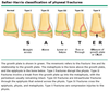
How should one treat Salter-Harris I and II fractures?
Closed reduction
[Good prognosis]
[UpToDate:Salter I (Ogden IA-B) — The fracture line extends through the zone of hypertrophic cartilage (zone 3), causing the epiphysis and physeal elements to separate from the metaphysis. Type I injuries can have normal radiographs and the diagnosis is therefore often made clinically when focal tenderness is found over the growth plate.
- A type IB Ogden fracture is characterized by the fracture line extending through the primary spongiosa bone layer resulting in a thin line of bone displaced with the epiphysis. Type IB fractures usually occur in children with systemic diseases such as myeloproliferative disorders. Subsequent growth is usually normal with Type IA and IB fractures.
- A Type IC Ogden fracture has an associated injury to the germinal portion of the physis. Type IC fractures can cause growth arrest and occur rarely after age two to three years.
Salter II (Ogden IIA-D) — The fracture line extends through the physis and then propagates across the physeal-metaphyseal junction into the metaphysis. Type II fractures are the most common physeal fractures. The resultant metaphyseal wedge in a Salter II or Ogden Type IIA fracture is called the Thurston Holland fragment.
- A type IIB involves further extension of the fracture line bidirectionally through the metaphysis creating a free metaphyseal fragment or multiple fragments.
- A type IIC fracture is a transverse physeal fracture that includes a thin layer of metaphysis along with the metaphyseal triangular corner segment.
- A type IID fracture is characterized by the angulation of the two segments resulting in the metaphyseal segment compressing the physis and creating an osseous bridge that leads to permanent growth arrest.]

What test should be ordered for a patient with suspected nerve root compression?
MRI
[UpToDate: For imaging of the lumbar spine, MRI, CT, and CT myelography (CT scan after intrathecal administration of contrast media) are equally sensitive for the diagnosis of disc herniation. For routine initial assessment, an MRI is more informative than CT because it can also identify other intraspinal pathologies, including inflammatory, malignant, and vascular disorders. In addition, MRI is not associated with ionizing radiation and is less invasive than CT myelography.
However, there is a high prevalence of abnormal neuroimaging findings in asymptomatic individuals, including some who have what appears to be frank nerve root compression by MRI. As an example, one study of 98 people without back pain found MRI evidence of disc herniation in 27%. Furthermore, lumbar spine abnormalities on MRI in asymptomatic patients do not appear to be predictive for the future development or duration of low back pain.
Although rarely indicated, CT myelography can visualize spinal nerve roots and their trajectory through the neural foramina. It is useful for patients with intolerance of or contraindications to MRI (eg, implanted electrical devices such as cardiac pacemakers or defibrillators) when standard CT fails to define the anatomic correlates of the clinical presentation. In addition, CT myelography is preferred for patients who have surgically placed spinal hardware that produces magnetic artifacts.
A CT scan can assess osseous structures better than either plain radiography or MRI and is therefore helpful in assessing for bony disease. However, CT alone is unable to visualize nerve roots, so it is not helpful in the direct imaging of a radicular process.]
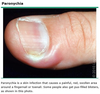
What is the treatment for Felon?
Incision over the tip of the finger and along the medial and lateral aspects to prevent necrosis of the tip of the finger
[UpToDate: A very early presentation of a pulp space infection without a fluctuant swelling may be treated with warm soaks, rest, elevation, and oral antibiotics. However, most patients with a pulp abscess require surgical intervention. A simple incision and drainage procedure may provide temporary relief; however, it is better to debride the abscess cavity in the operating room because the infection may be more extensive than the symptoms and clinical appearance suggest.]

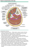
The posterior tibial artery lies in which leg compartment?
Deep posterior compartment

The anterior tibial artery lies in which leg compartment?
Anterior compartment

Which type of Salter-Harris fracture crosses the epiphysis and the growth plate (physis), but not the metaphysis?
Salter-Harris III
[UpToDate: Salter III (Ogden IIIA-D) — The fracture line extends through the physis and then spreads through the epiphysis into the intraarticular space. If the transverse fracture extends across the complete width of the physis, two epiphyseal segments may be formed.
- A Type IIIB fracture, similar to type IB, courses through the primary spongiosa physeal layer resulting in a thin bony metaphyseal line displaced with the epiphyseal segment.
- Type IIIC injuries involve epiphyses in mostly nonarticular areas.
- Type IIID fractures penetrate the germinal zone and interrupt the blood supply to the avulsed segment. These fractures are difficult to visualize on traditional radiographs.]

What is the treatment for slipped capital femoral epiphysis?
Surgical pinning
[UpToDate: The treatment of SCFE is operative. Children with SCFE, regardless of classification, should be referred promptly to an orthopedic surgeon; they must avoid bearing weight until they have undergone orthopedic evaluation.
Approximately 30% to 60% of patients with unilateral SCFE at presentation eventually have SCFE in the contralateral hip. To prevent delay in diagnosis of the second slip, all patients with unilateral involvement who do not undergo prophylactic repair of the contralateral hip should be followed closely by an orthopedic surgeon until after the child has finished growing. Patients and parents should be instructed to seek medical attention immediately if they experience symptoms of SCFE (eg, nonradiating, dull, aching pain in the hip, groin, thigh, or knee).]

What is the treatment for a clavicle fracture?
Application of a sling due to risk of vascular impingement
[UpToDate: Operative versus nonoperative treatment of displaced midshaft clavicle fractures is individualized and based on factors such as the degree of displacement, shortening, and comminution, as well as functional and cosmetic concerns. The data available to address this important question is limited, but studies do not show a clear benefit to surgery over nonoperative management in many cases.
Patients with nondisplaced or minimally displaced middle third fractures are treated with a sling, analgesics, and regular elbow range of motion exercises. For patients with complete displacement who decline surgery, immobilization using a figure of eight bandage may help to correct or prevent shortening, but a sling is acceptable.
Clinically, fractures of the distal clavicle are easily confused with acromioclavicular separations. Radiographs are necessary to differentiate between the two. Orthopedic referral is recommended for most distal clavicle fractures. An exception is type I fractures confirmed by normal stress views using plain radiographs. Confirmed type I fractures can be managed using a sling and early shoulder range of motion exercises, begun as soon as symptoms allow.
Acute fractures of the proximal clavicle should alert the physician to the possibility of serious internal injury. In most cases, evaluation is performed in the emergency department. If there are no associated injuries and the fracture is nondisplaced, treatment involves sling immobilization. Stress fractures develop insidiously from repetitive stress on the proximal clavicle related to a range of activities, including rowing and gymnastics. Conservative treatment is generally successful.
Among children, 90% of clavicle fractures occur in the middle third. In children 10 and under, the majority are nondisplaced; above age 10, the majority are displaced. Treatment generally does not differ from that recommended for adults, but healing occurs more quickly.]

What is the treatment for a talus fracture?
Closed reduction for most
[Open reduction and internal fixation (ORIF) for severe displacement]
[UpToDate: Most talar head fractures are managed by surgeons, and non-operative treatment of talar neck fractures should be performed only by primary care clinicians experienced in caring for patients with musculoskeletal problems, including the use of braces, casts, and foot orthoses. The treatment of talar head fractures is aimed at maintaining and preserving the articular surfaces of the talus and the stability of the talonavicular joint.
Non-surgical treatment is indicated for isolated, non-displaced impaction or avulsion fractures involving a small portion (<5 mm) of the talonavicular surface without extension into the anterior subtalar joint, as determined by CT. All other talar head fractures are referred for surgical consultation.
Non-displaced talar head fractures involving less than 5 mm of the talonavicular joint surface and not involving the subtalar joint are treated in a short-leg walking cast with a molded arch or in a removable cast boot with arch support for six to eight weeks. Repeat radiographs are obtained every two to three weeks to monitor healing. Casting is continued for six to eight weeks or until signs of healing are present. These signs include the absence of tenderness over the fracture site and radiographic evidence of healing (eg, filling of the fracture line). The patient is then transitioned to a longitudinal arch support for two to three months. There are no long term outcome studies of talar head fractures but these injuries are associated with an increased risk of talonavicular osteoarthritis.]

The sural nerve lies in which leg compartment?
Superficial posterior compartment
[Wikipedia: The sural nerve subserves a purely sensory function, and therefore its removal results in only a relatively trivial deficit. For this reason, it is often used for nerve biopsy, as well as the donor nerve when a nerve graft is performed.]

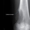
Buckling of the metaphyseal cortex that is seen in children is called what?
Torus fracture
[UpToDate: A buckle fracture occurs at the distal metaphysis, where the bone is most porous, usually in younger children. This injury is caused by buckling of the cortex due to compression failure. Torus fractures are stable, and treatment is aimed at pain relief, comfort, and protection of the bone from any further injury using a short arm cast or a splint.
Based on several small trials, we suggest that children with a torus (buckle) fracture receive a removable splint for immobilization rather than a below-elbow cast. However, the choice of splint versus cast is dependent on the degree of initial pain, the degree of anticipated activity of the child, and parental preference. A splint can be removed for showering and minor activities, and may be preferable by some caregivers, but a cast offers more protection of the fracture site in active children.
Before treating with a splint, it is crucial to radiographically distinguish a buckle fracture from a nondisplaced greenstick fracture. Patients with greenstick fractures warrant casting as volar splints may not prevent refracture during healing.
Current evidence does not identify the optimal type of removable splint (eg, premolded splint versus molded volar plaster or fiberglass splint). In the authors’ experience, a well-padded, molded volar splint (either plaster or fiberglass) is easy to apply and comfortable.
Taken together, the evidence suggests that children with buckle fractures may be safely treated with a removable splint and that a splint may encourage earlier return to normal activities than a short arm cast. Splinting may initially be more painful than casting and may not be ideal in children with higher baseline pain.]