30: Stomach Flashcards
What might be necessary to treat a Mallory-Weiss tear that will not stop bleeding despite EGD with hemo-clips?
Gastrostomy and over-sewing of the vessel
[If EGD with hemo-clips is not successful in stopping the bleeding, gastrostomy and oversewing the vessel may be neccessary.]
[SCORE: Initial management includes volume resuscitation, gastric lavage, and nasogastric decompression. Most patients with Mallory-Weiss tears stop bleeding spontaneously either before treatment or after these early measures. Once bleeding has stopped, rebleeding is rare. Nonoperative management consisting of endoscopic electrocoagulation or injection therapy has been successfully applied to these lesions. Esophageal balloon tamponade is contraindicated because it can convert a partial thickness tear into a full-thickness esophageal laceration. In cases not amenable to endoscopic therapy, operative management consists of oversewing the laceration through an anterior longitudinal gastrotomy in the middle third of the stomach. The mortality rate in recent series has been between 5% and 10%; deaths were related to associated disease, most notably cirrhosis.]
Epigastric pain radiating to the back that abates with eating but recurs 30 minutes after is typical for what?
Gastric or duodenal ulcer
[Healthline.com: Gastric ulcer symptoms are more likely to be felt immediately after eating. Duodenal ulcers are more likely to be felt a few hours after eating.]
[Medscape: Epigastric pain is the most common symptom of both gastric and duodenal ulcers. It is characterized by a gnawing or burning sensation and occurs after meals—classically, shortly after meals with gastric ulcer and 2-3 hours afterward with duodenal ulcer. Food or antacids relieve the pain of duodenal ulcers but provide minimal relief of gastric ulcer pain.
Duodenal ulcer pain often awakens the patient at night. About 50-80% of patients with duodenal ulcers experience nightly pain, as opposed to only 30-40% of patients with gastric ulcers and 20-40% of patients with nonulcer dyspepsia (NUD). Pain typically follows a daily pattern specific to the patient. Pain with radiation to the back is suggestive of a posterior penetrating gastric ulcer complicated by pancreatitis.]
[UpToDate: Upper abdominal pain or discomfort is the most prominent symptom in patients with peptic ulcers. Approximately 80% of patients with endoscopically diagnosed ulcers have epigastric pain. Occasionally the discomfort localizes to the right or left upper quadrants of the hypochondrium. Radiation of pain to the back may occur, but back pain as the primary symptom is atypical. In untreated patients, symptoms can last a few weeks followed by symptom-free periods of weeks or months. The “classic” pain of duodenal ulcers occurs two to five hours after a meal when acid is secreted in the absence of a food buffer and at night (between about 11 PM and 2 AM) when the circadian stimulation of acid secretion is maximal.
Patients with peptic ulcers, and particularly pyloric channel ulcers, may have food-provoked symptoms due to visceral sensitization and gastroduodenal dysmotility. These symptoms include epigastric pain that worsens with eating, postprandial belching and epigastric fullness, early satiety, fatty food intolerance, nausea, and occasional vomiting.
Approximately 70% of peptic ulcers are asymptomatic. Patients with silent peptic ulcers may later present with ulcer related complications. Between 43% and 87% of patients with bleeding peptic ulcers present without antecedent dyspepsia or other heralding gastrointestinal symptoms. Older adults and individuals on nonsteroidal anti-inflammatory drugs (NSAIDs) are more likely to be asymptomatic and later present with ulcer complications.]

Should a cholecystectomy be performed during bariatric surgery (Roux-en Y approach)?
If stones are present
[UpToDate: Cholelithiasis is a common complication of any type of weight loss surgery. In the Teen-LABS study, cholecystectomy was required within three years in 8.6% of patients (9.9% for RYGB and 5.1% for sleeve gastrectomy). An additional 5% of participants required other abdominal operations, including lysis of adhesions, gastrostomy, or ventral or internal hernia repair.]

Which condition is characterized by bile reflux into the stomach, combined with histologic evidence of gastritis?
Alkaline (bile) reflux gastritis
[UpToDate: Reflux of bile into the stomach is common after operations that remove or bypass the pylorus. In most patients there are no serious clinical sequelae. However, approximately 2% of patients develop alkaline reflux gastritis, a syndrome of persistent burning epigastric pain and chronic nausea that is aggravated by meals. The diagnosis is made primarily by excluding other causes of symptoms, although endoscopy may reveal gastritis, and technetium biliary scan can demonstrate excessive reflux of bile into the stomach.]
What is the surgical treatment for chronic gastric atony causing symptoms of delayed gastric emptying (nausea, vomiting pain, early satiety)?
Near-total gastrectomy with Roux-en Y
[UpToDate: Surgery is rarely indicated in patients with gastroparesis. Indications for surgery include placement of an enterostomy tube (eg, gastrostomy, jejunostomy) that cannot be placed endoscopically and completion or subtotal gastrectomy to relieve refractory nausea and vomiting in patients with a partial gastrectomy.
Surgical pyloroplasty and gastrojejunostomy have also been performed to treat refractory gastroparesis. As an example, in one series that included 28 patients with gastroparesis, pyloroplasty was associated with an improvement in symptoms, gastric emptying, and a reduction in the need for prokinetics at three months. However, long-term studies are needed before they can be recommended.]
How do you diagnose duodenal ulcers?
Endoscopy
[UpToDate: The diagnosis of peptic ulcer disease is suspected in patients with dyspepsia, especially in the setting of nonsteroidal anti-inflammatory drug (NSAID) use or a history of Helicobacter pylori infection. While contrast imaging is not needed to diagnose peptic ulcer disease, if performed, it may be supportive of the diagnosis. The diagnosis of peptic ulcer disease is definitively established by direct visualization of the ulcer on upper endoscopy. However, the need to pursue a definitive diagnosis in patients who have undergone contrast imaging depends upon the clinical setting. In patients with benign-appearing duodenal ulcers identified on radiologic imaging and no alarm features, endoscopy is not required to establish the diagnosis. In contrast, all patients diagnosed with gastric ulceration on radiologic imaging should undergo an upper endoscopy. However, the timing of the upper endoscopy may be deferred to 12 weeks after therapy in the absence of alarm features.
On upper endoscopy, benign gastric and duodenal ulcers have smooth, regular, rounded edges, with a flat, smooth ulcer base often filled with exudate. Endoscopy is the most accurate diagnostic test for peptic ulcer disease. The sensitivity of upper endoscopy in the detection of gastroduodenal lesions is approximately 90% but varies based on the location of the ulcer and the experience of the endoscopist.]

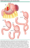
Why is Roux-en-Y gastro-jejunostomy the best method of reconstruction following antrectomy?
Less dumping syndrome and reflux gastritis compared to Billroth I (gastro-duodenal anastamosis) and Billroth II (Gastro-jejunal anastamosis)
[UpToDate: The choice of reconstruction following antrectomy for ulcer disease or distal gastrectomy for tumor depends upon the remnant anatomy available for reconstruction, taking into consideration the complications related to the specific postgastrectomy physiology that will result. However, based upon randomized trials, Roux-en-Y reconstruction appears to be tolerated better overall and leads to a better quality of life compared with Billroth reconstruction (Billroth I or Billroth II). Whether to preferentially perform a Roux-en-Y in patients whose anatomy supports a Billroth I or Billroth II, or convert to a Roux-en-Y only if complications occur remains controversial. For patients who have not undergone vagotomy, we suggest a primary Roux-en-Y reconstruction. Patients who have had a vagotomy have an increased risk of roux stasis syndrome.
A metaanalysis of 15 randomized trials comparing at least two of the gastric reconstruction techniques (ie, Billroth I, Billroth II, or Roux-en-Y reconstruction) following gastrectomy assessed postoperative morbidity and mortality, quality of life, and the incidence of postgastrectomy syndromes. Although complication rates were similar, patients with a Roux reconstruction had fewer complaints of reflux gastritis and better quality of life. A later trial also confirmed this finding.
The longest follow-up comparing techniques was reported for a trial that has followed 75 patients treated for duodenal ulcer for 12 to 21 years. Patients were randomly assigned to Billroth II (n = 39) or Roux-en-Y (n = 36) reconstruction between 1984 and 1993. Patients who received a Roux-en-Y reconstruction had overall better clinical outcomes with a significantly lower incidence of reflux esophagitis (3% vs 33%), fewer abnormal findings on upper endoscopy of the distal esophagus and esophagogastric junction (10% vs 82%), and a lower incidence of Barrett’s esophagus (3% vs 21%). The gastric remnant was also normal in significantly more Roux-en-Y patients (100% vs 18%). There were no differences between the groups in the incidence of Helicobacter pylori infection. It is interesting to note that the Roux limb in these patients was arbitrarily chosen to be 60 cm. Although the authors were concerned about the potential for the “Roux syndrome”, it was not clinically apparent in their series.]

What are the surgical options for treating alkaline reflux gastritis?
Conversion of Billroth I or Billroth II to Roux-en Y gastrojejunostomy with afferent limb 60 cm distal to gastrojejunostomy
[UpToDate: A variety of medical therapies for alkaline gastritis have been reported, but none have proven particularly effective. Surgical therapies aim at separating the remnant stomach from duodenal content by interposing a loop of jejunum between them. Examples include Roux-en-Y reconstruction (with a 45-60 cm Roux loop), Henley loop (interposition of a 40 cm isoperistaltic jejunal loop between the gastric remnant and the duodenum), Billroth II reconstruction with Braun enteroenterostomy (positioned 45 to 60 cm from the gastrojejunal anastomosis). The reoperative procedure is chosen based upon a patient’s existing anatomy and how much remnant stomach is left.]

What is Virchow’s node?
A lymph node in the left supraclavicular fossa that takes its supply from lymph vessels in the abdominal cavity. An enlarged, hard node (referred to as Troisier’s sign) is strongly indicative of the presence of cancer in the abdomen, specifically gastric cancer, that has spread through the lymph vessels.
[UpToDate: Supraclavicular lymphadenopathy is associated with a high risk of malignancy. In two studies, malignancy was found in 34% and 50% of patients with this presentation; the risk was highest in those over the age of 40. Right supraclavicular adenopathy is associated with cancer in the mediastinum, lungs, or esophagus. Left supraclavicular adenopathy (“Virchow’s node”) suggests abdominal malignancy (eg, stomach, gallbladder, pancreas, kidneys, testicles, ovaries, or prostate).]

What are 4 symptoms of afferent-loop obstruction in patients with Billroth II or Roux-en Y?
- RUQ pain
- Steatorrhea
- Nonbilious vomiting
- Pain relieved with bilious emesis
[UpToDate: Afferent and efferent loop syndromes develop after Billroth II reconstruction with a gastrojejunostomy. They are related to mechanical obstruction of the two loops by kinking, anastomotic narrowing, adhesions, or, rarely, anastomotic ulceration.
The afferent loop refers to the duodenojejunal loop proximal to the gastrojejunal anastomosis. Most afferent loop syndromes can be prevented if the distance from the ligament of Treitz to the gastrojejunostomy is no more than 12 to 15 cm. A patient with an acute afferent loop obstruction presents with acute onset of severe abdominal pain and vomiting, which requires immediate operation to prevent bowel necrosis or duodenal blowout. Chronic afferent loop syndrome is typically associated with postprandial epigastric pain and intermittent projectile bilious vomiting which leads to resolution of the pain for a period of up to several days. In patients suspected of having afferent loop syndrome based upon symptoms (eg, intermittent projectile bilious vomiting), the detection of a distended afferent loop on abdominal computed tomography is diagnostic.]

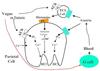
What inhibits gastrin production?
H+ in the duodenum
[Somatostatin also inhibits the release of gastrin, along with secretin, GIP (gastroinhibitory peptide), VIP (vasoactive intestinal peptide), glucagon and calcitonin.]
[UpToDate: Gastrin is released from specialized endocrine cells (G cells) into the circulation in response to a meal. The specific components of a meal that stimulate gastrin release include protein, peptides, and amino acids. Gastrin release is profoundly influenced by the pH of the stomach; fasting and increased gastric acid in the stomach inhibit its release, whereas a high gastric pH provides a strong stimulus for its secretion. The G cells are tightly regulated by two counterbalancing hormones, gastrin-releasing peptide and somatostatin, which exert stimulatory and inhibitory effects, respectively.]
What is the surgical treatment for blind-loop syndrome following a Billroth II or a Roux-en Y?
Re-anastomosis with shorter (40-cm) afferent limb

What must be ruled out in patients with complicated ulcer disease?
Gastrinoma
[Zollinger-Ellison syndrome: Gastric acid hyper secretion, peptic ulcers, and gastrinoma]
[UpToDate: Other causes of peptic ulcer disease should be considered when H. pylori and use of NSAIDs have been excluded (eg, gastrinoma in patients with multiple simultaneous ulcers, or those in unusual locations [second portion of the duodenum and into the proximal jejunum]).
In patients with gastric ulcers without a clear etiology, we perform an upper endoscopy 12 weeks after initiating medical therapy (with biopsies of the ulcer if still present). This upper endoscopy allows for additional biopsies of the ulcer to exclude neoplastic, infiltrative, or infectious causes of ulceration.]
What is the treatment for efferent-loop obstruction in patients with Billroth II or Roux-en Y?
Balloon dilation
[Surgical option is to find the site of obstruction and relieve it]
[UpToDate: The efferent loop refers to the jejunal segment distal to the gastrojejunostomy that drains sulcus entericus away from the stomach. Obstruction of the efferent loop causes gastric outlet obstruction manifested by symptoms of epigastric pain, distension, and bilious vomiting. When diagnosed by either computed tomography or upper gastrointestinal series, surgical correction is the treatment of choice for efferent loop syndrome.]
Which type of gastric cancer has a less favorable prognosis?
Diffuse gastric cancer
[25% 5-year survival]
[UpToDate: Diffuse type cancers are highly metastatic and characterized by rapid disease progression and a poorer prognosis than intestinal cancers. Diffuse carcinomas also have a greater tendency to invade the gastric wall, sometimes extending to the lower esophagus or to the duodenum. Occasionally, a broad region of the gastric wall or even the entire stomach is extensively infiltrated, resulting in a rigid thickened stomach, termed “linitis plastica”.
Histologically, individual tumor cells are seen to invade the surrounding tissues, and there is no gland formation. When intracellular mucin is abundant, it pushes aside the nucleus of the individual cells, resulting in the so-called signet ring carcinoma. It has long been thought that signet ring histology is an independent predictor of a worse prognosis as compared to other forms of gastric cancer. However, more recent studies have begun to question this notion. Some studies suggest that signet ring histology is associated with more advanced stage of disease at presentation, and that when adjusted for stage, signet ring cancer does not portend a worse prognosis.]
Where is the vagus nerve divided in a truncal vagotomy?
Both the left and right vagal nerve trunks are divided at the level of the diaphragmatic hiatus
[This decelerates gastric emptying of solids. Proximal vagotomy divides individual fibers and does not affect emptying of solids.]
[UpToDate: Truncal vagotomy is the simplest procedure to perform. Basal and stimulated acid secretions are reduced by 80% and 50%, respectively. However, truncal vagotomy sacrifices innervation to the pancreas, small intestine, proximal colon, and hepatobiliary tree, and alters gastric physiology requiring some form of gastric emptying procedure (pyloroplasty or gastroenterostomy). Highly selective vagotomy reduces basal and stimulated acid secretion by more than 75% and 50%, respectively, while minimizing the effects of vagotomy on gastric emptying. However, surgeons-in-training have less exposure to the more technically demanding procedures, like highly selective vagotomy (parietal cell vagotomy), because of the decrease in the hospitalization rate for peptic ulcer disease.]

Does central obesity have a better or worse prognosis in the general population?
Worse
[UpToDate: Both overall obesity, defined by BMI, and abdominal obesity or central obesity (assessed by measuring waist circumference, waist-to-hip ratio [WHR], or waist/height ratio), are associated with an excess risk of CVD. The WHR is infrequently used by clinicians and is not currently recommended as part of the routine obesity evaluation by the American Heart Association/American College of Cardiology/Obesity Society guideline, although it was in the previous version.
Data from the Third National Health and Nutrition Examination Survey (NHANES III) suggest that normal-weight central obesity (normal BMI with increased WHR) is associated with higher mortality than BMI-defined mortality, particularly when compared to individuals without central obesity. In a cross-sectional survey of over 15,000 individuals, men with a normal BMI (18.5 to 24.9 kg/m2) but central obesity (WHR ≥0.90) had the highest total mortality risk when compared to men without central obesity who were normal weight, overweight (25 to 29.9 kg/m2), or obese (≥30 kg/m2) (HR 1.87, 2.24, and 2.42, respectively). Normal weight women with central obesity (WHR ≥0.85) also had higher mortality risk compared to normal weight and obese women without central obesity (HR 1.48 and 1.32, respectively). A limitation of the study is that central obesity was determined by WHR only; no quantitative imaging studies of adipose tissue were performed. These data suggest that normal weight individuals with central obesity appear to have an increased mortality risk and should be targeted for lifestyle modification strategies.]
Is metastatic disease outside the area of resection a contraindication to resection of gastric cancer?
Yes
[Unless performing surgery for palliation]
[UpToDate: The only widely accepted criteria of unresectability for gastric cancer are the presence of distant metastases, invasion of a major vascular structure, such as the aorta, or disease encasement or occlusion of the hepatic artery or celiac axis/proximal splenic artery. Distal splenic artery involvement is not an indicator of unresectability; the vessel can be resected en bloc with a left upper quadrant exenteration: stomach, spleen, and distal pancreas.
The lymphatics around the stomach are rich, and the presence of locoregional lymph node metastases that are located geographically distant from the tumor (eg, celiac nodes with a primary tumor on the greater curvature of the stomach) should not necessarily be considered an indicator of unresectability. However:
- Patients who have bulky adenopathy fixed to the pancreatic head that might indicate the need for a Whipple procedure are at a high risk for occult metastatic disease. In these cases, it is probably best to consider staging laparoscopy or upfront chemotherapy rather than surgery initially. Performance of a Whipple for gastric cancer is an extremely rare occurrence.
- Lymph nodes behind or inferior to the pancreas, aorto-caval region, into the mediastinum, or in the porta hepatis are typically considered outside of the surgical field and thus evidence of unresectability. These nodes would fall into areas that would be defined as third or fourth echelon nodes in the Japanese nomenclature.
Since resection of the primary lesion also offers the most effective means of symptom palliation, exploration may also be considered in patients with known metastatic disease, if the severity of symptoms so dictates. The choice of operation for gastric cancer depends upon the location of the tumor within the stomach, the clinical stage, and the histologic type. The major surgical considerations include the extent of stomach resection (total versus partial gastrectomy) and the extent of lymph node dissection.]
What is the most common cause of leak following bariatric surgery (Roux-en Y approach)?
Ischemia
[UpToDate: The anastomotic leak remains the most dreaded technical complication of bariatric surgery, and is one of the most challenging complications of weight-loss surgery. The risk of a leak ranges from 0.8% to 6% depending on procedures chosen as well as technical and patient factors involved. As examples:
- A metaanalysis of 4888 patients who underwent laparoscopic sleeve gastrectomy revealed a leak rate of 2.4%. Body mass index (BMI) greater than 50 kg/m2 and using a bougie smaller than 40 French were the factors associated with increased leak rate.
- A retrospective analysis of 4444 patients in the longitudinal assessment of bariatric surgery (LABS) database revealed an anastomotic leak rate of 1.0% after RYGB (both open and laparoscopic). Open surgery, revision surgery, and routine drain placement were associated with an increased leak rate.
In revisional bariatric surgery, the risk of anastomotic leak approaches 35%.
Most leaks after bariatric surgery occur early, generally within the first week after surgery. Many post-sleeve-gastrectomy leaks can occur after patient discharge. Therefore, vigilant follow up during the first 30 days is recommended for that group of patients.
The clinical presentation of an anastomotic leak is subtle and requires vigilance for signs such as low-grade fevers, respiratory compromise, and/or unexplained sustained tachycardia greater than 120 bpm. These signs may also be present in the setting of pulmonary embolism.]

What is usually required to remove a trichobezoar (hairball)?
Gastrostomy and removal
[EGD generally inadequate]
[UpToDate: Therapy for gastric bezoars should be tailored to the composition of the concretion and to the underlying pathophysiologic process. In patients with pharmacobezoars, the toxicity of the underlying ingested pharmaceutical agent must be considered as decontamination may be required.
While the optimal strategy is controversial in the absence of studies comparing different modalities, for patients with mild symptoms due to bezoars, we initially attempt chemical dissolution. We use prokinetic metoclopramide as adjuvant therapy. For patients with bezoars that fail to dissolve or are resistant to chemical dissolution (trichobezoars), and patients with moderate to severe symptoms due to large bezoars, we suggest endoscopic therapy. We reserve surgery for selected patients with gastric bezoars if chemical dissolution and endoscopic fragmentation cannot be performed or fail and for patients with complications (eg, obstruction, significant bleeding).]
What are the 4 main symptoms of blind loop syndrome (small intestine bacterial overgrowth)?
- Pain
- Steatorrhea (due to bacterial deconjugation of bile)
- B12 deficiency (bacteria use it up)
- Malabsorption
[UpToDate: The majority of patients with small intestinal bacterial overgrowth (SIBO) present with nonspecific symptoms of bloating, flatulence, or abdominal discomfort, or they may be asymptomatic. Many patients diagnosed with severe SIBO have diarrhea. Although classic SIBO descriptions include steatorrhea with greasy or bulky stools, this is uncommon and occurs principally if the SIBO is caused by altered anatomy such as blind loop syndrome. Rarely, patients have weight loss due to severe diarrhea, malabsorption, or poor oral intake. Although diarrhea is a common symptom in children, they may present with chronic abdominal pain alone. Children may also have evidence of malnutrition and may fail to gain weight. Patients with hypoalbuminemia due to malabsorption may have peripheral edema on physical examination. Rarely, patients with SIBO may also present with symptoms and signs secondary to vitamin deficiencies. It is important to assess for these in subjects with severely altered anatomy causing SIBO, severe immunodeficiencies, or tropical sprue.]
What is the overall 5-year survival rate for gastric lymphomas?
50%
[UpToDate: GI diffuse large B cell lymphoma (DLBCL) includes lesions previously called “high-grade” MALT lymphoma. It can occur anywhere along the GI tract and is the most common histology for primary gastric lymphoma, representing approximately 50% of cases. Compared with patients with low-grade lymphoma, these patients tend to have more systemic symptoms, a more advanced stage at diagnosis, and a worse prognosis. In one series of 114 patients, for example, those with DLBCL had lower rates of complete remission (68% vs 92%) and five-year overall survival (46% vs 75%) when compared with those with low-grade MALT lymphoma.]
What is the effect of a truncal vagotomy on post-prandial bile flow?
Decreases post-prandial bile flow
[NCBI: The exocrine pancreatic and biliary secretion in response to vagal stimulation by insulin hypoglycemia was measured in preoperative patients with duodenal ulcer and in patients who underwent highly selective vagotomy, bilateral selective vagotomy with pyloroplasty, and truncal vagotomy with pyloroplasty. Significant stimulation of both biliary and pancreatic secretion occurred only in patients with an intact vagal nerve supply.]
What are the primary treatment modalities for gastric lymphoma?
Chemotherapy and XRT
[Surgery for complications: Only possibly indicated for stage I disease where the tumor is confined to the stomach mucosa - only partial resection is indicated]
[SCORE: When gastric lymphoma is first diagnosed by endoscopic means, evidence of systemic disease should be sought. Computed tomography of the chest and abdomen to detect lymphadenopathy, lymphangiography, bone marrow biopsy, and biopsy of enlarged peripheral lymph nodes may be appropriate. A multimodality program is used in most centers to manage primary gastric lymphomas. Gastrectomy is the first step in the therapeutic strategy. Increasing numbers of patients are treated with chemoradiation therapy alone. The risk of hemorrhage or perforation frequently alluded to in the past has probably been overstated. The risk of perforation if primary gastric lymphoma is managed with cytolytic agents and not resected approximates 5%.]
[UpToDate: Primary gastrointestinal (GI) non-Hodgkin lymphoma (NHL) is a heterogeneous group of B and T cell lymphoid malignancies. The management of these lymphomas may differ from lymphomas of lymph node origin.
For most patients with GI diffuse large B cell lymphoma (DLBCL), we recommend the use of combination chemotherapy plus immunotherapy (eg, R-CHOP) such as that used for other patients with DLBCL with or without involved-field RT rather than treatment with surgery or H pylori eradication therapy (Grade 1B).
For patients with enteropathy-associated T cell intestinal lymphoma who have a good performance status and chemotherapy sensitive disease, we suggest treatment with intensive chemotherapy followed by autologous HCT rather than chemotherapy alone.]