16: Critical Care Flashcards
Where is the ventilation/perfusion ratio the highest and lowest in the lung?
Highest in the upper lobes and lowest in the lower lobes
What is the most potent stimulus for systemic inflammatory response syndrome (SIRS)?
Endotoxin (lipopolysaccharide lipid A)
[Lipid A is a very potent stimulatory of TNF release. Inflammatory response is activated systemically (TNF-alpha and IL-1 are major components) and results in capillary leakage, microvascular thrombi, hypotension, and eventually end-organ dysfunction.]
What are the 2 primary determinants of myocardial oxygen consumption?
- Increased ventricular wall tension (#1 determinant)
- Heart rate
[Can lead to myocardial ischemia.]
What results from stimulation of Beta 1 and 2 receptors?
- Beta 1: Increased myocardial contraction and rate
- Beta 2: Vascular smooth muscle and bronchial smooth muscle relaxation
[Beta 2 receptor stimulation also increases insulin, glucagon, and renin.]
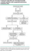
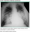
What are the 4 Berlin criteria for acute respiratory distress syndrome (ARDS)?
- Acute onset (within 1 week of known clinical insult or 1 week of worsening symptoms)
- Bilateral pulmonary infiltrates (consistent with pulmonary edema)
- Carrico index (PaO2/FiO2) < 300 (mild 200-300, moderate 100-200, severe < 100)
- Absence of heart failure (wedge pressure < 18 mmHg)
Which hormones are the rapid and sustained neurohormonal responses to hypovolemia mediated by?
Rapid: Epinephrine and norepinephrine (Vasoconstriction and increased cardiac activity)
Sustained: Renin (vasoconstriction and water resorption), ADH (water resorption), and ACTH (cortisol release)
What are the effects of Dopamine infusion at the below doses?
- Low dose (2-5 ug/kg/min):
- Medium dose (6-10 ug/kg/min):
- High dose (>10 ug/kg/min):
- Low dose (2-5 ug/kg/min): Dopaminergic - Increase in renal blood flow
- Medium dose (6-10 ug/kg/min): Beta-adrenergic - Increased heart contractility
- High dose (>10 ug/kg/min): Alpha-adrenergic - Vasoconstriction and increased BP
[UpToDate: dopamine dilates the interlobular arteries and both the afferent (preglomerular) and efferent (postglomerular) arterioles. The net effect is a relatively large increase in renal blood flow with a lesser or no elevation in glomerular filtration rate. At higher concentrations (above 5 mcg/kg per minute), dopamine induces renal vasoconstriction.]
What is the effect of hydralazine infusion?
Alpha-blocker; Lowers blood pressure
[UpToDate: Direct vasodilation of arterioles (with little effect on veins) with decreased systemic resistance.]
What do the following equations equal:
- Cardiac output (CO) x Systemic vascular resistance (SVR)
- Cardiac output (CO) x Body surface area (BSA)
- Cardiac output (CO) x Systemic vascular resistance (SVR) = Mean arterial pressure (MAP)
- Cardiac output (CO) x Body surface area (BSA) = Cardiac index (CI)
What is the effect of Vasopressin mediated by the below receptors?
- V-1:
- Intrarenal V-2:
- Extrarenal V-2:
- V-1: Vasoconstriction of vascular smooth muscle
- Intrarenal V-2: Water reabsorption at collecting ducts
- Extrarenal V-2: Mediation of release of factor VIII and von Wilebrand factor
[UpToDate: Vasopressin stimulates a family of arginine vasopressin (AVP) receptors, oxytocin receptors, and purinergic receptors (Russell 2011). Vasopressin, at therapeutic doses used for vasodilatory shock, stimulates the AVPR1a (or V1) receptor and increases systemic vascular resistance and mean arterial blood pressure; in response to these effects, a decrease in heart rate and cardiac output may be seen. When the AVPR2 (or V2) receptor is stimulated, cyclic adenosine monophosphate (cAMP) increases which in turn increases water permeability at the renal tubule resulting in decreased urine volume and increased osmolality. Vasopressin, at pressor doses, also causes smooth muscle contraction in the GI tract by stimulating muscular V1 receptors and release of prolactin and ACTH via AVPR1b (or V3) receptors.]
Which condition do the following signs/symptoms characterize?
- Nausea and vomiting
- Thirst
- Polyuria
- Increased ketones
- Decreased sodium
- Increased potassium
- Increased glucose
Diabetic ketoacidosis
[Initial treatment is normal saline and insulin.]
Why is blood in the left ventricle oxygen content 5 mmHg lower than blood in the pulmonary capillaries?
Unsaturated bronchial blood empties into the pulmonary veins
What is the effect of isoproterenol infusion?
Beta 1&2-adrenergic (increased heart rate and contractility, vasodilation)
[Side effects: extremely arrhythmogenic; Increased heart metabolic demand (rarely used); may actually decrease blood pressure.]
[UpToDate: Stimulates beta1- and beta2-receptors resulting in relaxation of bronchial, GI, and uterine smooth muscle, increased heart rate and contractility, vasodilation of peripheral vasculature.]
What is the advantage of using continuous venovenous hemofiltration (CVVH) over hemodialysis?
CVVH is slower and better for ill patients who cannot tolerate the volume shifts (septic shock patients, etc.)
[Hematocrit increases by 5-8 for each liter taken off with dialysis.]
[UpToDate: Continuous renal replacement therapies (CRRTs) involve either dialysis (diffusion-based solute removal) or filtration (convection-based solute and water removal) treatments that operate in a continuous mode. The major advantage of continuous therapy is the slower rate of solute or fluid removal per unit of time. Thus, CRRT is generally better tolerated than conventional therapy since many of the complications of intermittent hemodialysis are related to the rapid rate of solute and fluid loss.
There are many variations of CRRT. The different modalities are categorized according to the access characteristics: blood or peritoneal, venovenous (VV) or arteriovenous (AV). A large number of acronyms have been derived that describe the different continuous therapies.
The choice of modality is dependent upon several factors including availability, the expertise of the clinician, hemodynamic stability, vascular access, and whether the primary need is for fluid and/or solute removal.]
What will be seen on electroencephalogram (EEG) and magnetic resonance angiography (MRA) in a patient with brain death?
- EEG: Electrical silence
- MRA: No blood flow to brain

Which disease states decrease pulmonary compliance?
- ARDS
- Fibrotic lung disease
- Reperfusion injury
- Pulmonary injury
- Atelectasis
[Compliance = (change in volume) / (change in pressure). High compliance means lungs are easy to ventilate.]
Do the below factors cause pulmonary vasoconstriction or vasodilation?
- PGE1:
- Prostacyclin (PGI2):
- Hypoxia:
- Histamine:
- Nitric oxide:
- Bradykinin:
- Serotonin:
- TXA2:
- Alkalosis:
- Acidosis:
- PGE1: Vasodilation
- Prostacyclin (PGI2): Vasodilation
- Hypoxia: Vasoconstriction
- Histamine: Vasoconstriction
- Nitric oxide: Vasodilation
- Bradykinin: Vasodilation
- Serotonin: Vasoconstriction
- TXA2: Vasoconstriction
- Alkalosis: Vasodilation
- Acidosis: Vasoconstriction
What is the treatment for neurogenic shock?
- Give volume 1st
- Give Phenylephrine after volume resuscitation
What are the below characteristics of antidiuretic hormone released?
- Where is it released:
- What triggers its release:
- What is its mechanism of action:
- Where is it released: Posterior pituitary gland
- What triggers its release: Released in response to high osmolality
- What is its mechanism of action: Acts on collecting ducts for water resorption. Also a vasoconstrictor
[UpToDate: In normal subjects, the urine output is primarily determined by water intake. Changes in water intake lead to alterations in the plasma osmolality that are sensed by the osmoreceptors in the hypothalamus that regulate both ADH release and thirst. As an example, an increase in water intake sequentially lowers the plasma osmolality, decreases ADH secretion, and reduces collecting tubule permeability to water; the net effect is the rapid excretion of the excess water in a dilute urine.
What is the normal p50 (O2 level at which 50% of O2 receptors are saturated)?
27 mmHg

What are the below characteristics of atrial natriuretic peptide released?
- Where is it released:
- What triggers its release:
- What is its mechanism of action:
- Where is it released: From wall of atrium
- What triggers its release: Released in response to atrial distension
- What is its mechanism of action: Inhibits Na and water resorption in the collecting ducts. Also a vasodilator
[UpToDate: Atrial natriuretic peptide (ANP) is primarily released from the atria in response to volume expansion, which is sensed as an increase in atrial stretch. ANP release is increased in heart failure. Plasma ANP levels rise early in the course of the disease and have been used as a marker for the diagnosis of asymptomatic left ventricular dysfunction. With chronic and more advanced heart failure, ventricular cells can also be recruited to secrete both ANP and brain natriuretic peptide (BNP), an analogous peptide, in response to the high ventricular filling pressures. These relationships have allowed the plasma concentration of these peptides, particularly BNP, to be used to detect heart failure and to predict the outcome and perhaps guide therapy in patients with established disease.
Both ANP and BNP have diuretic, natriuretic, and hypotensive effects. They also inhibit the renin-angiotensin system, endothelin secretion, and systemic and renal sympathetic activity. Among patients with HF, increased secretion of ANP and BNP may partially counteract the effects of norepinephrine, endothelin, and angiotensin II, limiting the degree of vasoconstriction and sodium retention. In one study of patients with moderately severe HF, for example, administration of an orally active inhibitor of the endopeptidase that degrades ANP led to reductions in plasma aldosterone concentrations and right ventricular (RV) and left ventricular (LV) filling pressures, as well as a decrease in body weight presumed to be due to diuresis, natriuresis, or both. These effects were presumably mediated by enhanced natriuretic peptide activity.
An unexpected finding is that BNP may protect against collagen accumulation and the pathologic remodeling that contributes to progressive HF. Studies in BNP knockout mice reveal increased cardiac fibrosis in response to ventricular pressure overload.]

What is the formula for calculating arterial O2 (CaO2) content?
CaO2 = Hgb x 1.34 x O2 saturation + (Po2 X 0.003)
[UpToDate: The arterial oxygen content (CaO2) is the amount of oxygen bound to hemoglobin plus the amount of oxygen dissolved in arterial blood:
CaO2 (mL O2/dL) = (1.34 x hemoglobin concentration x SaO2) + (0.0031 x PaO2)
where SaO2 is the arterial oxyhemoglobin saturation and PaO2 is the arterial oxygen tension. In dyshemoglobinemias, the oxygen content is calculated with the same equation, although the saturations (and therefore the oxygen content) will be different for a specific PaO2. Normal CaO2 is approximately 20 mL O2/dL.
Similarly, the mixed venous blood oxygen content (CvO2) is the amount of oxygen bound to hemoglobin plus the amount of oxygen dissolved in mixed venous blood:
CvO2 (mL O2/dL) = (1.34 x hemoglobin concentration x SvO2) + (0.0031 x PvO2)
where SvO2 is the mixed venous oxyhemoglobin saturation and PvO2 is the mixed venous oxygen tension. Normal CvO2 is approximately 15 mL O2/dL. Mixed venous blood is drawn from the right atrium. Peripheral venous blood should not be substituted because it tends to overestimate venous oxygen content.]
What is the effect of an intraaortic balloon pump (IABP) on the the below measures?
- Diastolic blood pressure
- Systolic blood pressure
- Coronary blood flow
- Afterload
- Diastolic blood pressure: +
- Systolic blood pressure: -
- Coronary blood flow: +
- Afterload: -
[Improved diastolic blood pressure improves coronary perfusion.]
[The hemodynamic effects of an intraaortic balloon pump depend upon the volume of the balloon, its position in the aorta, heart rate, rhythm, the compliance of the aorta, and systemic resistance. The higher the arterial elastance, which is determined in part by compliance, the greater the hemodynamic improvement from intraaortic balloon pump counterpulsation (IABP). Despite this variability, expected changes in the hemodynamic profile in the majority of patients with cardiogenic shock include:
- A decrease in systolic pressure by 20%
- An increase in aortic diastolic pressure by 30%, which may raise coronary blood flow to territory perfused by a vessel with a critical stenosis.
- An increase in mean arterial pressure especially in patients with shock due to an acute mechanical abnormality such as mitral regurgitation (MR) or ventricular septal defect (VSD) or to improvement in perfusion of a territory resulting in overall improved ventricular function.
- A reduction of the heart rate by less than 20%
- A decrease in the mean pulmonary capillary wedge pressure by 20%
- An elevation in the cardiac output by 20%, especially in patients with MR, VSD, or a large territory of medically refractory ischemia that is improved by the addition of counterpulsation
In addition, intraaortic balloon pumping reduces mean systemic impedance and developed systolic pressure, and causes a 14% decline in calculated peak left ventricular wall stress. The reductions in afterload and wall stress lead to a fall in myocardial oxygen consumption, which is one of the goals of treatment of patients with myocardial ischemia.]
What is the best test for azotemia (elevated nitrogen-containing compounds in the blood)?
Fractional excretion of sodium (FeNa)
[FeNa = (Urine sodium x Serum creatinine) / (Serum sodium x Urine creatinine) x 100]
[UpToDate: The fractional excretion of sodium (FENa) measures the percent of filtered sodium that is excreted in the urine. The FENa is commonly used to assist in differentiating prerenal disease (a reduction in glomerular filtration rate [GFR] due solely to decreased renal perfusion) from acute tubular necrosis (ATN), the two most common causes of acute kidney injury (AKI).
Among patients with suspected prerenal disease or ATN, we recommend measuring the FENa. A value of the FENa below 1% commonly indicates prerenal disease; in comparison, a value between 1 and 2 percent may be seen with either disorder, while a value above 2% usually indicates ATN.]