21: Adrenal Flashcards
What are the following characteristics of hypocortisolism?
- Most common overall cause of hypocortisolism
- Most common cause of primary hypocortisolism
- Best test to diagnose hypocortisolism
- Most common overall cause of hypocortisolism: Withdrawal of exogenous steroids
- Most common cause of primary hypocortisolism: Autoimmune disease
- Best test to diagnose hypocortisolism: Cosyntropin test (ACTH given, urine cortisol measured -> Cortisol will remain low)
[Hypocortisolism can be treated with dexamethasone prior to cosyntropin test because it does not interfere with the test results.]
What is a rare, benign, and asymptomatic tumor of neural crest origin located in the adrenal medulla or sympathetic chain and what is the treatment?
- Ganglioneuroma
- Treatment: Resection
[UpToDate: Ganglioneuromas are rare, slow growing, large tumors that arise from sympathetic ganglion cells. They can be grouped among the peripheral neuroblastic tumors but consist of mature ganglion cells and as such are benign. The cell of origin is derived from embryonic neural crest cells and ganglioneuromas are thought to represent the final stage of maturation from neuroblastomas. They are large tumors, with an average size of 7 centimeters, and they are encapsulated.
Ganglioneuromas tend to occur more frequently in females, with 60% occurring before age 20. They occur anywhere along the sympathetic chain, with common locations being the mediastinum, retroperitoneum, and adrenal glands. They are asymptomatic except for local mass effect, such as causing constipation when located in the pelvis, or radicular pain if presacral.
Histopathology reveals large, mature ganglion cells (neurons), axons, satellite cells, Schwann cells, and fibrous stroma. The Schwann cells are not neoplastic but associate with the neurons though they do not elaborate any myelin. This separates them from schwannomas and neurofibromas in which the Schwann cells are neoplastic. Immunohistochemistry shows strong staining of the ganglion cells for neurofilaments, and strong staining of Schwann cells by S100.
Treatment is surgical excision, though given the large size, care must be taken for adjoining structures and nerves. The capsule may itself be adherent to important structures and total excision, though desirable, may not be possible. Postoperative autonomic dysfunction otherwise seems uncommon.]

What are the effects of the following hormones released from the adrenal gland?
- Aldosterone
- Cortisol
- Aldosterone: Stimulates renal resorption of sodium and renal secretion of potassium and hydrogen ions
- Cortisol: Inotropic, chronotropic, increases vascular resistance, promotes proteolysis and gluconeogenesis, decreases inflammation
How does venous drainage differ between the right and the left adrenal gland?
- The left adrenal vein goes to the left renal vein
- The right adrenal vein goes to the inferior vena cava

Adrenal metastases are most commonly from which primary?
Lung cancer
[Other common primaries to metastasize to the adrenal gland include breast cancer, melanoma, and renal carcinoma. Some isolated metastases to the adrenal gland can be resected with adrenalectomy.]
How does the presentation of congenital adrenal hyperplasia caused by a defect in the enzyme 11-hydroxylase differ from the more common congenital adrenal hyperplasia caused by a defect in 21-hydroxylase?
- 11-hydroxylase defect is a salt saving condition that can result in hypertension
- 21-hydroxylase defect is a salt wasting condition that can result in hypotension
[Both cause virilization in females and precocious puberty in males. Deoxycortisone acts as a mineralcorticoid in patients with a defect in 11-hydroxylase. Treatment for both conditions is cortisol and genitoplasty.]

In 90% of cases, congenital adrenal hyperplasia results from a defect in which enzyme and how does it present?
- Defective enzyme: 21-hydroxylase
- Presentation: Precocious puberty in males, virilization in females, salt wasting with hypotension
[A defect in 21-hydroxylase will prevent cholesterol from being converted into cortisol and aldosterone and instead will force it down the androgen pathway, leading to increased production of testosterone.]

Which 5 syndromes can be associated with pheochromocytomas?
- MEN IIa
- MEN IIb
- Neurofibromatosis type 1 (von Recklinghausen’s disease)
- Tuberous sclerosis
- Sturge-Weber disease
[UpToDate: Most catecholamine-secreting tumors are sporadic. However, some patients (approximately 30%) have the disease as part of a familial disorder; in these patients, the catecholamine-secreting tumors are more likely to be bilateral adrenal pheochromocytomas or paragangliomas.
There are several familial disorders associated with pheochromocytoma, all of which have autosomal dominant inheritance:
Von Hippel-Lindau (VHL) syndrome, associated with mutations in the VHL tumor suppressor gene.
Multiple endocrine neoplasia type 2 (MEN2), which is associated with mutations in the RET-proto-oncogene.
Pheochromocytoma is also seen, albeit rarely, with neurofibromatosis type 1 (NF1), due to mutations in the NF1 gene.
The approximate frequency of pheochromocytoma in these disorders is 10% to 20% in VHL syndrome, 50% in MEN2, and 0.1% to 5.7% with NF1.]
What do the following lab values indicate in the setting of hypercortisolism?
- High cortisol and low ACTH
- High cortisol and high ACTH
- High cortisol and low ACTH: Cortisol secreting lesion (adrenal adenoma or adrenal hyperplasia)
- High cortisol and high ACTH: Pituitary adenoma or an ectopic source of ACTH (eg small cell lung cancer)
[High dose dexamethasone suppression test can be used to differentiate between a pituitary adenoma and ectopic ACTH production.]

What are the 3 layers of the adrenal cortex (from outside to inside) and which hormones does each layer secrete?
- Zona glomerulosa: Aldosterone
- Zona fasciculata: Glucocorticoids
- Zona reticularis: Androgens/estrogens
[GFR: Salt, sugar, sex]

What is the treatment of hypercortisolism resulting from adrenal hyperplasia?
- Metyrapone (blocks cortisol synthesis) and aminoglutethimide (inhibits steroid production)
- Bilateral adrenalectomy if medical treatment fails
[Steroids must be given postoperatively when operating for Cushing’s syndrome.]
Which enzyme in the production of catecholamines is only found in the adrenal medulla and what does it catalyze?
- Enzyme: Phenylethanolamine N-methyltransferase (PNMT)
- Catalysis: Norepinephrine -> Epinephrine
[Epinephrine is exclusively produced in the adrenal medulla. For this reason, only adrenal pheochromocytomas will produce epinephrine.]

What is the most common site of extramedullary adrenal tissue?
Organ of Zuckerkandl (inferior aorta near the bifurcation)
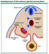
[UpToDate: The adrenal medulla and sympathetic nervous system develop in concert. The medullary elements, the sympathogonia, migrate forward from both sides of the neurogenic crest to the paraaortic and paravertebral regions and along the adrenal vein toward the medial aspect of the developing adrenal fetal cortex.
Most extra-adrenal chromaffin cells regress. However, some cells remain and form the organ of Zuckerkandl, which is located to the left of the aortic bifurcation near the origin of the inferior mesenteric artery.
Extra-adrenal chromaffin tumors are termed extra-adrenal pheochromocytomas, and are also known as paragangliomas. Extra-adrenal pheochromocytomas constitute 15% of adult and 30% of pediatric pheochromocytomas. They are situated most commonly in the organ of Zuckerkandl, which is the collection of paraganglia located anterolaterally to the distal abdominal aorta between the origin of the inferior mesenteric artery and the aortic bifurcation. The second most common location of extra-adrenal pheochromocytomas is at the left renal hilum. Extra-adrenal pheochromocytomas can be multiple and have also been reported in the neck, posterior chest, atrium, and bladder.]

What innervates the adrenal gland?
- The adrenal medulla receives innervation from the sympathetic splanchnic nerves
- The adrenal cortex receives no innervation

What is the diagnostic approach to a patient with hypercortisolism with a high ACTH?
- High-dose dexamethasone suppresion test
- Urine cortisol suppressed: Pituitary adenoma
- Urine cortisol not suppressed: Ectopic producer of ACTH (eg small cell lung cancer)
[NP-59 scintography can help localize tumors and differentiate adrenal adenomas from hyperplasia.]

What are the 3 most common non-iatrogenic causes of Cushing’s syndrome
- Pituitary adenoma is #1
- Ectopic ACTH production is #2
- Adrenal adenoma is #3
How does metyrosine work for treating pheochromocytoma?
It inhibits tyrosine hydroxylase, resulting a decrease in synthesis of catecholamines
[UpToDate: Metyrosine should be used with caution and only when other agents have been ineffective or in patients where tumor manipulation or destruction (eg, radiofrequency ablation of metastatic sites) will be marked. Although some centers advocate that this agent should be used routinely preoperatively, most reserve it primarily for patients who cannot be treated with the typical combined alpha and beta-adrenergic blockade protocol because of intolerance or cardiopulmonary reasons.
The protocol used at the Mayo Clinic with short-term preprocedure preparation is to start with metyrosine 250 mg every six hours on day 1, 500 mg every six hours on day 2, 750 mg every six hours on day 3, and 1000 mg every six hours on the day before the procedure, with the last dose (1000 mg) the morning of the procedure. With this short-course metyrosine therapy, the main side effect is hypersomnolence.
The side effects of metyrosine can be disabling and with long-term therapy they include sedation, depression, diarrhea, anxiety, nightmares, crystalluria and urolithiasis, galactorrhea, and extrapyramidal signs. Metyrosine may be added to alpha and beta-adrenergic blockade when the resection will be difficult (eg, malignant paraganglioma) or if destructive therapy is planned (eg, radiofrequency ablation of hepatic metastases or cryoablation of bone metastases).
The extrapyramidal effects of phenothiazines or haloperidol may be potentiated and their use concomitantly with metyrosine should be avoided. High fluid intake to avoid crystalluria is suggested for any patient taking more than 2 g daily. The metyrosine-phenoxybenzamine regimen has not been compared with the phenoxybenzamine-beta-adrenergic blocker regimen. The cost of metyrosine has increased dramatically recently and may make use of this medication prohibitive.]

What is the initial workup for a suspected pheochromocytoma?
Urine metanephrines and VMA
[VMA can be falsely elevated due to: Coffee, tea, fruits, vanilla, iodine contrast, labetalol, alpha- and beta-blockers.]
[UpToDate: The diagnosis of pheochromocytoma is typically made by measurements of urinary and plasma fractionated metanephrines and catecholamines. However, there are major regional, institutional, and international differences in the approach to the biochemical diagnosis of pheochromocytoma.
We suggest initial biochemical testing based upon the index of suspicion that the patient has a pheochromocytoma. If there is a low index of suspicion, we suggest 24-hour urinary fractionated catecholamines and metanephrines; if there is a high index of suspicion, we suggest plasma fractionated metanephrines. Our approach differs from the 2014 Endocrine Society clinical practice guideline, which suggests initial biochemical testing using 24-hour urinary fractionated metanephrines or plasma fractionated metanephrines (drawn supine with an indwelling cannula for 30 minutes). However, many clinicians do not measure plasma fractionated metanephrines under these ideal conditions and the test is associated with a high false positive rate.
Historically, many institutions relied upon measurements of 24-hour urinary excretion of catecholamines and total metanephrines. More recently, plasma fractionated metanephrines have been proposed by some as being a superior test. The majority of the metabolism of catecholamines is intratumoral, with formation of metanephrine and normetanephrine. Most laboratories now measure fractionated catecholamines (dopamine, norepinephrine, and epinephrine) and fractionated metanephrines (metanephrine and normetanephrine) by high-performance liquid chromatography (HPLC) with electrochemical detection or tandem mass spectroscopy (MS/MS). These techniques have overcome the problems with fluorometric analysis (eg, false positive results caused by alpha-methyldopa, labetalol, or sotalol, and false negative results caused by imaging contrast agents).
For any of the biochemical tests, sensitivity will be lower and specificity will be higher for hereditary compared with sporadic pheochromocytoma because tumors detected in patients with a familial disposition tend to be small tumors that release catecholamines in amounts that are often too low to be detected. In contrast, sporadic pheochromocytomas tend to be larger and present with typical signs and symptoms of catecholamine excess. Our suggested approach to testing based upon the patient’s clinical presentation is discussed here.
Measurement of plasma fractionated metanephrines is useful to rule out pheochromocytoma, but a positive test (ie, plasma normetanephrine above the upper limit of the reference range) only slightly increases suspicion of disease when screening for sporadic pheochromocytoma.]

What are the following characteristics of Conn’s syndrome (hyperaldosteronism)?
- Symptoms
- Diagnosis
- Treatment
- Symptoms: Hypertension secondary to sodium retention without edema, hypokalemia, weakness, polydipsia, and polyuria
- Diagnosis: Salt-load suppression test (urine aldosterone will stay high), aldosterone:renin ratio > 20
- Treatment: Resection if adenoma, medical therapy if hyperplasia (spironolactone, calcium channel blockers, potassium)
[UpToDate: The initial evaluation should consist of documenting that the plasma renin activity (PRA) or plasma renin concentration (PRC) is reduced (typically undetectable) and that the plasma aldosterone concentration (PAC) is inappropriately high for the PRA (typically >15 ng/dL [416 pmol/L]); the net effect is a PAC/PRA ratio greater than 20 (depending upon the laboratory normals).
An elevated PAC/PRA ratio alone is not diagnostic of primary aldosteronism. As a result, we recommend confirming the diagnosis by demonstrating inappropriate aldosterone secretion. For aldosterone suppression testing, we use oral sodium loading and measurement of urine aldosterone excretion. Some experts prefer intravenous sodium chloride loading and measurement of the PAC.
Many centers and experts, including the author of this topic, use oral sodium loading over three days. After hypertension and hypokalemia are controlled (hypokalemia suppresses aldosterone secretion) and avoiding spironolactone and eplerenone as described above, the patients should receive a high-sodium diet for three days.
On the third day of the high-sodium diet, serum electrolytes are measured and a 24-hour urine specimen is collected for measurement of aldosterone, sodium, and creatinine. The 24-hour urine sodium excretion should exceed 200 mEq (4600 mg) to document adequate sodium loading. Urine aldosterone excretion >12 mcg/24 hours (33 nmol/day) in this setting is consistent with hyperaldosteronism.]
When is surgery indicated for an adrenal incidentaloma?
- Ominous characteristics (non-homogenous)
- > 4-6 cm
- Functioning tumor
- Enlarging tumor
[If surgery is not pursued for an incidentaloma, repeat imaging every 3 months is needed for 1 year, then yearly imaging.]
[UpToDate: We recommend surgery for all patients with biochemical documentation of pheochromocytoma (Grade 1B). The preoperative management of patients with pheochromocytoma is reviewed elsewhere.
We suggest a surgical resection for patients with subclinical Cushing’s syndrome who are younger and who have disorders potentially attributable to excess glucocorticoid secretion (eg, recent onset of hypertension, diabetes, obesity, and low bone mass) (Grade 2C).
We suggest a surgical resection for patients with adrenal masses greater than 4 cm in diameter (Grade 2B). However, the clinical scenario, imaging characteristics, and patient age frequently guide the management decisions in patients who have adrenal incidentalomas that fall on either side of the 4 cm diameter cutoff.
In a patient with a known primary malignancy elsewhere who has a newly discovered adrenal mass that has an imaging phenotype consistent with metastatic disease, performing a diagnostic CT-guided fine-needle aspiration (FNA) biopsy may be indicated, but only after excluding pheochromocytoma with biochemical testing. Adrenal biopsy would not be needed if the patient was already known to have widespread metastatic disease.
We recommend excision of a tumor if the initial imaging phenotype is suspicious (Grade 1B).
For all adrenal masses larger than 10 cm, including those masses with benign imaging phenotypes, we recommend an open adrenalectomy rather than a laparoscopic procedure (Grade 1B).
For incidentalomas with a benign appearance on imaging, we suggest a repeat imaging study at 6 to 12 months after initial discovery (Grade 1C). The rationale is that many malignant lesions will grow in this interval, leading to earlier intervention. Whether to obtain additional images (eg, at 6, 12, and 24 months after initial discovery) and the type of image obtained (eg, CT, MRI, or ultrasound) should be guided by clinical judgment and imaging phenotype. The yield and cost-effectiveness of such a strategy are not known.
We suggest removal of any tumor that enlarges by more than 1 cm in diameter during the follow-up period (Grade 2C).]
What is the rate-limiting step in catecholamine synthesis from the adrenal medulla?
Tyrosine -> dopa (catalyzed by tyrosine hydroxylase)

What is the treatment for a pheochromocytoma?
Adrenalectomy
[Ligate the adrenal veins first to avoid spilling catecholamines during tumor manipulation.]
What are the following characteristics of adrenocortical carcinoma?
- Percent functioning tumors
- Percent with advanced disease at time of diagnosis
- Age distribution
- Treatment
- Percent 5-year survival
- Percent functioning tumors: 50% (cortisol, aldosterone, sex steroids)
- Percent with advanced disease at time of diagnosis: 80%
- Age distribution: Bimodal (Before age 5 years, 5th decade of life)
- Treatment: Radical adrenalectomy (debulking helps symptoms and prolongs survival), Mitotane (adrenalytic drug) for residual/recurrent/metastatic disease
- Percent 5-year survival: 20%
What is the 10% rule for pheochromocytomas?
- 10% are malignant (Extra-adrenal tumors are more likely to be malignant)
- 10% are bilateral (Right sided predominance)
- 10% occur in children
- 10% are familial
- 10% are extra-adrenal


