18: Plastics, Skin, and Soft Tissue Flashcards
What cell type originates from the bone marrow and act as antigen-presenting cells in the skin and mucosa?
Langerhan cell
[They have a role in contact hypersensitivity reactions (type IV).]

Which type of melanoma is the least aggressive, exhibits minimal invasion, and usually grows radially first?
Lentigo maligna melanoma
[UpToDate: Lentigo maligna is a slowly evolving type of melanoma in situ that typically occurs in the sun-damaged skin of the face and neck of older individuals. The risk of progression to invasive lentigo maligna melanoma ranges from 5 to 20 percent. The development of darker pigmentation, sharper borders, or elevated or nodular areas are clinical signs of progression. There are no randomized trials evaluating treatment for lentigo maligna. We suggest surgical excision with margins of 5 to 10 mm, depending upon lesion size and location (Grade 2C).]
Are bone sarcomas typically found early or late?
Late
[Most are metastatic at the time of diagnosis.]
[UpToDate: At the time of presentation, between 10 and 20 percent of patients have demonstrable macrometastatic disease and are classified as stage III according to the staging system used by the Musculoskeletal Tumor Society. Distant metastases most commonly involve the lungs, but can also involve bone.
Occult micrometastases are presumed to be present in the majority of those who appear to have clinically localized disease, since before the era of adjuvant chemotherapy, over 80 percent of patients with osteosarcoma developed metastatic disease despite achieving local tumor control. It was postulated that these patients had subclinical metastases that were present at the time of diagnosis. With routine use of systemic adjuvant chemotherapy, at least two-thirds of children and adolescents with nonmetastatic osteosarcoma will be long-term survivors, implying the success of chemotherapy in eradication of micrometastases. Prognosis is worse in adults with osteosarcoma, particularly those over the age of 65.]
Match the risk factor for the type of sarcoma:
- Asbestos
- PVC and arsenic
- Chronic lymphedema
- Asbestos: Mesothelioma
- PVC and arsenic: Angiosarcoma
- Chronic lymphedema: Lymphangiosarcoma
What is the treatment for disseminated Kaposi’s sarcoma?
Interferon-alpha
[HAART is the best treatment for AIDS-related Kaposi’s sarcoma. Surgery may be required for severe intestinal hemorrhage.]
[UpToDate: Recombinant interferon alfa (IFNa) is approved for treatment of AIDS-associated KS in the US. While the mechanism of antitumor action of IFNa in KS is not known, it may involve direct antiproliferative effects, antiviral effects, inhibition of angiogenesis, and modulation of host cellular and humoral immune responses
Intralesional injection of interferon alfa, alone or in combination with interleukin-2, has also been reported to induce regression of classical KS lesions. The studies cited evaluated the effects of twice- or thrice-weekly injections over a period of four to six weeks, which is unlikely to be practical for routine treatment of individuals with multiple cutaneous lesions.]
What is the most common subtype of childhood rhabdomyosarcoma (The most common soft tissue sarcoma in kids)?
Embryonal subtype
[alveolar subtype has the worst prognosis.]
[UpToDate: The embryonal subtype is the most common, accounting for 59 percent of all RMS cases. Most (50 percent) are of the classic subtype, and the botryoid and spindle cell variants comprise 6 and 3 percent, respectively. Alveolar RMS represents 21 percent of all cases, while the remainder are classified as undifferentiated (8 percent), pleomorphic/anaplastic (1 percent), or NOS (11 percent).
The morphologic appearance of the tumor cells comprising alveolar and embryonal RMS is nonspecific. The cells have scant cytoplasm and a centrally placed round nucleus that occupies the majority of the cell. It is the organizational architecture of the tumor that distinguishes alveolar from embryonal subtypes.
Classic embryonal RMS is composed of typical rhabdomyoblasts arranged in sheets and large nests, with infrequent intermixed fusiform cells, and no suggestion of an alveolar architectural pattern. The typical rhabdomyoblast has moderate to deeply eosinophilic cytoplasm, representing poorly-formed myofilaments. Myofilaments with cross-striations are usually present only in the well-differentiated spindle cell subtype, which is so named because the cells have a characteristic elongated spindle-like appearance. Spindle cell RMS often presents in a paratesticular location.]

What is the description of a stage III pressure sore?
Full-thickness skin loss with subcutaneous fat exposure
[Treatment: Sharp debridement and it will likely need a myocutaneous flap.]

Arsenical keratosis is associated with which type of skin cancer?
Squamous cell carcinoma
[UpToDate: Different types of arsenic-related skin lesions have been described in the West Bengal and Bangladesh chronic poisonings. Hyperpigmentation or hypopigmentation can be an early manifestation. Hyperkeratoses and scaling, particularly diffusely on the palms and soles, also are quite characteristic. Eczematous lesions have also been described. Skin manifestations, particular maculopapular eruptions, have been described following an acute curry-poisoning incident, and included maculopapular eruptions (sometimes in intertriginous areas), nail changes (Mee’s or Beau’s Lines), and periungual pigmentation.
Ingestion of inorganic arsenic increases the risk of developing skin cancers. Lesions commonly described are multiple squamous cell carcinomas, arising from the arsenic hyperkeratotic warts, as well as basal cell carcinomas arising from cells not associated with hyperkeratinization.]

What 2 treatments can be given for systemic melanoma?
- IL-2
- Tumor vaccines
[UpToDate: High-dose interleukin-2 (IL-2) was the first treatment to modify the natural history of patients with metastatic melanoma and may have resulted in cure in a small fraction of patients. However, its severe toxicity limited its application to carefully selected patients treated at centers with experience in managing the side effects of treatment.
More recent research led to the development of immunotherapy using checkpoint inhibitors (the anti-programmed cell death 1 [PD-1] antibodies [pembrolizumab, nivolumab] and the anti-cytotoxic T-lymphocyte-associated protein 4 [CTLA-4] antibody [ipilimumab]) and targeted therapy (inhibition of the BRAF and/or MEK genes). Both checkpoint inhibitor immunotherapy and targeted therapy prolong progression-free and overall survival compared with chemotherapy, which has not been proven to increase overall survival.]

What percent of basal cell carcinoma of the skin occurs on the head and neck?
80%
[UpToDate: Approximately 70 percent of BCCs occur on the face, consistent with the etiologic role of solar radiation. Fifteen percent present on the trunk, and only rarely is BCC diagnosed on areas like the penis, vulva, or perianal skin.]

Which 2 non-cytotoxic agents can be used for desmoid tumors if surgery is not a viable option?
- Sulindac
- Tamoxifen
[UpToDate: Options for systemic therapy include noncytotoxic therapy (a nonsteroidal antiinflammatory agent [NSAID], tamoxifen), radiation therapy, targeted therapy with imatinib, or cytotoxic chemotherapy.]

What is the treatment for a glomus cell tumor?
Tumor excision
[Glomus cell tumors are benign.]
[UpToDate: Treatment of glomus tumors is surgical excision. The location of the glomus tumor under the nail plate must be marked before anesthetic injection or tourniquet application, since exsanguination precludes the visualization of the tumor. To expose the tumor, a partial nail plate avulsion, such as a trap door avulsion or a lateral curled nail avulsion, is preferred.
In the trap door avulsion, the nail plate remains attached proximally over the proximal nail matrix, with full access to the underlying hyponychium, nail bed, and distal nail matrix. This avulsion technique is less frequently associated with postoperative complications, such as paronychia and pterygium than complete avulsion.
When complete exposure of the proximal matrix and eponychium is necessary, the lateral curled nail plate avulsion is ideal. The lateral curled avulsion is best performed using a hemostat, first to undermine the isolated lateral portion of the nail apparatus and then to clamp and roll the loosened nail plate away from the nail sulcus.
Following nail plate avulsion, the tumor is dissected from the surrounding tissues with blunt curved scissors. The nail bed defect can be closed with absorbable sutures if larger than 3 to 4 mm. The avulsed portion of the nail plate is often replaced and secured to the lateral and distal nail folds to protect the surgical site. Although the nail plate will not reattach permanently, it will protect the wound for several weeks until it is pushed out by a newly growing nail plate. The entire glomus tumor must be enucleated to prevent recurrence. Glomus tumors that occur in or under the nail matrix are the most difficult to excise and have the highest risk of recurrence because of incomplete removal.
A nail bed margin approach has also been proposed. A nail bed margin incision on the side of the tumor is performed under an operating microscope and the nail bed is dissected and elevated to expose the tumor. The tumor is carefully enucleated and resected completely using microsurgical scissors to minimize damage to the nail bed. The nail bed flap is then placed back into its original position and sutured.]

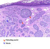
Which type of sweat gland produces milky sweat?
Apocrine sweat glands
[Highest concentration of glands in palms and soles; most sweat is the result of sympathetic nervous system via acetylcholine.]

What are the recommended negative margins for resection of basal cell carcinoma of the skin?
0.3-0.5 cm margins
[XRT and chemotherapy may be of limited benefit for inoperable disease, metastases, or neuro/lymphatic/vessel invasion.]
[UpToDate: Because surgical excision of truncal, extremity, or small facial BCCs on the head or neck with 4 to 5 mm margins has been associated with five-year cure rates exceeding 95 percent, 4 mm surgical margins are commonly used for the excision of these lesions.
However, the results of a 2010 meta-analysis of 89 studies on conventionally excised BCCs that excluded studies of previously excised or irradiated lesions and data on morpheaform BCCs (all features associated with increased risk for tumor recurrence) suggest that 3 mm surgical margins may be only slightly less efficacious. Rates of pathologically confirmed complete excisions were similar for surgical margins between 3 and 5 mm, and mean recurrence rates for BCCs excised with 5 mm, 4 mm, 3 mm, and 2 mm surgical margins were 0.4, 1.6, 2.6, and 4 percent, respectively.]
What is the treatment for a desmoid tumor?
Surgery if possible
[Chemotherapy (sulindac, tamoxifen) if vital structures are involved or too much bowel would need to be resected.]
[UpToDate:
Extraabdominal and abdominal wall tumors
- Observation is an appropriate option in asymptomatic patients who may be reliably followed. If desmoids remain unchanged or shrink, observation may be continued. If the tumor increases in size or becomes symptomatic, if there is imminent risk to adjacent structures, or if the desmoid creates cosmetic concerns, treatment should be pursued.
- We suggest complete surgical excision as the treatment of choice for a potentially resectable extraabdominal (extremity, trunk, breast) or abdominal wall desmoids in a patient who is medically able to tolerate surgery and if resection can be accomplished without major functional or cosmetic deficit (Grade 2B). Controversy remains as a number of patients will fare well without surgical intervention, so a more conservative approach appears rational for relatively static lesions.
- Primary radiation therapy (RT) is an appropriate option for patients who need treatment but are not good surgical candidates, those who decline surgery, and those for whom surgical morbidity would be excessive. An alternative approach is initiation of systemic treatment in these patients, especially if they are young and there are concerns about the potential for late toxicity from RT.
- For a recurrent desmoid tumor, our preference is observation or surgical resection. However, systemic therapy or RT alone are reasonable alternative treatments in selected patients who are thought to have a higher morbidity from repeat operation and an increased probability of positive margins. For patients who undergo surgery for a recurrent desmoid, we suggest observation or postoperative RT if the margins are positive (Grade 2C).
Intraabdominal desmoid, Gardner’s syndrome
- For patients who have large intraabdominal desmoid tumors, particularly in the setting of Gardner’s syndrome, surgery is still a standard approach for resectable tumors. However, the infiltrative nature of the desmoid in this situation often precludes surgery, and the surgical margins are often positive. Medical therapy in lieu of surgery is a viable option for patients with more difficult tumors such as those involving the mesentery, major vessels, or other critical structures.
- Surgery with or without RT is an appropriate option if there is no response to medical therapy. In this situation, consideration should be given to use of intraoperative electron beam therapy as a component of the treatment.
- Management of recurrence of an intraabdominal tumor in patients with Gardner’s syndrome is challenging because recurrences tend to become more frequent and aggressive with each surgical intervention. Most of these patients are managed with systemic therapy rather than additional local measures.]

What should be done about clinically positive nodes in basal cell carcinoma of the skin?
Regional adenectomy
What resection margins are required for melanoma in situ or thin lentigo maligna (Hutchinson’s Freckle)
0.5 cm margins are ok
[This type of melanoma is just in the superficial papillary dermis.]
[UpToDate: Lentigo maligna is a slowly evolving type of melanoma in situ that typically occurs in the sun-damaged skin of the face and neck of older individuals. The risk of progression to invasive lentigo maligna melanoma ranges from 5 to 20 percent. The development of darker pigmentation, sharper borders, or elevated or nodular areas are clinical signs of progression. There are no randomized trials evaluating treatment for lentigo maligna. We suggest surgical excision with margins of 5 to 10 mm, depending upon lesion size and location (Grade 2C). If available, surgical techniques that allow complete margin control such as staged excision with permanent sections (“slow” Mohs) are a preferred option. Among nonsurgical treatments for lentigo maligna, options include radiation therapy with multifractionated high voltage regimens or grenz rays or soft x-rays for large lesions in older patients for whom surgical removal and reconstruction would be difficult.]

Which epidermal cell type is of neuroectodermal (neural crest cell) origin?
Melanocytes

Which cells types are contained in a xanthoma?
Lipid-laden cells and histiocytes
[Treatment is excision]
[UpToDate: For xanthomas occurring in association with hyperlipidemia, it is hypothesized that when serum levels of lipoproteins are substantially elevated, extravasation of lipoproteins through dermal capillary blood vessels with subsequent engulfment by macrophages leads to the lipid-laden cells found in xanthomas, which has been demonstrated using electron microscopy.
Cutaneous xanthomas associated with hyperlipidemia often improve with treatment of the underlying lipid abnormality. Surgical and destructive treatments are the primary therapeutic options for xanthelasma in normolipidemic patients and for verruciform xanthomas.]

Xeroderma pigmentosum is a risk factor for which form of skin cancer?
Melanoma

Rank the following types of skin cancer in order of most to least frequently metastatic?
- Squamous cell carcinoma
- Basal cell carcinoma
- Melanoma
- Melanoma
- Squamous cell carcinoma
- Basal cell carcinoma
What is the treatment for hidradenitis?
Antibiotics and improved hygiene
[May need surgery to remove skin and associated sweat glands.]
[UpToDate: The level of disease severity strongly influences the approach to the treatment of Hidradenitis suppurativa/acne inversa (HS/AI). Patients with Hurley stage I HS/AI present with single or multiple nodules and abscesses without associated sinus tracts or scarring. For these patients, we suggest daily treatment of the involved areas with topical clindamycin (Grade 2B). We manage acute, inflamed lesions with punch debridement (mini-unroofing). If punch debridement is not feasible, topical resorcinol is an alternative patient-administered treatment that we often employ for the treatment of acute lesions. Intralesional corticosteroids and short courses of systemic antiinflammatory antibiotic therapy also can be beneficial for reducing symptoms.
Patients with Hurley stage II HS/AI exhibit recurrent inflamed nodules and abscesses, some of which lead to sinus tracts and scarring. We approach treatment with a combination of medical and surgical therapy. For the medical treatment of the inflammatory component of Hurley stage II disease, we suggest treatment with systemic antibiotic therapy (Grade 2C). We usually treat with doxycycline and continue treatment for two to three months or more. Combination therapy with clindamycin and rifampin is typically reserved for patients who fail to respond to other antibiotic regimens. Concurrent use of anti-androgenic agents may provide additional benefit.
Incision and drainage does not alter the clinical course of HS/AI and should only be performed when immediate relief of pain from a tense abscess is required. Incision and drainage should not be used for routine management of HS/AI. Punch debridement (for small acute, nodular lesions) and unroofing (for larger areas of involvement) are our preferred surgical interventions.]
What is the most common type of benign skin cyst?
Epidermal inclusion cyst
[Has completely mature epidermis with creamy keratin material.]
[UpToDate: Epidermoid cysts, also called epidermal cysts, epidermal inclusion cysts, or, improperly, “sebaceous cysts,” are the most common cutaneous cysts. They can occur anywhere on the body and typically present as skin-colored dermal nodules, often with a clinically visible central punctum. The size ranges from a few millimeters to several centimeters in diameter. Infected, fluctuant cysts tend to be larger, more erythematous, and more painful than sterile inflamed cysts, although an intense inflammatory response to cyst rupture may also present as a fluctuant nodule.
Epidermoid cysts unusual in number and location (extremities rather than face, base of ears, and trunk) may be seen in the setting of Gardner syndrome, a rare inherited condition characterized by familial adenomatous polyposis of the colon associated with a number of extracolonic abnormalities.
The cyst wall consists of normal stratified squamous epithelium derived from the follicular infundibulum. The cyst may be primary or may arise from the implantation of the follicular epithelium in the dermis as a result of trauma or comedone. Lesions may remain stable or progressively enlarge. Spontaneous inflammation and rupture can occur, with significant involvement of surrounding tissue. There is no way to predict which lesions will remain quiescent and which will become larger or inflamed.]

Which mechanoreceptor (sensory nerve) in the skin is responsible for sensing pressure?
Pacinian corpuscles