Week 6 - D - Regional Adult Trauma (3) - Humeral shaft, elbow, forearm, distal radius fractures Flashcards
Humeral shaft fractures can be caused by direct trauma (such as during an RTA) resulting in transverse or comminuted fractures, or by fall with or without twisting injury resulting in oblique or spiral fractures.
What nerve may be injured?
The radial nerve may be injured as it runs through the radial (spiral) groove
If there was a radial nerve palsy in a humeral shaft fracture How would this show?
There would be a loss of sensation in the first dorsal web space and a wrist drop would be present (loss of innervation to the extensors)

Most cases are treated non-operatively with a functional humeral brace which compresses the fragments into acceptable alignment and provides some stability. What will be used if there is non-union?
Use internal fixation with an intramedullary nail or plates
What age group does supracondylar fractures usually occur in? What are complications?
Usually occurs in childen
Can result in injury to the brachial artery and the median nerve
Brachial artery rupture can lead to haemorrhage causing compartmnet syndrome and this can lead to volkmann’s ischaemic contracture
How is an intra-articular fracture of the distal humerus treated?
Treat with open reduction and internal fixation
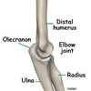
Olecranon – common and occur when falling onto the point of the elbow with a contracted triceps Where to the triceps insert distally?
What is the treatment?
Onto the olecranon process of the ulna
Treatment is open reduction internal fixation
Radial head and neck fractures usually occur due to a fall onto the outstretched arm. Some undisplaced fractures may not show up on x‐ray other than a posterior fat pad sign on the lateral x‐ray (a triangle like a sail anterior to the distal humerus - due to a raised anterior fat pad) with lateral elbow pain on supination / pronation.
What is the treatment of the undisplaced radial head and neck fractures?

Conservative treatment with a sling for comfort
Patients often lose 10‐15° of terminal extension
What is given if it is an intraarticular fracture of the radial head?
Would give ORIF
The forearm consists of the radius and ulna bones which are connected proximally and distally by strong ligaments around the proximal and distal radio‐ulnar joints where supination and pronation occur. Because of these strong ligaments, the forearm acts as a ring
If there is one fracture therefore, what happens?
There tends to be another fracture also or a dislocation
Isolated fractures of the ulna can occur after a direct blow. What is a nightstick fracture properly known as?
Ulnar shaft fracture
What must you make sure there is no associated of in an isolated ulnar shaft fracture?
No associated of a Monteggia fracture dislocation
What is the management of an isolated ulnar shaft fracture?
Many cases are dealt with by conservative management however ORIF may afford earlier return to function and may reduce the risk of non‐union.
What is a monteggia fracture dislocation?
This is where there is a fracture of the ulna with a dislocation of the radial head at the elbow MUGGER
MU - ulnar fracture monteggiA (A for proximal where the radial head is disocated at elbow)

When the radius dislocates from the humerus at the elbow joint, what is the joint?
This is the radio-capitellar joint - supination and pronation

Which of the elbow joints cause flexion and extension?
This would be the ulnohumeral joint - the trochlea of the humerus inserts into the trochlear notch of the ulna
What is a galeazzie fracture dislocation?
This is where there is a radial fracture with an ulnar dislocation at the distal radioulnar joint MUGGER
GR - radial fracture galeaZZi (Z for distal - dislocation at distal radioulnar joint)

It is advisable to form elbow xrays in monteggia fractures to make sure the radius is dislocated What is the mandatory xray view for galeazzia fractures?
Mandatory to have a lateral xray view
What is the treatment of monteggia and galeazzia fractures? How is an isolated ulnar fracture treated? Describe both fractures again?
Monteggia and galeazzi fractures are treated with open reduction internal fixation
Isolated ulnar fracture - conservative management
Monteggia - fracture of the ulnar accompanied by dislocation of the radius at the elbow
Galeazzi - fracture of the radius accompanied by dislocation of the ulnar at the distal radioulnar joint
Describe a Colle’s fracture
Extraarticular fracture of the distal radius within an inch of the articular surface with dorsal angulation or displacement of the radius due to a fall on outstrectched hand (FOOSH)
How does a colle’s fracture usually occur?
Usually occurs due to FOOSH (fall on out-stretched hand)
Fractures which heal with less angulation and shortening tend to have a better outcome in terms of pain, grip strength, ROM(range of motion), and function How will minimally displaced colles fractures be treated?
Reduce and splint
In a Colle’s fracture, any angulation past neutral (the distal radius articular surface is normally 10° volarly angulated) is usually corrected by manipulation. How is it held together?
Held together by a plaster cast
If the colle’s fracture is comminuted or particularly unstable after reduction What is carried out?
ORIF + Kwiring (percutaneous pins) may be required
What nerve can be compressed in colle’s fracture and what are signs of this?
Can have median nerve compression, causes an ape like hand deformtiy and loss of sensation /sometimes pain in 1st,2nd and 3rd digits

