Week 4 - H - Knee (2) - Meniscal Tears, Cruciate ligament ruptures and collateral ligament tears / ruptures, disclocation Flashcards
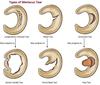
As with most clinical presentations in medicine, the diagnosis of knee injuries can usually be found by detailed history taking with confirmation by clinical examination and subsequent investigation. Which meniscus is more likely to tear than the other and why?
Medial mensical tears are approximately 10 times more common than lateral meiscus tears
This is due to the fact that the medial mensicus is more fixed and less movile than the lateral mensicus and the force from pivoting is centred on the medial compartment
How do meniscal injuries classically occur? What are the symptoms and when does swelling occur?
Meniscal injuries classically occur with a twisting force onto a loaded knee (eg turning at football, squatting)
The patients localises pain to the medial or lateral joint line
An effusion usually develops the following day
After the initial localised pain to the medial (majority) or lateral joint line, with an effusion following the next day, what do patients complain off?
The patient then complains of pain and usually has mechanical symptoms
Either a catching sensation or ‘locking’ where they have difficulty straightening the knee with a 15 degree or so block to full extension
Patients knees may feel about to give way if a loose meniscal fragment is caught in the knee when walking.
True knee “locking” is defined as a mechanical block to full extension and is caused by the significantly torn meniscus flipping over and becoming stuck in the joint line What is the tear known as when the meniscus locks like this? How is this diagnosis confirmed?
This type of tear is known as a Bucket handle meniscal tear -
the locked knee will have a 15 degree or so springy block to full extension
MRI will confirm the clinical suspicion

It is possible to have a meniscal tear without “locking” if the torn meniscus isn’t sufficiently unstable to flip and become caught in the joint. Patients with other knee pathologies, such as arthritis, will describe that their knee can “become stuck” with temporary difficulty in straightening the joint.
Why does this usually occur and what is the term given to these patients?
This usually occurs after rising from sitting and it will either spontaneously resolve or the patient will describe a trick manoeuver that relieves the issue
This sign is known as ‘pseudo-locking’ and should be distinguished from locking
How would a menscal tear appear on examination?
Clinical examination may revea
l * An effusion
* medial gutter sweep test - small effusion
* patellar tap - visible effusion
Joint line tenderness pain on tibial rotation localizing to the affected compartment -
Steinmann’s test
uneven heel height test if displaced buckethandle meniscal tear
Degenerate meniscal tears can occur as the meniscus weakens with age. Degenerate tears are probably the first stage in many cases of knee osteoarthritis. How do degenerate meniscal tears differ from acute meniscal tears on examination? Why is this important?
Degenerate meniscal tears are Steinmann’s negative and are likely to be associated with early symptoms and signs of OA
This is important are degenerate meniscal tears are managed differently from acute tears

Which part of the meniscus has an arterial blood supply? Which type of meniscal tears are the only type that should be considered for a meniscal repair?
Only the outer 1/3rd of the meniscus has an arterial blood supply
Only reasonably fresh longitudinal tears involving the outer 1/3rd of the meniscus in a younger patient (above 25/30 and healing rates are poor) should be considered for meniscal repair
What is involved in meniscal repair? What is carried out should the meniscal repairs fail?
Meniscal repair involves suturing the meniscus to its bed
Even with careful patient selection a relatively high proportion (around 25%) of meniscal repairs fail requiring arthroscopic menisectomy.
More than 90% of meniscal tears are not suitable for repair. Whilst meniscal tears do not usually heal, the pain and inflammation may settle with time, particularly with degenerate meniscal tears. The knee can also “smooth off” its own meniscus given time * What may help degenerate meniscal tears symptoms in the early period? * If pain or mechanical symtpoms do not settle in 3 months of a meniscal tear, what operation can be performed?
Steroid injection in degenerate tears may help symptoms in the early period In acute tears, if the pain or mechanical symptoms do not settle within around 3 months, then arthroscopic partial menisectomy can be performed
How do anterior cruciate ruptures tend to occur? Give sporting examples
ACL ruptures usually occur with higher rotation force (than in meniscal tears) , turning the upper body laterally on a planted foot leading to internal rotation force on the tibia -
often football, rugby, skiing
What is typically heard on ACL rupture? How long after does swelling occur and what is the swelling? What other symptoms are there?
A ‘POP’ sound is usually felt or heard
Within an hour of the injury the patient develops a haemarthrosis (effusion due to bleeding in the joint - in this cases from the vascular supply within the ACL) and there is also a deep pain in the knee
What is the principal complain that the patient with an ACL rupture may complain of chronically?
Chronically, the patient may then complain of rotatory instability with their knee giving way when turning on a planted foot (due to excessive internal rotation of the tibia).
What would clinical examination of a patient with an ACL rupture reveal?
Clinical examination will reveal
* Knee swelling
* Excessive anterior translation of the tibia on the anterior drawer test (knee and hip flexed to 90 degrees and tibia pulled forward)
* Excessive anterior translation of the tibia on Lachman’s test (knee at 20 degrees flexion and tibia pulled forward)
ACL ruptures may cause little or no problems in some, whilst in others they can give substantial problems with function. What is the rule of 1/3rds in ACL rupture?
* Approximately 1/3rd of patients will compensate and do whatever they please (including sports) (usually it is patients with big muscle bulk that can compensate)
* Approximately 1/3rd will manage by avoiding certain movements but may not be able to do high impact sports
* Approximately 1/3rd will do poorly with frequent giving way even with normal daily activities
What may help patients compensate with the ACL rupture?
Physiotherapy to strengthen and give proprioceptive training to the quadriceps and hamstrings muscle bulk may help comepnsation
Primary repair of the torn ACL is not effective and overall around 40% of patients with ACL rupture end up having a reconstruction What is the difference between an ACL repair and an ACL reconstruction?
In an ACL repair, small holes are drilled into the tibia and femur and then sutures are athroscopically tied to both ends of the ACL drawing them together (A)
In an ACL reconstruction, a tendon graft is passed though tibial and femoral tunnels and held in place with staples or screws (B)

What are the tendons usually used in an ACL reconstruction? - is it from the person or from a donor usually How long can it take, if possible, to get back to high impact sports?
Tendons usually used are patellar tendon, semitendinsous or gracilis tendon autografts
* (Tissue taken from your own body is called an autograft. Tissue taken from a donor is called an allograft)
Intensive rehabilitation is required and it may take up to a year to get back to high impact sports
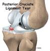
How may a PCL injury occur?
A direct blow to the anterior tibia (aka the dashboard injury or motorcycle crash) or hypterextension may cause rupture of the PCL due to the tibia being translated psoteriorly to the femur
What may a patient complain off symptoms wise after a PCL rupture?
Initially, pain and swelling
Positive posterior drawer test
Chronically, the patient may complain of
* recurrent instability with frequent hyperextension
* feeling unstable descending stairs - due to anterior subluxation of the femur
PCL rupture in isolation is not common When are PCL reconstructions usually carried out?
PCL reconstruction is usually performed for reconstruction of the multiple ligament injured knee
When are PCL reconstructions carried out for isolated injuries? What is the tendon graft used for PCL reconstruction?
With isolated PCL rupture only those with severe laxity and recurrent instability with frequent hyperextension or feeling unstable descending stairs (with anterior subluxation of the femur) are considered for surgical reconstruction (usually with cadaveric achilles tendon allograft).
How do MCL injuries typically occur? What is at risk of being damaged with MCL injuries?
MCL injuries occur typically due to valgus stress injuries (eg rugby tackle to lateral aspect of the knee causing a valgasing stress on the knee)
MCL injuries are associated with potnetialy damage to the ACL and medial meniscus - unhappy triad
MCL injuries are fairly common however the MCL is a fairly forgiving knee ligament with healing expected in the majority of partial and complete tears and little or no instability What are the features of an MCL injury on examination? How are acute tears usually treated?
Patient may have laxity and pain on valgus stress with tenderness over the origin or insertion of the MCL
Acute tears are usually treated in a hinged knee brace
How do lateral collateral ligament injuries typically occur? What other ligament may be injured with this?
Lateral colalteral ligaments typically occur when a varus stress injury takes place - there may or may not be damage to the PCL
What are the features on examination of a LCL ligament injury?
Patient may have laxity and pain on varus stress
Patients also have marked instability on external rotatory movement of the knee
What is the treatment of lateral collateral ligament ruptures? What nerve can be injured during a LCL rupture?
Treatment of LCL ruptures is usually surgical with early repair or late reconstruction (tendon graft)
The hyperextension and varus giving rise to the injury also gives a high incidence of common peroneal nerve injury from excessive stretch
With higher degrees of force, some injuries can involve rupture of more than one of the four knee ligaments. Multi‐ligament knee injuries often require surgical reconstruction due to the degree of instability. What does a complete knee dislocation involve? What is at risk here?
Complete knee dislocations result in rupture of all four of the knee ligaments (ACL, PCL, MCL, LCL) and have a high incidence of neurovascular injury

How are complete knee dislcoations managed?
Complete knee dislocations are reduced as an emergency and may require external fixation for temporary stabilisation
Patients usually require mutliple ligament reconstruction
