Tumors Flashcards
1
Q

A
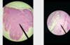
Fibroma: fibrous and connective tissue tumor
- benign tumors of fibrocytes produced large amounts of collagen
- hari follicles, sebaceous glands(dilated by the surrounding tumor)
- tumor cells: elongated nuclei, which shows strong basophillia
- stroma is made up by collagen CT
- Blue: nuclei of well differentiate elongated neoplastic fibroblasts
2
Q

A
Fibroma
3
Q

A
Fibrosarcoma:
= malignant CT tumor of fibroblast
4
Q

A
Fibrosarcoma:
- see the mitotic figures
- intraluminal blood vessels
- necrosis
- lymphocytes
5
Q

A
Sarcoma gigantocellulare:
= pleomorphic sarcoma / anaplastic sarcoma with giant cells
- malignant fibrous histocytoma
- vascular intratumoral areas
- multinucleated giant cells
- mitotic figures(metaphase)(anaphase)
6
Q

A
Haemangioma cavernosum:
=benign tumors of vascular endothelium (blood vessel producing tissue)
- large vascular spaces filled with blood
- very pink material= fibrin
- in between CT = edema/serous infiltration.
*cavernousous(large)
*capillary(small)
- the differences is the size of vascular spaces.
- see mast cells, plasma cells as well
7
Q

A
Simple adenoma(adenom simplex):
- mature tumor of glandular tissue
- well demarcated
- One cell type( tubular)= so tumor is peri-tubular
- arises from the milk ducts and not from the lobuli
- simple cuboidal
- simple columnar
*neoplastic tubuler are separeted by fibrovascular CT stroma.
8
Q

A
Simple Adenoma(adenoma simplex).
- look at the cells
- columnar
- cuboidal
9
Q

A
Adenocarcinoma:
10
Q

A
Adenocarcinoma:
- in SI( villi) or LI( no villi)
- intact parts of the intestines (Villi, goblet cells, glands/crypts)
- tumoral part( propria glands are like bubles, lack of villi stucture)
- mitotic and apoptoic cells
- lumen filled w/ proliferating cells
- necrosis, polymorphism and hypo-hyper chromatic cells
11
Q
A
Papilloma (oral mucosa):
- identify . the epidermal hyperplasia
- Ghost cells
- Kilocytes= due to papillomavirus
- mitotic figures
- Inclusion bodies
- Keratocytes
- Finger like processes
12
Q

A
Papilloma (oral mucosa):
- identify . the epidermal hyperplasia
- Ghost cells
- Kilocytes= due to papillomavirus
- mitotic figures
- Inclusion bodies
- Keratocytes
- Finger like processes
13
Q

A
Squamous cell carcinoma (skin):
- finger like projections going inwards
- identify dysplastic epidermis(enlarged and broken epidermis w/ inflammatory cells) and hyperkeratosis on top
- hair follicles
- tumor islets and stroma
- keratin pearl in middle (like foreign body) = concentric lamellae of keratin (keratosis)
- outer mitotic cells and outer flattened cells
