Lung and Kidney Flashcards

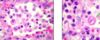
Serous desquamative bronchopneumonia:
- the alveoli, alvolar ducts, bronchi are filed w/ exudate
- exudate contains desquamative tissue(lymphocyte, neutrophils)
- Bronchitis( squamous metaplasia, mucous gl. hyperplasia)
- Bronchiolitis obliterans( small structure w/ capsule like structure, purple in center)
- Desquamated type 2 pneumocyte
- chronic demarcation area
!! Mycoplasma, Pasturella, Streptococcus
- Desquamated alveolar epithelial cells
- Chronic dermacation areas
Desquamated type 2 pneumocyte

Fibrinous (Kruposa) pneumonia:
- diff to req pga little amount of alvoli
- fibrin rich exudate in alveoli/bronchi
- marked hyperaemia due to dilated blood vessels( elongated w/fibrin)
- thrombosis in lymphvessels
1) Stage congestion= exudate(firbrin)
2) stage hepatization= fresh RBC to destroyed RBC to neutrophils

Fibrinous( Kruposa) pneumonia:
- fibrin rich exudate in alveoli
- hyperaemia
Actinobacillus pleuropneumonia
seros-hemorrhagic-necrotic-pneumonia
- hyperaemia
- alveolar edema
- macrophage proliferation
- necrosis + demarcation zone
- dilated lymphvessels(thrombosis)

Actinobacillus pleuropneumonia:
- hyperaemia
- alveolar edema
- macrophage proliferation (inflam cells in the alveolar lumen)=blurry, degenerating by toxins
- necrosis with macrophages and inflam cells, demercation zone
- dilated lymph vessels( thrombus)

Interstitial pneumonia(subacute):
- more narrow than normal
- no exudate
- cell proliferation= in wall of alveoli/a ducts, consist of lymphocytes, neutrophills,histocytes and plasma cells
- closed bronchi( Atelectasis)(no gas exchange, functional loss)
- peribronchial interstitial pneumonia
!!Lungworms, toxic gas, influenza, herpes, distemper, salmonella, streptococcus

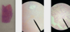
Oxalate crystals (kidney)
- see the glomerular(kidney structures)
- calcium oxalate crystals(look in the cortex, outermost widest part)
- degenerated: karyopycnosis, karyolysis + desquamated epithelial cells)

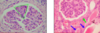
Chronic (diffuse) glomerulonephritis:
- glomeral structure
- enlarged and shrunken glomerus
- mesangial cells proliferate and lead to enlargement= segmented glomerulus
- Capsule thickened= CT proliferaion
- Protein rich exudate in Tubules
*subacute–> exudate in glomerulus, not tubules
*chronic
!! SLE + Amyloidosis
Chronic diffuse glomerulonephritis:
- mesangial cell proliferation
- protein rich exudate in tubules( exudate in glomerulus= subacute)

Interstitial nephritis:
- periglomerular + interglomerular spaces is infiltrated foccally by inflammation cells
- identify the cells ( plasma cells, lymphocytes, Histocytes, neutrophils)
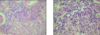
Interstitial nephritis:
-recognice the inflammatory cells around the glomerulus aparatus

Kidney fibrosis

Kidney fibrosis:
-see the fibrin/fibrous due to all the collagen fibers
(collagen fibrous proliferation and replace the damaged tubules ad glomerulus)
- thickening of the bowmans capsule
- hyaline like material (pink) deposition in glomerulus (exudate)
- sec. Calcium deposits in capsule and basal membrane
Oxalate nephritis
