The nervous system Flashcards

1
Q

Flatworms have a brain consisting of a cluster of nerve cell bodies which is concentrated where?

A

In their head or cephalic region

2
Q

In worms, the cell bodies are no restricted to the head. where do they also occur?

A

In fused pairs known as glanglia, along a nerve cord

3
Q

Whats a spinal reflex?

A

A simple, involunatary movement that can be integrated within the spinal cord without input from the brain

4
Q

Whats is included in the CNS

A

The brain and spinal cord

5
Q

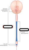
Name the 5 main sections of the spinal cord, which is each divided into smaller sections ?

A
  1. cervical
  2. thoracic
  3. lumbar
  4. sacral
  5. coccygeal
6
Q

What are the nerves coming off each section of the spinal cord known as?

A

the spinal nerves/ peripheral nerves

7
Q

Tell me what the following are

  1. cranial nerves
  2. spinal nerves
  3. ganglia
A

cranial nerves: nerves coming off the brain stem

Spinal nerves: nerves coming off the spinal cord

ganglia: a structure in the PNS. it houses the cell bodies of afferent and efferent nerves

8
Q

What are afferent nerves?

A

sensory neurons that carry nerve impulses from the sensory stimuli towards the CNS and brain

9
Q

What are efferent nerves?

A

These are motor neurons that carry neural impulses away from the CNS and towards muscle and cause movement

10
Q

What are neurones and what does this include ?

A

Neurones are the powerhouse of the nervous system. this includes; axons, dendrites and cell bodies

11
Q

Whats are dendrites?

A

The branch off of the cell body and they recieve information and deliver it to the cell body.

There are many denrites in a neurone

12
Q

Whats an axon?

A

This takes information away from the cell body

Theres only one axon in a neurone

13
Q

Whats an axon hillock?

A

This is the very first part of an axon as it leaves the cell body, it has lots of Na+ channels and is the start of the action potential (trigger zone)

14
Q

Whats myelin sheath?

A

this helps to insulate the axon and help speed up electrical conduction

15
Q

label this neurone

A
16
Q

Whats the name of the type of cells that provide support and insulation for neurones?

A

glial cells

17
Q

Name the glial cells found in the CNS/PNS and the glial cells found in only the PNS

A

CNS/PNS

  1. Astrocyte
  2. oligodendrocyte
  3. microglia
  4. ependymal cells

PNS only

  1. Schwann cells
18
Q

What are astrocytes role?

A

They help to regulate the transmission of electrical impulses

19
Q

What’s the oligodendrocytes role?

A

responsible for making myelin

20
Q

Whats the role of the microglia cells?

A

Immuce cells, they provide some immune surveillance but not as much as a macrophage

21
Q

Whats the role of ependymal cells?

A

cells that provide cerebral spinal fluid that line the open spaces in the brain and spinal cord

22
Q

Whats the role of the schwann cells in the PNS?

A

They take up the role of the astrocyte (transmission of electrical impulses) and oligodendrocytes (responsible for making myelin). they’re the powerhouse of the PNS

23
Q

Identify the glial cells

A
24
Q

What is grey matter and what resides in it?

A

Grey matter is greyish nerve tissue of the CNS which is mainly composed of nerve cell bodies and dendrites

25
Q

Whats white matter?

A

white matter is whiteish nerve tissue of the CNS that is mainly composed of myelinated nerve fibres (or axons)

26
Q

What parts of neurones are found in the white matter and whats found in the grey matter?

A

White matter

  • Myelinated axon
  • axon hillock

Grey matter

  • cell bodies
  • unmyelinated axons
  • dendrites
27
Q

What glial cells are found in the white matter and/or in the grey matter

A

white matter

  • astrocytes
  • oligodendrocytes
  • microglia
  • ependyma cells
  • schwann cells

Grey matter

  • astrocytes
  • oligodendrocytes
  • microglia
28
Q

Whats the nucleus/ nuclei and its role?

A

a group of nerve cell bodies within the CNS, consisting of neurones with relating functions and having defines input and output tracts (bundles of axons connected to different regions of the CNS)

29
Q

Whats the ganglion?

A

nerve cell bodies outside the CNS, often visible as a swelling in a nerve or at the junction of a group of nerves e.g. dorsal root ganglia

30
Q

Whats an interneurone?

A

this is a neurone completely contained within the CNS

31
Q

Name the 4 lobes of the brain?

A
  1. frontal lobe
  2. parietal lobe
  3. temporal lobe
  4. occipital lobe
32
Q

whats meant by sulci?

A

infoldings of the cerebral hemispheres that form ‘valleys’ between the gyri

(singlular= sulcus)

33
Q

Whats gyri?

A

ridges of the infolded cerebral cortex

singular= gyrus

34
Q

Name the 4 functional areas in the cerebrum and what lobes they are located in?

A

1. motor

Primary motor and premotor in frontal lobe

2. sensory

Primary somatosensory and somatosensory association areas in paritetal lobe

3. visual

primary visual and visual association areas in occipital lobe

4. auditory

primary auditory and auditory association areas in temporal lobe

35
Q

What are 2 areas on the left side of the brain and what are they responsible for?

A
  1. Broca’s area: responsible for producing language. it controls motor functions with speech
  2. Wernicke’s area: important for language development and important for the comprehension of speech
36
Q

what does the diencephalon overarch?

A
  1. thalamus
  2. hypothalamus
37
Q

whats the thalamus’ role?

A

a relay station for information coming into the cortex from either sensory impulses (travelling to sensory cortex) or inputs for subcortical motor nuclei and cerebellum (travelling to the cerebral motor cortex)

38
Q

Whats the hypothalamus important for?

A

its an important autonomic control centre e.g. homeostasis

39
Q

Whats does the brain stem overarch?

A
  1. midbrain
  2. pons
  3. medualla oblongata
40
Q

what are the 3 centres in the midbrain and what are these centres involved in?

A

1. superior and inferior colliculi: visual and auditory reflex centres

2. red nucleus: subcortical motor centre

3. substantia nigra: involved in reward-seeking, motor learning and others

41
Q

Whats does the pons contain and what does this connect?

What do the nuclei in the pons do?

A

The pons cintains a conduction area which connects the forebrain and cerebellum

the nuclei contribute to the regulation of respiration as well as hearing and balance

42
Q

What 2 centre does the medulla oblongata contain and what are they involved in?

A

1. Vital centres: regulating respiratory rhythm, heart rate, blood pressure

2. non-vital centres: regulating coughing, sneezing, swallowing, vomiting

43
Q

the 2 helispheres in the cerebellum contains what? and are surrounded by what?

A

the cerebellum has 2 hemispheres which contains internal grey matter nuclei surrounded by white matter and an outer cortex of grey matter

44
Q

Whats the cerebellum important for?

A

balance and coordinating motor activity

45
Q

Whats the spinal cord and what is it protected by?

A

The spinal cord is a two-way impulse conduction pathway and reflex centre. it is protected by the meninges and CSF

46
Q

Where are the meninges located and what’s their role?

A

The meninges lies between the bones and tissues of the CNS.

These membranes help stabilise the neural tissue and protect it from bruising against bones

47
Q

What are the 3 protective layers in the meninges from the outside in?

A

outside

Dura mater

arachnoid membrane

pia mater

inside

48
Q

Tell me about the dura mater?

A
  • thickest of the 3 membranes
  • associated with veins that drain blood from the brain through vessels or cavities known as sinuses
49
Q

Tell me about the Arachnoid membrane?

A
  • closely ties to the inner memrbane
  • these is a subarachnoid space between the 2 layers
  • CNS tissue
50
Q

Tell me about the pia mater?

A
  • thin membrane that adheres to the surface of the brain and spinal cord
  • arteries that supply blood to the brain are associated with this layer
51
Q

Whats CSF? Whats it produced by?

A

CSF is the cerebrospinal fluid. it is a clear, cell-free fluid produced by the choroid plexus (ependymal cells) that circulates the subarachnoid space

52
Q

Whats the flow of the CSF, starting at the ventricles? Where is it absorbed back into the blood

A

ventricules –> subarachnoid space –> flows around neural tissue –> absorbed back into blood by villi on the arachnpid membrane in the cranium

53
Q

What are thr 2 main roles of the CSF?

A

chemical and physicla protection

54
Q

What are the subdivision of the nervous system and what do all of these branch off to?

A
55
Q

Whats the meaning of somatic?

A

relating to the body wall and limbs (muscles, skin, bones, joints)

56
Q

Whats the meaning of visceral?

A

relating to the internal organs (heart, lungs, digestive system, kidneys, reproductive system etc.)

REMEMBER: respiratory muscles and skeletal are controlled by the somatic NS

57
Q

Is both the CNS and PNS both voluntary and involuntary?

A

yes

58
Q

what are synapses?

A

electrochemical communication between two or more neurones via synapses between axons and dendrites

59
Q

label this synapse?

A
60
Q

What are the branches from the visceral motor divison in the subdivision of the motor system?

A
61
Q

whats the parasympathetic nervous system?

Where are the parasympathetic neurones located?

A

“rest and digest”/ craniosacral

Parasympathetic neurones are located carnially and sacrially ie. brain stem and in the sacral cords S2-S4

62
Q

Whats the sympathetic nervous system.

Where are the sympathetic neurones located?

A

“fight or flight”/ thoracolumbar

the sympathetic neurones are located in the spinal cord ie. T1-L2 in the thoracic cord and upper lumbar cord

63
Q

what controls the parasympathetic and sympathetic nervous systems?

A

the hypothalamus with the exception of some function which operate in a reflex action

64
Q

Identify the parasympathetic and sympathetic NS?

A
65
Q

What does the ANS and endocrine system control?

A
  1. innervates smooth muscle, cardiac muscle and glands of internal organs (involuntary)
  2. the two neurone chain (1st in CNS and 2nd in peripheral)
66
Q

What are the name of the neurones in the two-neurone chain? where do they originate and they myelination ?

A

1. preganglionic neurone: originate in the CNS; preganglionic efferent axons are lightly myelinated

2. postganglionic neurone: originate in the ganglion located outside the CNS; postganglionic neurones are not myelinated

67
Q

What are the responses caused when the sympathetic system is in control?

A
  1. pupils dilate
  2. increase heart and resiratory rates
  3. increases BP and blood glucose levels
  4. dilation of bronchioles
  5. induces sweating
68
Q

during exercise there is forced sympathetic activation. what does this cause?

A

it causes sympathetic vasoconstriction which moves blood from the skin and digestive viscera to the heart, brain and skeletal muscles

69
Q

What are the responses when the parasympathetic NS is in control?

A
  1. pupil constriction
  2. glandular secretion to help with digestion and other smooth muscle functions
  3. increases digestive tract mobility
  4. smooth muscle activity; leads to elimination of faeces and urine
70
Q

Tell me some effects that the sympathetic NS causes in the body/ helps with?

A
  • increases arterial BP and HR
  • inhibition of pancreatic enzyme secretion
  • inhibition of pancreatic insulin secretion
  • dilation of iris (eye)
  • ejaculation in men
  • decrease in gut mobility and secretion
71
Q

Tell me some effects of the parasympathetic NS on the body

A
  • decrease in arterial BP and HR
  • stimulation of pancreatic enzyme secretion
  • stimulation of pancreatic insulin secretion
  • pupil constriction
  • increased gut mobility and secretion
  • penile erection in men
72
Q

do both the sympathetic and parasympathetic neurones both have a two chain neurone?

A

yes

73
Q

in the parasympathetic and sympathetic neurones, compare…

  1. the location of the postganglionic neurone
  2. length of the preganglionic neurone
  3. length of postganglionic neurone
  4. Neurotransmitter from preganglionic neurone
  5. types of receptors on the postganglionic neurone
  6. Neurotransmitters from the postganglionic neurone
  7. receptors on the target cells
A

1. Location of postganglionic neurone

​Sympathetic: close to spinal cord

Parasympathetic: close to organ it innervates

2. Length of preganglionic neurone

Sympathetic: short

Parasymapthetic: long

3. length of postganglionic neurone

Sympathetic: long

parasympathetic: short

4. Neurotransmitter released from preganglionic neurone

Sympathetic: ACh

Parasympathetic: ACh

5. Receptors on the postganglionic neurone

Sympathetic: Nicotinic receptors

Parasympathetic: Nicotinic receptors

6. Neurotransmitters released from postganglionic neurone

Sympathetic: Norephinephrine (NE) and ACh

Parasympathetic: ACh

7. Receptors on the target cells

Sympathetic: Alpha or beta- adrenergic receptors and muscarinic receptors

Parasympathetic: Muscarinic receptors

74
Q

Comparitive features between the autonomic and somatic motor systems…

  1. The length of the motor fibre
  2. Is there a synapse present
  3. if yes, then is the preganglionic fibre and postganglionic fibre myelinated or unmyelinated and what neurotransmitter is released at the first synapse, what type of ganglion is this?
  4. The neurotransmitter released at the end of the fibre
  5. what effector it targets
A

1. The length of the motor fibre

Somatic: long

Autonomic: short

2. Is there a synapse present in the middle of the fibre

Somatic: No

Autonomic: Yes

3. If yes to #2, then is the preganglinic fibre/ postganglionic fibre myelinated or unmyelinated, what neurotransmitter is released at the synapse, what type of ganglion is it

Autonomic:

Myelinated preganglionic fibre and unmyelinated postganglionic fibre

ACh released at the synapse

Autonomic ganglion

4. The neurotransmitter released at the end of the fibre

Somatic: ACh

Autonomic: ACh or NE

5. what effector it targets

Somatic: Somatic effectors (skeletal muscle)

Autonomic: Visceral effectors (cardiac muscle, smooth muscle, glands)

75
Q

How many pairs of spinal nerves are there? What are they and their locations?

A

31 pairs of spinal nerves

8 cervical nerves (C1-C8)

12 thoracic nerves (T1-T12)

5 lumbar nerves (L1-L5)

5 sacral nerves (S1-S5)

1 coccygeal nerves (Co)

76
Q

During development the vertebral column elongates more than the spinal column, do the nerves still exit from the correct locations?

A

yes

77
Q

The more posterior (caudal) nerves run down inside the vertebral canal forming a bundle known as what before exiting?

A

cauda equine

78
Q

A number of nerves fuse to form what? give an example…

A

Fuse to form plexi such as the brachial plexus innervating the upper limbs; lumbar sacral plexus

79
Q

Label the spinal nerve…

A
80
Q

What are the name of the 2 roots that each spinal nerve has? What occurs there?

A

1. Dorsal root (posterior)

  • afferent fibres enter
  • contains the dorsal root ganglion which the cell bodies of the afferent fibres are

2. Ventral root (anterior)

  • efferent fibres leave
  • their cell bodies are within the spinal cord
  • motor information goes via this root
81
Q

Can roots be either motor or sensory?

A

yes

82
Q

Each segment of the spinal cord gives rise to a pair of spinal nerves that consits of what 3 things? whats their role?

A
  1. axons of motor neurones: innervate a group of muscles
  2. axons of sensory neurones: innervate a defined area of skin
  3. axons of sympathetic neurones: structures in the body wall that control body temperature
83
Q

Name the two areas that are supplied by single spinal nerves

A

Dermatomes and Myotomes

84
Q

What is a myotome and dermatome?

A

Myotome: provides sensory and motor supply of an adjacent muscle mass

Dermatome: the cutaneous supply of an area of skin, sensory

85
Q

Are dermatomes and myotomes identical?

A

no

86
Q

Define the following?

  1. reflex
  2. reflex arc
  3. spinal reflex
A
  1. Reflex: rapid, involuntary response to a stimulus
  2. Reflex arc: receptor, sensory neurone, integration centre, motor neurone and effector
  3. spinal reflex: somatic spinal reflexes provide information on integrity of the relfex pathway and degree of excitability of the spinal cord
87
Q

What does a simple monosynaptic neural circuit only require to go through?

A

only requires going through one sensory neurone to a motor neurone

88
Q

Whats an action potential?

A

A brief change in electrical potential across the cells membrane (neurone’s cell membrane) involving the movement of ions (sodium and potassium) across the membrane

The other wise negatively charged cells become positively charged

89
Q

Whats the resting membrane potential of a neurone?

A

-70 mv

90
Q

What do ion pumps maintain?

A

A concentration gradient

91
Q

Other than the ions inside and outside of the cell, what else contributes to the overall resting membrane potential?

A

Anions (proteins) which are also found within the cell

92
Q

At rest, what is the cell membrane more permeable to?

A

K+

93
Q

What is the Na+/K+ ATPase channel blocked by?

A

the toxin Ouabain

94
Q

Whats the K+/Na+ ATPase pump also known as?

A

The active sodium/potassium pump

95
Q

For every single ATP consumed, whats the ratio of sodium and potassium that pumped in/out of the cell via the ATPase pump and what direction do the ions move?

A

For every 3Na+ pumped out of the cell, 2K+ are pumped into the cell

96
Q

Tell me the steps to the generation of an action potentials and the recovery after?

A
  1. The membrane is at rest (RMP) at around -70mv

PK+>> PNa+
membrane is highly permeable to K+

2. The membrane is passively depolarised

PK+>>PNa+

shifted to a slightly more positive membrane potential

Doesn’t require any energy

3. The membrane reaches threshold

Voltage- gates Na+ channels are activated
An action potential has been generated

Na+ current depolarises the membrane

PNa+>>PK+

Em –> ENa+

4. Positive feedback cycles of depolarisation

Na+ channels open

depolarisation caused, which opens more Na+

Em overshoots 0 and peak AP is reached

5. Em approaches ENa+

the driving force for the current decreases

Na+ channels close

K+ start to open

PNa+ decreases and PK+ increases

6. All Na+ inactivate (close)

all K+ channels open

K+ current repolarises the membrane

Em returns to resting value

7. Hyperpolarisation occurs

PK+ >>> PNa+

Em moves towards EK

8. Em returns to resting value

Na+ channels deactivate and return to resting state

K+ channels close

9. The absolute refractory period

membrane is not excitable as all Na+ are inactivated

No additional APs can be generated

97
Q

What kind of response are action potentials?

A

The ‘all-or-nothing’ response. which means all action potentials have the same magnitude. The strength of a stimulus is known by the frequency of the action potentials

98
Q

Action potentials are known to be quite efficitent as they only require a few ions to generate an action potential, why is this?

A

The cell’s lipid membrane is a capacitor which stores electrical charge which helps to generate ATP

99
Q

What properties of a neuron allow for rapid conduction of signals and why?

A

1. rapidly activated voltage-dependant Na+ channels:

increase inward depolarising current in response to depolarisation (positive feedback)

2. Propagation of voltage-change over membrance surface

3. Myelination:

(lipid insulation) increases the ratio of Rout/Rin resulting in increased condution velocity of an axon

increased resistance = faster action potential

R=radius

100
Q

What is myelin formed by in PNS and CNS?

A

PNS: schwann cells

CNS: oligodendrocytes

101
Q

In unmyelinated neurones, the action potentials move through the neurons via local currents, explain these

A
  • adjacent active and inactive regions exchnage charge
  • this depolarises the next section of the axon to threshold
  • AP doesn’t travel in reverse direction due to repulsion of charges
102
Q

Whats the name of the conduction in which action potentials move in myelinated nuerones?

A

saltatory conduction

103
Q

Explain the process of saltatory conduction

A
  • rapid AP transmission
  • zero signal loss
  • small axon diameters
  • signal jumps from node to node
  • myelination increases the ratio of Rout/Rin
104
Q

Explain how neurotransmitters are released at synapses?

A
  1. The action potential arrives at the synapse
  2. The AP depolarises the nerve terminal and opens voltage-gated Ca2+ channels (requires ATP which is gained from mitochondria located in presynaptic neuron)
  3. This triggers the fusion of the neurotransmitter,which contains vesicles, with the plasma membrane
  4. The contents are released into the synapse
105
Q

What happens in the post synaptic membrane?

A
  • The post synaptic current depolarises the post synaptic membrane
  • the postsynaptic potentials summate until the membrane reaches the threshold for action potential generation
106
Q

Neurotransmitters can be excitatory or inhibituary, give an example for each and what they do?

A

Excitatory e.g Glutamate

EPSPs: miniture responses, only 1-2 vesicles released

EPSCs current through the ligand-gates channels

membrane depolarises (Na+ moves into the cell)

Inhibituary e.g. GABA

IPSPs: can summate to reduce excitability, hyperpolarise the cell (K+ moves out of the cell, Cl- move into the cell)

IPSCs current flows through inhibitatory neurotransmitter activated channels

107
Q

What two things make a neuron excitable?

A
  1. Resting membrane potentials
  2. Voltage-gates Na+ channels
108
Q

What properties of a neuron allow for rapid conduction of a signal?

A
  1. large diameter
  2. myelination
109
Q

How are signals transmitted across a synapse?

A
  1. by calcium-dependant neurotransmitter release
  2. activation of postsynaptic receptors which modify potential of postsynaptic neuron or muscle
110
Q

What is a pure lipid membrane not permeable to?

A

ions

111
Q

Say you had a pure lipid membrane and on one side of the cell there was an equal number of K+ and Cl- ions. If a K+ selective ion channels were then added to the membrane what would happen and what happens to the RMP?

A

When there’s a pure lipid membrane, the charges are in eqilibrium and don’t move anywhere.

When K+ channels are added, the K+ diffuse out of the cell via the channels

A diffusion potenial is generated due to the charge imbalance across the membrane

the RMP becomes negative

112
Q

When there are selective ion channels in the cell, what does the electrochemical gradient establish in the cell?

A

An equilibrium between the electrical charge gradient and the concentration gradient

113
Q

How can one measure the membrane potential experimentally?

A

using a volt meter

114
Q

Whats the Nernst equation and what does each symbol stand for?

A
115
Q

In the Nenst eqaution, what should Em (E) be directly proportional to?

A

any chnage in log[K+]out

116
Q

What ions effect the RMP?

A

K+, Na+ and Cl-

117
Q

Whats the Goldman constant field equation?

A
118
Q

The Em redistributes Cl-, is this done passively or actively? and why?

A

It’s done passively, as if the Cl- channels were open then this would lead to hyperpolarisation

119
Q

What are the 3 key points to how the membrane potential is generated?

A
  1. selective permeability to K+ and other ions
  2. difference in ionic concentration inside to outside
  3. ion pumps help maintain concentration gradients (K+ and Na+)
120
Q

What type of pump is the ATPase pump?

A

An electrogenic pump

121
Q

For every cycle through the ATPase enzyme, why is the cell becoming more negative?

A

2K+ are pumped into the cell for every 3Na+ pumped out using ATP (ATP–> ADP + Pi), therefore a positive charge is being lost every time

122
Q

An experiment by Hodgkin and Katz determined what?

A

The depolarisation phase is dependant on extracellular sodium ions