Skeletal muscles Flashcards
What are the three types of muscles found in our bodies?
Are they voluntary or involuntary?
Where are they found/ help with?
1. Skeletal muscles
voluntary
help with movement, posture and regulating body temperature
2. Smooth muscle
Involuntary
line organs such; stomach, bladder, blood vessels
3. Cardiac muscle
Involuntary
found only in the heart
Whats do all muscles convert?
All muscle types convert chemical energy (ATP) into mechanical energy
Define the following…
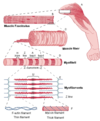
- muscle fasciculus
- muscle fibre
- myofibril
- myosin filament
- F-actin filament
- Titin filaments
- Muscle fasciculus: a bundle of muscle fibres
- Muscle fibre: cylindrical, multinucleated cells composed of many myofibrils (a myocyte)
- Myofibril: The basic rod-like unit of a muscle cell
- Myofilaments: the myofibruls thick, thin and elastic filaments
- Myosin filament: the thick filament
- F-actin filament: the thin filament
- Titin filaments: the elastic filaments that run through the core of each thick filament and anchor it to the Z-line

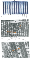
What do the following light/ electron micrographs show. Label the A/I and Z bands


What is there an abundance of in muscles and why?
There is an abundance of mitochondria and also glycogen granules. These help provide energy but are also energy strores (GG)
Label the myofibril…


What band is the mitochondria usually located in, in the myofibril. Why is this the case?
It is usually located in the I band close to the parts of the actin and myosin filament that interact during contractions
Whats the sarcomere?
A basic unit of striated muscle tissue. It is repeating usually between two Z lines
Whats the sarcoplasmic and endoplasmic reticulums main function?
to store calcium
Label the sarcomere…


Tell me the steps to the sliding filament theory aka. muscle contraction?
- ATP binds to myosin. This breaks the cross-bridge between the actin and myosin (ATP-binding decreases the actin-bonding affinity of myosin)
- Myosin hydrolyses ATP –> ADP + Pi (the products stay bound to the myosin). Energy from ATP rotates the myosin head to the cocked position. Myosin then binds weakly to actin via a cross-bridge
- The Ca2+ signal initiates the power stroke. Here tropomyosin moves off the binding site and allows myosin to bind more strongly. Myosin releases Pi
- Myosin releases ADP at the end of the power stroke. The myosin head is bound tightly to the actin in rigor state. The cycle is ready to begin again when more ATP is available
During contraction, what happens to the H, Z, I and A bands?
- H zone becomes smaller
- Z lines get closer together
- I band gets smaller
- A band stays the same length
What two binding sites does a myosin head have?
The actin- binding site
The myosin ATPase site (here ATPase and ATP cand bind)
Whats the rough length of a myosin tail?
100 nm
Tell me the steps to the initiation of contraction?
- Ca2+ levels increase in cytosol. One protein of the complex-troponin C- binds reversibly to Ca2+
- Ca2+ binds to troponin (TN). The calcium-troponin complex pulls tropomyosin completely away from the actin’s myosin-binding site
- Troponin-Ca2+ complex pulls tropomyosin away from actin’s myosin binding site. This enable the myosin heads to form strong, high-force cross-bridges and carry out power strokes
- Myosin binds strongly to actin and completes power stroke. This moves the actin filament
- Actin filament moves. Contractile cycle repeats as long as binding sites are uncovered


