The cardiovascular system Flashcards

1
Q

What’s the role of the plasma membrane?

A

To seperate the intracellular and extracellular components

2
Q

What in the memrbane provides homeostatic control of the intracellular properties?

A

The transmembrane proteins

3
Q

Tell me some advantages of being multicellular?

A
  1. calls can be specialised
  2. organisms can create their own internal environment
  3. Homeostatic control
  4. complexity
4
Q

Tell me some disadvantages of being multicellular?

A
  1. Necrosis
  2. diffusion is slow (high energy use to substances in)
  3. cannot individual sense external environment
  4. many cells need to be coordinated in order to effect a change
  5. communication, intercellular signalling is needed
5
Q

Why do we need a heart?

A
  • diffusion is too slow over great distances to survive on that alone
  • to supply O2 and substrates and remove CO2 and waste products
  • heart helps transport these substances by allowing bulk flow movement of blood
6
Q

What can the process of oxidative phosphorylation produce a lot of? what does it depend on though?

A

ATP from energy and substrates

Provided that oxygen is available as an electron acceptor for mitochondria

7
Q

What has a higher affinity for oxygen, adult or fetal haemoglobin?

A

fetal

8
Q

When does the human heart first start beating?

A

day 20 from conception until death. It beats continuously whilst being formed

9
Q

Tell me the ciruclation of blood through the heart starting at the right atria

A
  • left atrium
  • left AV valve
  • Left ventricle
  • Aortic SL valve
  • arteries of each organ
  • arterioles of each organ
  • capillaries of each organ
  • venules of each organ
  • veins of each organ
  • vena cava
  • right atrium
  • right AV valve
  • right ventricle
  • pulmonary SL valve
  • pulmonary artery
  • lungs arteries
  • lungs arterioles
  • lungs capillaries
  • lungs venules
  • lungs veins
  • pulmonary veins
  • left atrium
10
Q

What are the three major types of cardiac muscle that the heart is composed of?

A
  1. atrial muscles
  2. ventricular muscles
  3. excitatory and conductive muscle fibres
11
Q

What is the tricuspid valve also known as?

A

The atria-ventricular valve

12
Q

Label the heart and its internal structures

A
13
Q

Tell me some key features of the cardiac muscle?

A
  • myogenic
  • inherent pacemaker activity
  • coordinated pattern of contraction
  • striated and has branching
  • syncytial- intercalated discs (gap junctions)
14
Q

What drives the heart rhythm?

A

The sino-artial node in the right atrium

15
Q

What do the gap junctions between cells allow>

A

the spread of excitation

16
Q

What are the steps to the depolarisation of the sarcolemma and then how this leads to muscular contraction?

What stages are known as the E-C coupling stage (excitation-contraction coupling)

A
  1. An impulse travels to the neuromuscular junction on a muscle cell
  2. ACh is released from the axon to receptors located on the sarcolemma
  3. The binding ACh causes depolarisation of the sacolemma by opening ion channnels and allowing Na+ ions into the muscle cells
  4. Na+ ions diffuse into the muscle fibre and depolarisation occurs
  5. depolarisation creates a wave of AP across the sacolemma
  6. AP travels across the sarcolemma and down the T-tubules which triggers the sarcoplasmic reticulium to release Ca2+ through ryanodine receptor channels (RyR)
  7. Ca2+ binds to troponin which removes the blocking action of the tropomyosin from the actin binding sites
  8. myosin is now ready to bind with the actin and from cross-bridges which begins the contraction process
  9. in order to contract, ATP binds to the myosin
  10. ATP is hydrolysed to ADP + Pi, which gives the myosin the energy to move its head to the high-energy position
  11. actin and myosin bind together to form a cross-bridge
  12. the myosin heads then pull the actin filaments inward and release the SDP and Pi and return to a low energy position

The myosin is now ready for more ATP to bind and repeat the cycle (it will continue as long as theres CA2+ ions and ATP available)

The steps in italic are the E-C coupling stages

17
Q

All muscles have an intrinsic rhythm, but why is rhythm said to be controlled by the SA node?

A

the SA node has a faster rhythm so ends up gouverning the entire rhythm of the heart and masks the other cells intronsic rhythm

18
Q

What can modify the rate at the SA node?

A

The nervous system

19
Q

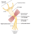
Label this conductive fibre containing the AV node ?

What do the number represent?

A

The numbers represent the interval of time (secs) from the origin of the impulse in the sinus node?
20
Q

Name and conductive fibre and it’s role within the heart?

A

The bundle of His spreads the excitation rapidly through the heart, producing a co-ordinated, bio-mechanically efficient beating of the heart

21
Q

Whats positive charge movement called?

A

An inward current

22
Q

Whats the name of the current that makes the heart beat reguarly>

A

the pacemaker current

23
Q

On an ECG, what does the following represent?

  1. P wave
  2. QRS wave
  3. T wave
A
  1. P wave: where the atria depolarises
  2. QRS wave: when the ventricles depolarise
  3. T wave: the hyperpolarisation of the ventricles
24
Q

Why are valves present and what determines when they open/close?

A

Valves are present so backflow doesnt occur and a change in pressure determines when they open or close.

The presence of valves allow biomechanical efficiency

25
Q

Whats a murmur?

A

When you can hear blood flow through the valves (turbulent flow)

26
Q

Whats meant by isovolumic contraction?

A

When the volume isn’t changing there’s just contraction happening

27
Q

Whats the reason ventricle fill?

A

due to venous return

28
Q

What do the following mean?

  1. End-diasotlic volume
  2. Stroke volume output
  3. End-systolic volume
  4. Ejection fraction
A
  1. End-diastolic volume: normal filling of the ventricles from venous return increases the volume of each ventrcile to about 110 to 120 mls
  2. Stroke volume output: Ventricles contract and empty during systole, and volume decreases ca. 70 mls
  3. End-systolic volume: The remaining volume in each ventricle, about 40 to 50 ml (can’t make volume 0)
  4. Ejection fraction: the fraction of the EDV that is ejected ca. 60%
29
Q

When the heart is contracting strongly, what can the ESV decrease to as little as?

A

10-20 mls

30
Q

Whats the formula to caluclate cardiac output?

A

CO= (EDV - ESV) x HR

31
Q

Why is regulation of the heart required?

A
  1. save enrgy
  2. matching the tissue demand
  3. ventilation perfusion mathcing (lung): blood must flow through the lungs at a rate that the lungs are able to oxygenate it
32
Q

What are the ‘global controls’ of the heart and their subsets?

A

1. ANS

  • parasympathetic NS
  • Sympathetic NS

2. circulatory systems

  • Pre load/ Frank-starling law
  • after-load
33
Q

What are the ‘local contorls’ that can effect the heart rate?

A
  1. Tissue PH
  2. nitric oxide
34
Q

For Autonomic regulation, where is the cardiovascular control centre located?

A

in the medulla oblongata

35
Q

In the ANS, tell me the stages for how the heart rate is increased? and the neurotransmitters involved?

A
  1. The sympathetic input into the heart is via the postganglionic fibres, these innervate the SAN and AVN
  2. the postganglionic fibres release noradrenaline
  3. The noradrenaline acts on Beta1 adrenoreceptors to increase the slope of the pacemaker potential
  4. therefore increasing the heart rate (a positive chronotropic effect)
  5. as well as increasing the force of contraction (a positive inotropic effect)
36
Q

Tell me the steps to how the ANS decreases the heart rate and the neurotransmitters involved in this process?

A
  1. The parasympathetic input into the heart is via the vagus nerve
  2. The vagus nerve forms synapses with postganglionic cells in the SAN and AVN
  3. when stimulated, ACh binds on to M2 receptors (muscarinic receptors)
  4. This decreases the slope of the pacemaker potential leading to a decreased heart rate (a negative chronotropic effect)
37
Q

Whats meant by a chronotropic effect?

A

Something which effects heart rate. either by increasing or decreasing it

38
Q

Whats considered to be ‘normal’ blood pressure?

Which part is the systolic and diastolic?

A

‘normal’ blood pressure is: 120/80 mmHg

systolic (peak) = 120 mmHg

diastolic (minimum)= 80 mmHg

39
Q

In blood vessles, what are the strucutures from inside –> out and what does this include ?

A

inside

Tunica Intima

  • endothelium
  • basement membrane

Tunica media

  • circular smooth muscle
  • elastic fibres
  • connective tissue

Tunia externa

  • connective tissue

Outside

40
Q

What are the 3 types of ciruclation and some facts about pressure and where its found?

A

1. Pulmonary

  • Low pressure
  • Right ventricle –> lung –> left atria

2. peripheral

  • High pressure
  • Left ventricle –> capillary beds –> right atria

3. ​cardiac

  • High pressure
  • Left ventricle –> aorta –> through cardiac muscle –> right atria
41
Q

The pulmonary system has a limited external hydrostatuc pressure and a low pressure, why?

A

so as not to damage the alveoli

42
Q

In the capillary beds, where is the hydrostatic pressure from ?

Where does fluid loss have to be returned to?

A

Hydrostatic pressure from extracellular fluid

Fluid loss has to be returned via the lymphatic system

43
Q

What supplies the following parts of the heart with blood?

  1. Coronary arteries
  2. Right atrium
A
  1. Coronary arteries: from aorta
  2. Right atrium: Coronary vein
44
Q

What is the velocity of blood flow inversely proportional to?

A

The vascular cross-sectional area

45
Q

What does venous return require?

A

musclar activity

46
Q

Whats the mechanism of the Frank-starling law?

What does the degree of ventricular filling determine?

A

The degree of ventricular filling determines the stroke volume

Mechanism: alteted overlap of actin-myosin leading to greater force of contraction- similar to length- tension curve of skeletal muscles

47
Q

Whats meant by the afterload?

A

The effects of aortic pressure on cariac output is minimal in ‘normal range’ but can be significant in hypertension

48
Q

Whats the main thing the ANS effects in the nervous system?

A

It allows the constriction/ relaxation of vasculature

49
Q

What controls the blood flow through a capillary network?

A

it is regulated by the relative contraction of precapillary sphincter surrounding arterioles and metaarterioles

50
Q

Whats Poiseuille’s law and what do the symbols represent?

A
51
Q

Whats the main message of Poiseuille’s law?

A

small changes in arteriole radius produce large changes in flow

52
Q

How does PH effects perfusion?

A

it alters the activity of arterioles which leads to a coupling activity?

53
Q

What does the presence of NO means about perfusion?

A

Its a signal that the tissue isn’t being well perfused

54
Q

What two receptors detect changes in BP/PH?

A

Baroreceptors: detect change in BP

Chemoreceptors: detects chnage in PH (changes in oxygen and carbon dixoide concentration)

55
Q

What stimulates the aortic or carotic baroreceptors?

What effect does this cause?

A

If the BP wihtin the aorta or carotid sinus suddenly increase beyond the set point.

stimulation of these stretch receptors causes the cardiac control centre to increase vagal inhibition of heart, thus slowing down HR and returning the BP to set point

56
Q

What things can cause an increase in BP?

A
  1. exercise
  2. stress
  3. haemorrhage (hypovolemic shock)
57
Q

explain how chemoreceptors detect changes in concentration levels in the blood and the effect it then causes in the body?

A